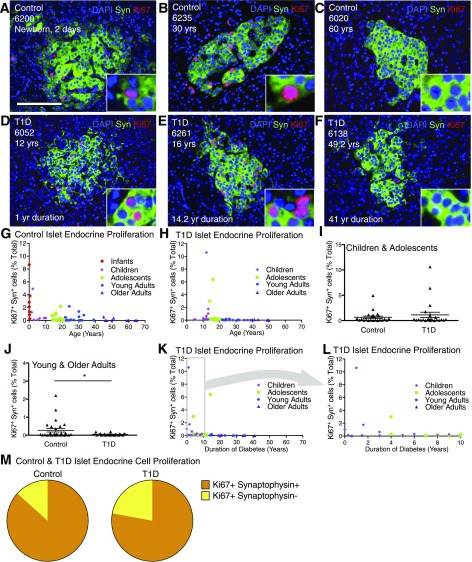
Figure 2.
Islet endocrine cell proliferation is greatly increased in some adolescent and young-adult pancreata. Analysis of islet endocrine cells in control and T1D pancreata. Islet images for control (A–C) and T1D (D–F) stained for DAPI (blue), synaptophysin (Syn) (green), and Ki67 (red). Scale bar: 100 μm. G and H: Quantification of islet endocrine cell proliferation in control (G) and T1D (H) represented by age (years). Data points represent the mean value for each of 59 control and 46 T1D pancreata. Ki67+ synaptophysin+ cells (% total) in control and T1D versus age (years) demonstrates that islet endocrine cell proliferation generally declines with age with notable proliferation in adolescents and young adults. I and J: Ki67+ synaptophysin+ (% total) in T1D was very similar to control child and adolescent pancreata (I) but reduced compared with control young- and older-adult pancreata (J). Results expressed as mean ± SEM for 23 control and 23 T1D children and adolescent subjects and 28 control and 23 young- and older-adult subjects. *P < 0.05. K and L: Islet endocrine cell proliferation vs. T1D duration (years) reveals that most pancreata from T1D individuals exhibited low rates of islet endocrine cell proliferation. M: Ki67+ islet endocrine cells represent a small fraction of the total intra-islet Ki67+ cells in a subset of highly proliferating control (n = 12) and T1D (n = 6) pancreata. yr, year; yrs, years.

