



Abstract
Previous preclinical studies have suggested a close relationship between cerebrovascular disease (CVD) and Alzheimer’s disease. However, a direct correlation between CVD and amyloid burden has not yet been shown in humans. If there is a relationship between CVD and amyloid burden, it is possible that the apolipoprotein E4 (APOE4) genotype may have an effect on this relationship because APOE4 is a risk factor for the development of AD. We therefore evaluated the effects of APOE4 on the relationship between white matter hyperintensities (WMH), a marker of CVD, and amyloid burden, measured by 11C-Pittsburgh compound B (PiB) PET. We recruited 53 patients with subcortical vascular cognitive impairments, who had both WMH on MRI and amyloid deposition assessed by PiB PET. Twenty-two of these patients were APOE4 carriers (41.5%). In the APOE4 non-carriers, a significant positive correlation was shown between the volume of WMH and PiB retention (β = 7.0 10−3, p = 0.034) while no significant correlation was found in APOE4 carriers (β = −9.0 × 10⁻³, p = 0.085). Statistical parametric mapping analyses in APOE4 non-carriers showed that WMH were associated with PiB retention in the bilateral medial occipitotemporal gyrus, cuneus, and superior cerebellum. Our results suggested that WMH are correlated with amyloid burden especially in the posterior brain regions in APOE4 non-carriers. However, this correlation was not observed in APOE4 carriers, perhaps because in these subjects the influence of APOE4 overrides the effect of CVD.
Keywords: Alzheimer’s disease, amyloid burden, apolipoprotein E4, cerebrovascular disease
INTRODUCTION
Subcortical vascular cognitive impairment (SVCI) refers to cognitive impairments due to cerebrovascular disease (CVD), which consists of subcortical vascular dementia (SVaD) and subcortical vascular mild cognitive impairment (svMCI) [1]. Pathological studies, however, demonstrated that patients who had been clinically diagnosed with SVaD often proved to have comorbid Alzheimer’s disease (AD) pathologies [2–4]. A study by our group using Pittsburgh compound-B (PiB) PET, a sensitive method to detect amyloid plaque burden during life [5], also showed that over 30% of SVaD patients turned out to be PiB-positive [6].
In epidemiologic studies, there is increasing evidence that CVD and AD dementia are strongly associated [7–10]. Some preclinical studies also suggest that CVD may directly induce amyloid burden [11]. Therefore, it has been suggested that CVD may accelerate the development of AD [12]. There are two possible mechanisms that could explain the growing evidence linking CVD and AD dementia. The first is that CVD accelerates the deposition of brain amyloid, which leads to AD dementia. The second possible interpretation is that CVD produces brain damage, which would reduce cognitive reserve and thus make the subjects more susceptible to the effects of AD pathology. One recent study of human subjects was unable to demonstrate any direct correlation between CVD and amyloid burden [13]. This result would be most consistent with the second mechanism (described above) that CVD lowers cognitive reserve without having a direct effect on development of brain amyloid pathology. However, this previous study did not consider the effect of the apolipoprotein E ε4 (APOE4) genotype on the relationship between CVD and brain amyloid. The APOE4 genotype is considered a major risk factor for AD and is thought to lower the age of onset for the development of AD and accelerate the aggregation of amyloid plaques [14]. Some studies also showed that APOE4 is associated with CVD, although their results were inconsistent [15–18]. Thus, it is possible that the APOE4 genotype might have an effect on the relationship between CVD and amyloid burden.
The current study aimed to examine the effects of APOE4 on the relationship between CVD, which was quantified as white matter hyperintensities (WMH) on fluid-attenuated inversion recovery (FLAIR) MRI, and brain amyloid, which was measured by PiB PET imaging. We evaluated the relationship between WMH and amyloid burden in APOE4 carriers and non-carriers, respectively. We hypothesized that there may be a correlation between the two pathologies affected by the APOE4 genotype status.
MATERIALS AND METHODS
Participants
A total of 136 SVCI patients were consecutively recruited at Samsung Medical Center from September 2008 to August 2011. Patients with SVaD met the diagnostic criteria for vascular dementia as determined by the Diagnostic and Statistical Manual of Mental Disorders-Fourth Edition (DSM-IV). DSM-IV criteria include the presence of focal signs suggestive of CVD; we defined the presence of focal signs as at least two focal neurologic signs out of corticobular, corticospinal, extrapyramidal signs and gait abnormalities. All SVaD patients had a significant ischemia on their MRI scans, which was defined as a volume of WMH ≥15 cm3. This cutoff value of 15 cm3 was the smallest volume of WMH in patients who met grade 3 modified Fazekas ischemia criteria. A prior study suggested that WMH volume measurement might be more sensitive than visual scores [19]. Patients with svMCI were diagnosed using the Petersen criteria [20] with the following modifications: 1) there was subjective cognitive complaint by the patient or his/her caregiver; 2) normal Activity of Daily Living (ADL) score by clinically and ADL scale (Seoul Instrumental ADL score below seven, which is a modified version of Lawton’s 1969 Instrumental ADL) [21, 22]; 3) the patient showed an objective memory decline below the 16th percentile on neuropsychological tests [1, 23, 24]; 4) not having dementia; and 5) presence of a subcortical vascular feature defined as both focal neurological symptom or sign and the significant ischemia on MRI, as in the SVaD. We excluded patients with other structural lesions on brain MRI such as territorial infarction, intracranial hemorrhage, traumatic brain injury, hydrocephalus, or WMH associated with radiation, multiple sclerosis, or vasculitis. The number of patients with svMCI and SVaD were 59 (43.4%) and 77 (56.6%), respectively.
Patients with SVCI were classified as PiB-positive (PiB+) or PiB-negative (PiB−) if they had a global PiB retention ratio greater than or less than 1.5, respectively [6]. Among the 136 patients with SVCI, 53 (39.0%) were positive for PiB retention, while 83 (61.0%) were negative. Since our aim was to investigate the relationship between the two pathologies, we considered that subjects that have only one of the two pathologies may be inappropriate for our study goal. Therefore, we only included, from our SVCI cohort, the SVCI patients with PiB-positive scan. Among the 53 patients who were PiB+, 58.5% were APOE4 non-carriers (n = 31: ε2/ε3, n = 6; ε3/ε3, n = 25), and 41.5% were APOE4 carriers (n = 22: ε3/ε4, n = 18; ε4/ε4, n = 4).
All patients completed a clinical interview and neurological examination, blood tests, and APOE genotype as described previously [6], and MRI scans at Samsung Medical Center using the same scanner. All patients also completed a three-step diagnostic process. First, patients completed a medical interview conducted by an experienced neurologist, who obtained a medical history, a history of cognitive, behavioral, and functional impairments, and performed neurological examinations, including the Mini-Mental Status Exam (MMSE), Clinical Dementia Rating Sum of Boxes (CDR-SOB), and Geriatric Depression Scale (GDS). The interview also included items to assess patients’ abilities to engage in activities of daily living. Second, the neuropsychology team performed a number of neuropsychological tests and conducted a clinical interview for cognitive, behavioral, and functional impairments using semi-structured questionnaires. The scales for Neuropsychiatric Inventory and Activities of Daily Living (ADL) scales were completed. The ADL scale used in this study was Seoul Instrumental ADL score, which is a modified version of Lawton’s Instrumental ADL [21, 22]. Third, patients were diagnosed based on the results of all the diagnostic tests (neuropsychological reports, blood tests, and MRIs). Blood tests included a complete blood count, blood chemistry test, vitamin B12/folate measure, syphilis serology, thyroid functioning tests, and APOE genotyping. We obtained a written consent from each patient and the Institutional Review Board of the Samsung Medical Center approved the study protocol.
Neuropsychological tests
All patients underwent neuropsychological testing using the Seoul Neuropsychological Screening Battery (SNSB) [25, 26]. This battery contains assessments of attention, language abilities, praxis, four elements of Gerstmann syndrome, visuospatial functioning, verbal and visual memory, and frontal/executive functioning. Among these subtests, the quantitatively scorable tests, including digit span (forward and backward), the Korean version of the Boston Naming Test (K-BNT) [27], the Rey-Osterrieth Complex Figure Test (RCFT; copying, immediate and 20-min delayed recall, and recognition), Seoul Verbal Learning Test (SVLT; three learning-free recall trials of 12 words, a 20-minute delayed recall trial for these 12 items, and a recognition test), phonemic and semantic Controlled Oral Word Association Test (COWAT), and a Stroop Test (word and color reading of 112 items during a 2-min period) were used in current study.
MR imaging and analysis
T2, T1, three-dimensional (3D) FLAIR, and T2 fast field echo (FFE) images were acquired for all 53 subjects at Samsung Medical Center using the same 3.0 T MRI scanner (Philips 3.0T Achieva). The MRI scanning protocol was the same as described previously [24].
Measurement of regional WMH volume
We quantified WMH volume (in cm3) on FLAIR images using an automated method as previously described [28]. First, we extracted the WMH candidate regions on FLAIR images applying classification method and morphological operation to T1-weighted images. Second, in order to extract WMH, a threshold method was applied to the FLAIR images within the WMH candidate regions. Even though the threshold value was selected considering the range of image intensities, segmented results could contain false positive or false negative regions depending on the extent of WMH. If the results contained an error, the threshold value was reselected through visual inspection by two raters, and they reached a consensus in the case of discrepancy. The rate of agreement between two neurologists was 92.3%.
[11C] PiB-PET imaging
All patients completed a standardized [11C] PiB-PET scan spanning the entire brain at Samsung Medical Center or Asan Medical Center using a Discovery STe PET/CT scanner (GE Medical Systems, Milwaukee, WI) in 3-dimensional scanning mode that examined 35 slices, each 4.25-mm thick. First, the 11C-PiB was injected into an antecubital vein as a bolus with a mean dose of 420 MBq (range 259–550 MBq). Sixty minutes after the injection, a CT scan was performed for attenuation correction. Afterwards, the 30-minute emission static PET scan was then initiated [6].
Data analysis of [11C] PiB-PET images
PiB PET images were co-registered to each individual’s MRIs, which were normalized to a T1-weighted MRI template. Using these parameters, MRI co-registered PiB PET images were normalized to the MRI template. The quantitative regional values of PiB retention on the spatially normalized PiB images were obtained by an automated volume of interest (VOI) analysis tool using the automated anatomical labeling atlas. Data processing was performed using SPM version 8 (SPM8) through Matlab 6.5 (Mathworks, Natick, MA, USA).
To measure PiB retention, we used the cerebral cortical region to cerebellum uptake ratio which is identical to the standardized uptake value ratios (SUVRs). The cerebellum was used as a reference region as it did not show group differences. We selected 28 cortical VOIs from left as well as right hemispheres using the automated anatomical labeling atlas. The cerebral cortical VOIs which were chosen for this study consisted of the following areas: bilateral frontal (superior and middle frontal gyri, medial part of superior frontal gyrus, opercular part of inferior frontal gyrus, triangular part of inferior frontal gyrus, supplementary motor area, orbital part of superior, middle, and inferior orbital frontal gyri, rectus and olfactory cortex), posterior cingulate gyri, parietal (superior and inferior parietal, supramarginal and angular gyri, and precuneus), lateral temporal (superior, middle and inferior temporal gyri, and heschl gyri), and occipital (superior, middle, and inferior occipital gyri, cuneus, calcarine fissure, and lingual and fusiform gyri). Regional cerebral cortical SUVRs were calculated by dividing each cortical VOI’s SUV by the mean SUV of the cerebellar cortex (cerebellum crus1 and crus2). The global PiB retention ratio was calculated from the volume-weighted averages of the SUVRs of the bilateral cerebral cortical VOIs. We defined the PiB retention ratio as a continuous variable.
Statistical analysis
Comparisons of demographic and clinical data between APOE4 non-carriers and carriers were conducted using Student’s t test for normally distributed variables, and the Mann-Whitney U test for non-normally distributed variables. Categorical variables were evaluated using a χ2 test. In order to examine the relationship between WMH and PiB retention ratio, multiple linear regression analyses were performed using WMH as an independent factor and the global PiB retention ratio as a dependent variable with adjustment for age and gender. Assumptions of residual about normality, homoscedasticity, and independence were confirmed. Statistical significance was set at p < 0.05. Statistical analyses were conducted using PASW Statistics 18 (SPSS Inc, Chicago, IL, USA) software.
In order to examine the relationship between WMH and PiB retention and to determine the regions where this relationship was significant, a voxel-based statistical analysis of the PiB images was performed using the Statistical Parametric Mapping program, version 8 (SPM8), and Matlab 6.5 for Windows (Math Works, Natick, MA, USA). An SPM regression analysis was performed without global normalization, since the 11C-PiB PET images had been normalized to the cerebellar ROI PiB binding. Multivariate regression analysis was performed using WMH as a predictor and the PiB retention ratio at each voxel as a dependent variable after adjusting for age and gender. Red or yellow colored area showed the statistically significant region with increased global PiB retention as the volume of WMH increased with adjustment for age and gender. We defined statistical significance as p < 0.001 (uncorrected for multiple comparisons).
We generated group-specific gray matter mask using T1-weighted images in order to exclude non-gray matter areas from the PiB images. We used classification method and chose the threshold of 0.5 to binarize gray matter probability map for the mask. Results were overlaid to a T1 weighted MRI of a representative male individual template provided by SPM.
RESULTS
Baseline characteristics of the subjects
Demographic characteristics and imaging findings of the 53 patients are presented in Table 1. There were no differences in demographics or severity of cognitive impairment between APOE4 non-carriers and carriers. There were also no differences in the median volume of WMH or mean global PiB SUVR between the two APOE groups.
Table 1.
Demographic characteristics and imaging findings in study population
| APOE4 Non-carriers | APOE4 Carriers | p value‖ | |
|---|---|---|---|
| Number | 31 | 22 | |
| Mild cognitive impairment* | 11 (35.5%) | 8 (36.4%) | >0.999 |
| Age† | 78.39 ± 5.14 | 76.41 ± 5.83 | 0.197 |
| Male, N (%)* | 13 (41.9%) | 6 (27.3%) | 0.420 |
| Education (year)† | 9.65 ± 6.21 | 9.91 ± 5.15 | 0.871 |
| K-MMSE† | 20.87 ± 5.78 | 22.59 ± 5.70 | 0.288 |
| CDR SOB‡ | 3.50 (1.50–7.00) | 3.50(1.50–6.00) | 0.950 |
| Hypertension* | 17 (54.8%) | 18 (81.8%) | 0.080 |
| Diabetes mellitus* | 7 (22.6%) | 6 (27.3%) | 0.946 |
| Dyslipidemia* | 6 (19.4%) | 7 (31.8%) | 0.475 |
| Coronary artery disease§ | 5 (16.1%) | 3 (13.6%) | >0.999 |
| Stroke history§ | 5 (16.1%) | 3 (13.6%) | >0.999 |
| WMH volume (cm3)‡ | 36.02 (24.34–49.93) | 33.77 (21.94–45.94) | 0.626 |
| Microbleed prevalence* | 18(58.1%) | 11 (50.0%) | 0.561 |
| Microbleed number‡ | 1.0 (0–4.0) | 0.5 (0–4.0) | 0.812 |
| Global PiB retention ratio† | 2.07 ± 0.38 | 2.10 ± 0.38 | 0.760 |
Data are presented as mean ± standard deviation for normally distributed and median (25th–75th percentile, interquartile range) for non-normally distributed variables.
χ2 test with Yates’ continuity correction was used.
Student’s t test was used for normally distributed variables.
Mann-Whitney U test was used for non-normally distributed variables.
Fischer exact test was used.
p-value for the comparison of the APOE4 non-carrier group and APOE4 carrier group. K-MMSE, Korean version of the Mini-Mental State Examination; CDR SOB, Clinical Dementia Rating Sum of Box; WMH, white matter hyperintensities.
Correlations between WMH and global PiB retention ratio in APOE4 non-carriers and carriers
In the whole PiB+cohort, there was no correlation between WMH volume and cortical PiB retention (unstandardized coefficient β = 4.0 × 10⁻³, SE (β) = 3.0 × 10⁻³, p = 0.175).
In the APOE4 non-carriers, a significant positive correlation was shown between the volume of WMH and cortical PiB retention (β = 7.0 × 10⁻³, SE(β) = 3.0 × 10⁻³, p = 0.034). However, no positive correlation was found in APOE4 allele carriers (β = −9.0 × 10⁻³, SE (β) = 5.0 × 10⁻³, p = 0.085) (Fig. 1).
Fig. 1.

Scatter plot graph between the volume of white matter hyperintensities (WMH) (cm3) and global PiB retention ratio. Multiple linear regression analyses were performed using WMH as an independent factor and the global PiB retention ratio as a dependent variable with adjustment for age and gender. A) Positive correlation with statistical significance was shown between WMH volumes and PiB retention areas in APOE4 non-carrier (β = 7.0 × 10⁻³, SE(β) = 3.0 × 10⁻³, p = 0.034). B) Significant correlation was not shown between WMH volumes and PiB retention in APOE4 carrier (β = −9.0 × 10⁻³, SE(β) = 5.0 × 10⁻³, p = 0.085).
To evaluate the effect modification of APOE4 status and WMH volume on amyloid burden, multiple linear regressions were performed in all APOE4 carriers and non-carriers (n = 53 for total APOE4 carriers and non-carriers). We used age, gender, WMH, APOE4 status, and the interaction terms (APOE4 status*WMH volume) as independent variables, and PiB retention ratio as the dependent variable. There tended to be interactions between APOE4 status and WMH volume (p = 0.090).
Regional correlation between WMH and cortical PiB retention in APOE4 non-carriers and carriers
In APOE4 non-carriers, WMH volume was positively correlated with PiB retention in the bilateral medial occipitotemporal gyrus, cuneus, and the superior part of the cerebellum such as culmen, declive, anterior quadrangular lobule (uncorrected p < 0.001) (Fig. 2). However, after false discovery rate (FDR) correction, the statistical significance disappeared in these regions.
Fig. 2.
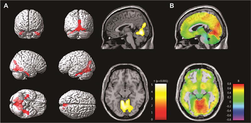
SPM multiple regression analysis of PiB retention in the APOE4 non-carriers. A) t value map: The red or yellow colored regions represents the significant positive correlation between white matter hyperintensities volumes and PiB retention ratio with adjustment for age and gender (uncorrected p < 0.001). B) Beta map: Strength of associations is illustrated with color-labeled standardized beta-values.
In APOE4 carriers, there were no regions showing the significant correlation between WMH volumes and PiB retention ratio. However, WMH volumes tend to have a negative correlation with PiB retention ratio in the right dorsolateral frontal, precuneus, and bilateral parietotemporal areas (uncorrected p < 0.01) (Fig. 3).
Fig. 3.

SPM multiple regression analysis of PiB retention in the APOE4 carriers. There were no regions showing the significant correlation between WMH volumes and PiB retention ratio. A) t value map: The blue or sky blue colored regions represent the negative correlation between WMH and PiB retention ratio with adjustment for age and gender (uncorrected p < 0.01). B) Beta map: Strength of associations is illustrated with color-labeled standardized beta-values.
DISCUSSION
Our major findings were as follows. First, there was a significant positive relationship between the volume of WMH and PiB retention in the APOE4 non-carriers, whereas no significant analogous relationship was seen in APOE4 carriers. Second, the brain regions where WMH correlated with PiB retention were in the temporal, occipital, and superior cerebellar regions. Our findings suggest that WMH is associated with amyloid burden, especially in the posterior brain regions of APOE4 non-carriers with PiB (+) SVCI. These findings raise the possibility that APOE4 interacts with the pathogenesis of amyloid deposition in the presence of CVD. Possible mechanisms are discussed below.
Our first major finding of a positive correlation between the volume of WMH and PiB retention ratio in APOE4 non-carriers is consistent with many epidemiologic studies demonstrating an association between CVD and AD pathology [7–9, 12, 29]. Furthermore, a recent preclinical study showed the direct relationship between ischemic lesion and amyloid burden [11]. A previous study also suggested that patients with stroke had increased PiB retention in the peri-infarct region [30]. In contrast, one PiB PET study, based on healthy subjects with normal cognition, showed there was no correlation between the two pathologies [13], but this study did not factor in the effect of the APOE4 genotype. Indeed, in the present study, we found no correlation between WMH and amyloid burden in the carriers and non-carriers combined. Another reason for the discrepancy between previous findings and our current findings may be the difference in the subject population. Our study included MCI and dementia patients with greater amounts of WMH and brain amyloid in comparison with the previous asymptomatic community subjects [13] with lower amounts of WMH and brain amyloid. Our finding might be supported by a more recent study showing that baseline WMH was positively correlated with the progression of amyloid burden over 2 years [31]. The mechanisms underpinning the positive correlation between WMH and PiB retention remain unclear. However, it is possible that these two factors interact with each other (interactive hypothesis). One hypothesis is that CVD, reflected by WMH can block the clearance of amyloid via perivascular lymphatic drainage [31–33]. Alternatively, amyloid deposition in small or medium sized arterioles can lead to amyloid angiopathy, which may also result in severe WMH [34, 35]. However, since both AD and vascular pathologies usually occur in the elderly and share risk factors such as hypertension, diabetes, and hyperlipidemia, it is also possible that both pathologies can occur independently by chance [12, 29, 36].
Our second major finding was that WMH were associated with amyloid burden predominantly in the temporal, occipital and superior cerebellar regions, which are not typically affected by amyloid burden in AD [37]. This finding is similar to a recent report that amyloid deposition occurred predominantly in the occipital areas and cerebellum in patients with carotid stenosis [38]. Our finding is also consistent with another previous study showing that baseline WMH was associated with progression of amyloid burden in the parieto-occipital region [31]. The reason why WMH were correlated with amyloid burden in the posterior brain region in APOE4 non-carriers remains unclear. We suggest that there are at least two possibilities to explain the relationship between WMH and amyloid: (1) the distribution corresponds to the posterior circulation, which is supplied by vertebrobasilar system; [39] or (2) it is related to the topography of cerebral amyloid angiopathy (CAA). The posterior circulation may be vulnerable to injury and dysfunction of the endothelium leading to blood-brain barrier (BBB) disruption [40]. For example, patients with hypertensive encephalopathy have vasculopathic changes predominantly in the posterior circulation areas. Furthermore, emerging evidence suggests that BBB disruption may contribute to the development of AD [33, 41–44], possibly through increased blockage of amyloid clearance. Therefore, it is possible that CVD may result in preferential BBB disruption in the posterior circulation, which may induce increased amyloid deposition in these areas. Alternatively, the association between WMH and amyloid burden might be related to CAA, which involves predominantly the occipital and temporal regions [34, 45, 46].
We found no correlation between WMH and PiB retention in APOE4 carriers, similar to a previous report that did not account for APOE4 [13]. Although animal studies have shown that APOE4 is associated with enhanced amyloid-β aggregation [47, 48] and reduced amyloid clearance [14, 42, 49], resulting in amyloid-β deposition, the mechanism by which APOE4 promotes earlier age of amyloid deposition in humans is not known. Amyloid imaging studies have repeatedly shown that age and APOE4 status are the primary predictors of brain amyloid load. Therefore, we suggest that the powerful effects of APOE4 on brain amyloid override the effects of CVD (WMH) on brain amyloid in APOE4 positive subjects. Alternatively, it might be related to the explanation that SVCI patients with severe WMH have less amyloid burden than SVCI patients with moderate WMH because both WMH and amyloid burden causes cognitive impairments. Indeed, there was a trend displaying negative correlation between WMH and amyloid burdens in APOE4 carriers.
There are some limitations to our study. First, we were unable to test the hypothesis of a causal relationship between ischemia and amyloid burden because this was a cross-sectional study. Longitudinal studies with serial MRI and PiB PET would be able to examine this issue. Second, all of the patients in this study had cognitive impairment, which may limit the generalizability of the results. However, since both CVD and amyloid pathology are related to cognitive impairment, our population is representative of an important clinical manifestation of these processes. Third, we were unable to differentiate the parenchymal amyloid from vascular amyloid using PiB-PET. However, in this study, there were no subjects who met the clinical criteria for CAA [50] or showed restricted lobar microbleeds. Finally, after corrections for multiple comparisons with FDR, significant associations in the SPM analysis disappeared. However, to reduce the chances of missing important associations during the early stage of analysis, we used a statistical significance level of 0.001 without correction for multiple comparisons.
Nevertheless, this study is noteworthy because it is the first human study demonstrating the correlation of severity of small vessel related ischemia and amyloid burden in APOE4 non-carriers, especially in the posterior cortical regions. Further investigation on the interaction of CVD and amyloid deposition may provide clues to treatment or prevention strategies for dementia.
Acknowledgments
This study was supported by Basic Research Program through the National Research Foundation of Korea (NRF) funded by the Ministry of Education (NRF-2013R1A1A2065365), the Korean Healthcare Technology R&D Project Ministry for Health & Welfare Affairs, Republic of Korea (HI10C2020 & HIC120713), by the KOSEF NRL program grant (MEST; 2011-0028333), by Samsung Medical Center (CRL-108011&CRS110-14-1), and by the Converging Research Center Program through the Ministry of Science, ICT and Future Planning, Korea (2013K000338).
Footnotes
Handling Associate Editor: Josephine Barnes
Authors’ disclosures available online (http://www.j-alz.com/disclosures/view.php?id=2075).
References
- 1.Seo SW, Ahn J, Yoon U, Im K, Lee JM, Tae Kim S, Ahn HJ, Chin J, Jeong Y, Na DL. Cortical thinning in vascular mild cognitive impairment and vascular dementia of subcortical type. J Neuroimaging. 2010;20:37–45. doi: 10.1111/j.1552-6569.2008.00293.x. [DOI] [PubMed] [Google Scholar]
- 2.Schneider JA, Arvanitakis Z, Bang W, Bennett DA. Mixed brain pathologies account for most dementia cases in community-dwelling older persons. Neurology. 2007;69:2197–2204. doi: 10.1212/01.wnl.0000271090.28148.24. [DOI] [PubMed] [Google Scholar]
- 3.Chui HC, Zarow C, Mack WJ, Ellis WG, Zheng L, Jagust WJ, Mungas D, Reed BR, Kramer JH, Decarli CC, Weiner MW, Vinters HV. Cognitive impact of subcortical vascular and Alzheimer’s disease pathology. Ann Neurol. 2006;60:677–687. doi: 10.1002/ana.21009. [DOI] [PMC free article] [PubMed] [Google Scholar]
- 4.Jagust WJ, Zheng L, Harvey DJ, Mack WJ, Vinters HV, Weiner MW, Ellis WG, Zarow C, Mungas D, Reed BR, Kramer JH, Schuff N, DeCarli C, Chui HC. Neuropathological basis of magnetic resonance images in aging and dementia. Ann Neurol. 2008;63:72–80. doi: 10.1002/ana.21296. [DOI] [PMC free article] [PubMed] [Google Scholar]
- 5.Klunk WE, Engler H, Nordberg A, Wang Y, Blomqvist G, Holt DP, Bergstrom M, Savitcheva I, Huang GF, Estrada S, Ausen B, Debnath ML, Barletta J, Price JC, Sandell J, Lopresti BJ, Wall A, Koivisto P, Antoni G, Mathis CA, Langstrom B. Imaging brain amyloid in Alzheimer’s disease with Pittsburgh Compound-B. Ann Neurol. 2004;55:306–319. doi: 10.1002/ana.20009. [DOI] [PubMed] [Google Scholar]
- 6.Lee JH, Kim SH, Kim GH, Seo SW, Park HK, Oh SJ, Kim JS, Cheong HK, Na DL. Identification of pure subcortical vascular dementia using 11C-Pittsburgh compound B. Neurology. 2011;77:18–25. doi: 10.1212/WNL.0b013e318221acee. [DOI] [PubMed] [Google Scholar]
- 7.Hofman A, Ott A, Breteler MM, Bots ML, Slooter AJ, van Harskamp F, van Duijn CN, Van Broeckhoven C, Grobbee DE. Atherosclerosis, apolipoprotein E, and prevalence of dementia and Alzheimer’s disease in the Rotterdam Study. Lancet. 1997;349:151–154. doi: 10.1016/S0140-6736(96)09328-2. [DOI] [PubMed] [Google Scholar]
- 8.Roher AE, Tyas SL, Maarouf CL, Daugs ID, Kokjohn TA, Emmerling MR, Garami Z, Belohlavek M, Sabbagh MN, Sue LI, Beach TG. Intracranial atherosclerosis as a contributing factor to Alzheimer’s disease dementia. Alzheimers Dement. 2011;7:436–444. doi: 10.1016/j.jalz.2010.08.228. [DOI] [PMC free article] [PubMed] [Google Scholar]
- 9.Shah NS, Vidal JS, Masaki K, Petrovitch H, Ross GW, Tilley C, DeMattos RB, Tracy RP, White LR, Launer LJ. Midlife blood pressure, plasma beta-amyloid, and the risk for Alzheimer disease: The Honolulu Asia Aging Study. Hypertension. 2012;59:780–786. doi: 10.1161/HYPERTENSIONAHA.111.178962. [DOI] [PMC free article] [PubMed] [Google Scholar]
- 10.Schmidt R, Ropele S, Enzinger C, Petrovic K, Smith S, Schmidt H, Matthews PM, Fazekas F. White matter lesion progression, brain atrophy, and cognitive decline: The Austrian stroke prevention study. Ann Neurol. 2005;58:610–616. doi: 10.1002/ana.20630. [DOI] [PubMed] [Google Scholar]
- 11.Garcia-Alloza M, Gregory J, Kuchibhotla KV, Fine S, Wei Y, Ayata C, Frosch MP, Greenberg SM, Bacskai BJ. Cerebrovascular lesions induce transient beta-amyloid deposition. Brain. 2011;134:3697–3707. doi: 10.1093/brain/awr300. [DOI] [PMC free article] [PubMed] [Google Scholar]
- 12.Petrovitch H, White LR, Izmirilian G, Ross GW, Havlik RJ, Markesbery W, Nelson J, Davis DG, Hardman J, Foley DJ, Launer LJ. Midlife blood pressure and neuritic plaques, neurofibrillary tangles, and brain weight at death: The HAAS. Honolulu-Asia aging Study. Neurobiol Aging. 2000;21:57–62. doi: 10.1016/s0197-4580(00)00106-8. [DOI] [PubMed] [Google Scholar]
- 13.Marchant NL, Reed BR, DeCarli CS, Madison CM, Weiner MW, Chui HC, Jagust WJ. Cerebrovascular disease. beta-amyloid, and cognition in aging. Neurobiol Aging. 2012;33:1006 e1025–e1036. doi: 10.1016/j.neurobiolaging.2011.10.001. [DOI] [PMC free article] [PubMed] [Google Scholar]
- 14.Castellano JM, Kim J, Stewart FR, Jiang H, DeMattos RB, Patterson BW, Fagan AM, Morris JC, Mawuenyega KG, Cruchaga C, Goate AM, Bales KR, Paul SM, Bateman RJ, Holtzman DM. Human apoE isoforms differentially regulate brain amyloid-beta peptide clearance. Sci Transl Med. 2011;3:89ra57. doi: 10.1126/scitranslmed.3002156. [DOI] [PMC free article] [PubMed] [Google Scholar]
- 15.Altamura C, Squitti R, Pasqualetti P, Tibuzzi F, Silvestrini M, Ventriglia MC, Cassetta E, Rossini PM, Vernieri F. What is the relationship among atherosclerosis markers, apolipoprotein E polymorphism and dementia? Eur J Neurol. 2007;14:679–682. doi: 10.1111/j.1468-1331.2007.01714.x. [DOI] [PubMed] [Google Scholar]
- 16.Lahoz C, Schaefer EJ, Cupples LA, Wilson PW, Levy D, Osgood D, Parpos S, Pedro-Botet J, Daly JA, Ordovas JM. Apolipoprotein E genotype and cardiovascular disease in the Framingham Heart Study. Atherosclerosis. 2001;154:529–537. doi: 10.1016/s0021-9150(00)00570-0. [DOI] [PubMed] [Google Scholar]
- 17.Graner M, Kahri J, Varpula M, Salonen RM, Nyyssonen K, Jauhiainen M, Nieminen MS, Syvanne M, Taskinen MR. Apolipoprotein E polymorphism is associated with both carotid and coronary atherosclerosis in patients with coronary artery disease. Nutr Metab Cardiovasc Dis. 2008;18:271–277. doi: 10.1016/j.numecd.2007.01.003. [DOI] [PubMed] [Google Scholar]
- 18.Kim HJ, Ye BS, Yoon CW, Cho H, Noh Y, Kim GH, Choi YS, Kim JH, Jeon S, Lee JM, Kim JS, Choe YS, Lee KH, Kim ST, Kim C, Kang DR, Ki CS, Lee JH, Werring DJ, Weiner MW, Na DL, Seo SW. Effects of APOE epsilon4 on brain amyloid, lacunar infarcts, and white matter lesions: A study among patients with subcortical vascular cognitive impairment. Neurobiol Aging. 2013;34:2482–2487. doi: 10.1016/j.neurobiolaging.2013.05.009. [DOI] [PubMed] [Google Scholar]
- 19.van Straaten EC, Fazekas F, Rostrup E, Scheltens P, Schmidt R, Pantoni L, Inzitari D, Waldemar G, Erkinjuntti T, Mantyla R, Wahlund LO, Barkhof F. Impact of white matter hyperintensities scoring method on correlations with clinical data: The LADIS study. Stroke. 2006;37:836–840. doi: 10.1161/01.STR.0000202585.26325.74. [DOI] [PubMed] [Google Scholar]
- 20.Petersen RC. Mild cognitive impairment as a diagnostic entity. J Intern Med. 2004;256:183–194. doi: 10.1111/j.1365-2796.2004.01388.x. [DOI] [PubMed] [Google Scholar]
- 21.Ku HM, Kim JH, Kwon EJ, Kim SH, Lee HS, Ko HJ, Jo S, Kim DK. A study on the reliability and validity of Seoul-Instrumental Activities of Daily Living(S-IADL) J Korean Neuropsychiatr Assoc. 2004;43:189–199. [Google Scholar]
- 22.Lawton MP, Brody EM. Assessment of older people: Self-maintaining and instrumental activities of daily living. Gerontologist. 1969;9:179–186. [PubMed] [Google Scholar]
- 23.Chin J, Seo SW, Kim SH, Park A, Ahn HJ, Lee BH, Kang SJ, Na DL. Neurobehavioral dysfunction in patients with subcortical vascular mild cognitive impairment and subcortical vascular dementia. Clin Neuropsychol. 2012;26:224–238. doi: 10.1080/13854046.2012.658865. [DOI] [PubMed] [Google Scholar]
- 24.Seo SW, Lee JM, Im K, Park JS, Kim SH, Kim ST, Ahn JH, Kim MJ, Kim GH, Kim JH, Roh JH, Cheong HK, Na DL. Cardiovascular risk factors cause cortical thinning in cognitively impaired patients: Relationships among cardiovascular risk factors, white matter hyperintensities, and cortical atrophy. Alzheimer Dis Assoc Disord. 2012;26:106–112. doi: 10.1097/WAD.0b013e31822e0831. [DOI] [PubMed] [Google Scholar]
- 25.Ahn HJ, Chin J, Park A, Lee BH, Suh MK, Seo SW, Na DL. Seoul Neuropsychological Screening Battery-dementia version (SNSB-D): A useful tool for assessing and monitoring cognitive impairments in dementia patients. J Korean Med Sci. 2010;25:1071–1076. doi: 10.3346/jkms.2010.25.7.1071. [DOI] [PMC free article] [PubMed] [Google Scholar]
- 26.Ahn HJ, Seo SW, Chin J, Suh MK, Lee BH, Kim ST, Im K, Lee JM, Lee JH, Heilman KM, Na DL. The cortical neuroanatomy of neuropsychological deficits in mild cognitive impairment and Alzheimer’s disease: A surface-based morphometric analysis. Neuropsychologia. 2011;49:3931–3945. doi: 10.1016/j.neuropsychologia.2011.10.010. [DOI] [PubMed] [Google Scholar]
- 27.Kim H, Na DL. Normative data on the Korean version of the Boston Naming Test. J Clin Exp Neuropsychol. 1999;21:127–133. doi: 10.1076/jcen.21.1.127.942. [DOI] [PubMed] [Google Scholar]
- 28.Jeon S, Yoon U, Park JS, Seo SW, Kim JH, Kim ST, Kim SI, Na DL, Lee JM. Fully automated pipeline for quantification and localization of white matter hyperintensity in brain magnetic resonance image. Int J Imaging Syst Technol. 2011;21:193–200. [Google Scholar]
- 29.Pappolla MA, Bryant-Thomas TK, Herbert D, Pacheco J, Fabra Garcia M, Manjon M, Girones X, Henry TL, Matsubara E, Zambon D, Wolozin B, Sano M, Cruz-Sanchez FF, Thal LJ, Petanceska SS, Refolo LM. Mild hypercholesterolemia is an early risk factor for the development of Alzheimer amyloid pathology. Neurology. 2003;61:199–205. doi: 10.1212/01.wnl.0000070182.02537.84. [DOI] [PubMed] [Google Scholar]
- 30.Ly JV, Rowe CC, Villemagne VL, Zavala JA, Ma H, Sahathevan R, O’Keefe G, Gong SJ, Gunawan R, Churilov L, Saunder T, Ackerman U, Tochon-Danguy H, Donnan GA. Subacute ischemic stroke is associated with focal 11C PiB positron emission tomography retention but not with global neocortical Abeta deposition. Stroke. 2012;43:1341–1346. doi: 10.1161/STROKEAHA.111.636266. [DOI] [PubMed] [Google Scholar]
- 31.Grimmer T, Faust M, Auer F, Alexopoulos P, Forstl H, Henriksen G, Perneczky R, Sorg C, Yousefi BH, Drzezga A, Kurz A. White matter hyperintensities predict amyloid increase in Alzheimer’s disease. Neurobiol Aging. 2012;33:2766–2773. doi: 10.1016/j.neurobiolaging.2012.01.016. [DOI] [PubMed] [Google Scholar]
- 32.Weller RO, Subash M, Preston SD, Mazanti I, Carare RO. Perivascular drainage of amyloid-beta peptides from the brain and its failure in cerebral amyloid angiopathy and Alzheimer’s disease. Brain Pathol. 2008;18:253–266. doi: 10.1111/j.1750-3639.2008.00133.x. [DOI] [PMC free article] [PubMed] [Google Scholar]
- 33.Zlokovic BV. Neurovascular pathways to neurodegeneration in Alzheimer’s disease and other disorders. Nat Rev Neurosci. 2011;12:723–738. doi: 10.1038/nrn3114. [DOI] [PMC free article] [PubMed] [Google Scholar]
- 34.Viswanathan A, Greenberg SM. Cerebral amyloid angiopathy in the elderly. Ann Neurol. 2011;70:871–880. doi: 10.1002/ana.22516. [DOI] [PMC free article] [PubMed] [Google Scholar]
- 35.Gurol ME, Viswanathan A, Gidicsin C, Hedden T, Martinez-Ramirez S, Dumas A, Vashkevich A, Ayres AM, Auriel E, van Etten E, Becker A, Carmasin J, Schwab K, Rosand J, Johnson KA, Greenberg SM. Cerebral amyloid angiopathy burden associated with leukoaraiosis: A positron emission tomography/magnetic resonance imaging study. Ann Neurol. 2013;73:529–536. doi: 10.1002/ana.23830. [DOI] [PMC free article] [PubMed] [Google Scholar]
- 36.de la Monte SM, Wands JR. Alzheimer’s disease is type 3 diabetes-evidence reviewed. J Diabetes Sci Technol. 2008;2:1101–1113. doi: 10.1177/193229680800200619. [DOI] [PMC free article] [PubMed] [Google Scholar]
- 37.Joachim CL, Morris JH, Selkoe DJ. Diffuse senile plaques occur commonly in the cerebellum in Alzheimer’s disease. Am J Pathol. 1989;135:309–319. [PMC free article] [PubMed] [Google Scholar]
- 38.Huang KL, Lin KJ, Ho MY, Chang YJ, Chang CH, Wey SP, Hsieh CJ, Yen TC, Hsiao IT, Lee TH. Amyloid deposition after cerebral hypoperfusion: Evidenced on [(18)F]AV-45 positron emission tomography. J Neurol Sci. 2012;319:124–129. doi: 10.1016/j.jns.2012.04.014. [DOI] [PubMed] [Google Scholar]
- 39.Caplan L. Posterior circulation ischemia: Then, now, and tomorrow. The Thomas Willis Lecture-2000. Stroke. 2000;31:2011–2023. doi: 10.1161/01.str.31.8.2011. [DOI] [PubMed] [Google Scholar]
- 40.Feske SK. Posterior reversible encephalopathy syndrome: A review. Semin Neurol. 2011;31:202–215. doi: 10.1055/s-0031-1277990. [DOI] [PubMed] [Google Scholar]
- 41.Bell RD, Zlokovic BV. Neurovascular mechanisms and blood-brain barrier disorder in Alzheimer’s disease. Acta Neuropathol. 2009;118:103–113. doi: 10.1007/s00401-009-0522-3. [DOI] [PMC free article] [PubMed] [Google Scholar]
- 42.Deane R, Du Yan S, Submamaryan RK, LaRue B, Jovanovic S, Hogg E, Welch D, Manness L, Lin C, Yu J, Zhu H, Ghiso J, Frangione B, Stern A, Schmidt AM, Armstrong DL, Arnold B, Liliensiek B, Nawroth P, Hofman F, Kindy M, Stern D, Zlokovic B. RAGE mediates amyloid-beta peptide transport across the blood-brain barrier and accumulation in brain. Nat Med. 2003;9:907–913. doi: 10.1038/nm890. [DOI] [PubMed] [Google Scholar]
- 43.Ujiie M, Dickstein DL, Carlow DA, Jefferies WA. Blood-brain barrier permeability precedes senile plaque formation in an Alzheimer disease model. Microcirculation. 2003;10:463–470. doi: 10.1038/sj.mn.7800212. [DOI] [PubMed] [Google Scholar]
- 44.Zlokovic BV, Ghiso J, Mackic JB, McComb JG, Weiss MH, Frangione B. Blood-brain barrier transport of circulating Alzheimer’s amyloid beta. Biochem Biophys Res Commun. 1993;197:1034–1040. doi: 10.1006/bbrc.1993.2582. [DOI] [PubMed] [Google Scholar]
- 45.Johnson KA, Gregas M, Becker JA, Kinnecom C, Salat DH, Moran EK, Smith EE, Rosand J, Rentz DM, Klunk WE, Mathis CA, Price JC, Dekosky ST, Fischman AJ, Greenberg SM. Imaging of amyloid burden and distribution in cerebral amyloid angiopathy. Ann Neurol. 2007;62:229–234. doi: 10.1002/ana.21164. [DOI] [PubMed] [Google Scholar]
- 46.Weller RO, Nicoll JA. Cerebral amyloid angiopathy: Both viper and maggot in the brain. Ann Neurol. 2005;58:348–350. doi: 10.1002/ana.20622. [DOI] [PubMed] [Google Scholar]
- 47.Bales KR, Liu F, Wu S, Lin S, Koger D, DeLong C, Hansen JC, Sullivan PM, Paul SM. Human APOE isoform-dependent effects on brain beta-amyloid levels in PDAPP transgenic mice. J Neurosci. 2009;29:6771–6779. doi: 10.1523/JNEUROSCI.0887-09.2009. [DOI] [PMC free article] [PubMed] [Google Scholar]
- 48.Holtzman DM, Bales KR, Tenkova T, Fagan AM, Parsadanian M, Sartorius LJ, Mackey B, Olney J, McKeel D, Wozniak D, Paul SM. Apolipoprotein E isoform-dependent amyloid deposition and neuritic degeneration in a mouse model of Alzheimer’s disease. Proc Natl Acad Sci U S A. 2000;97:2892–2897. doi: 10.1073/pnas.050004797. [DOI] [PMC free article] [PubMed] [Google Scholar]
- 49.Deane R, Sagare A, Hamm K, Parisi M, Lane S, Finn MB, Holtzman DM, Zlokovic BV. apoE isoform-specific disruption of amyloid beta peptide clearance from mouse brain. J Clin Invest. 2008;118:4002–4013. doi: 10.1172/JCI36663. [DOI] [PMC free article] [PubMed] [Google Scholar]
- 50.Knudsen KA, Rosand J, Karluk D, Greenberg SM. Clinical diagnosis of cerebral amyloid angiopathy: Validation of the Boston criteria. Neurology. 2001;56:537–539. doi: 10.1212/wnl.56.4.537. [DOI] [PubMed] [Google Scholar]