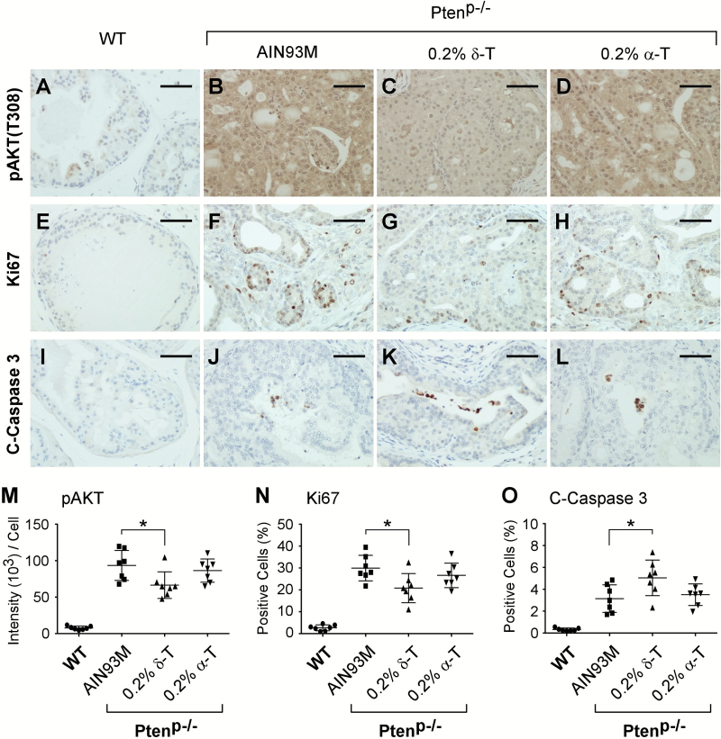
Figure 2.
Dietary δ-T reduced pAKT, suppressed proliferation and increased apoptosis in prostate tissues. Ptenp−/− mice fed either an AIN93M (n = 8), a 0.2% δ-T diet (n = 10) or a 0.2% α-T diet (n = 8) and the WT mice fed an AIN93M diet (n = 7) were analyzed for the active AKT, proliferation and apoptosis using IHC staining for pAKT (T308), Ki67 and cleaved-Caspase 3 (C-Caspase 3), respectively. Images of representative IHC staining for these mouse prostate samples are shown in (A–D, pAKT; E–H, Ki67; I–L, C-Caspase 3). The scale bar in the figures represents 50 µm. Quantified results of pAKT (T308), Ki67 and C-Caspase 3 IHC staining for the four groups of mice were determined using the Aperio ScanScope and summarized in (M), (N) and (O), respectively. Data are presented as mean ± SD. One-way ANOVA followed by Tukey’s post hoc test is used to compare the IHC staining results in the groups (*P-value = 0.037, 0.018 and 0.031 in M, N and O, respectively).

