Garnets record subduction earthquakes.
Abstract
Large earthquakes occur in rocks undergoing high-pressure/low-temperature metamorphism during subduction. Rhythmic major-element zoning in garnet is a common product of such metamorphism, and one that must record a fundamental subduction process. We argue that rhythmic major-element zoning in subduction zone garnets from the Franciscan Complex, California, developed in response to growth-dissolution cycles driven by pressure pulses. Using electron probe microanalysis and novel techniques in Raman and synchrotron Fourier transform infrared microspectroscopy, we demonstrate that at least four such pressure pulses, of magnitude 100–350 MPa, occurred over less than 300,000 years. These pressure magnitude and time scale constraints are most consistent with the garnet zoning having resulted from periodic overpressure development-dissipation cycles, related to pore-fluid pressure fluctuations linked to earthquake cycles. This study demonstrates that some metamorphic reactions can track individual earthquake cycles and thereby opens new avenues to the study of seismicity.
INTRODUCTION
Earthquakes in subduction zones can produce devastating tsunamis [for example, the 2004 moment magnitude (Mw) 9.2 Sumatra-Andaman and 2011 Mw 9.0 Tōhoku “megathrust” earthquakes] and may discharge subducted volatiles (1, 2) and trigger arc magmatism (3, 4). Although subduction seismicity can persist to depths of >600 km, it is most frequent and energetic in the uppermost part of the subducting slab, at depths of 30 to 70 km (5–8). The record of such seismicity might be visible in high-pressure/low-temperature (HP/LT) metamorphic rocks recovered from these depths. More so than for other tectonic environments, links between metamorphism and seismicity are proposed for subduction zones. For example, seismicity at anomalous (>50 to 70 km) depths in the subduction setting has been related to embrittlement due to metamorphic dehydration reactions and associated pore fluid pressure development and/or volume change (6, 9–14). Geologic records that contribute to our understanding of the mechanisms and magnitude recurrence rates for subduction seismicity come chiefly from Quaternary tsunami and turbidite deposits (15–20), subsidence/uplift histories from tidal wetlands and coral reefs (21, 22), and pseudotachylytes and breccias (23–25). Here, we show that garnet—one of the more common metamorphic minerals in HP/LT metamorphic rocks—may provide new insights into earthquake magnitude recurrence rates.
The blueschist- and eclogite-facies assemblages produced in subduction metamorphism record uniquely low geothermal gradients, typically <600°C GPa−1. Subduction metamorphism can be incredibly rapid, in cases involving time scales <104 to 106 years (26–29). Rhythmic major-element zoning in garnet is a common feature in HP/LT metabasic rocks (30–36), suggesting that it is a manifestation of a common process (or set of processes) in the subduction setting. To explore the nature of these processes and the origin of the HP/LT garnet zoning, we apply several modern and novel techniques to investigate metamorphism and paleoseismicity in the Jurassic Franciscan Complex, California.
RESULTS
Sample CA13-01 is a garnet-bearing eclogite-blueschist from Ring Mountain, Tiburon Peninsula, San Francisco Bay. Sample CA13-05A is a garnet-rich amphibolite from the same locality. Detailed rock descriptions are provided in Materials and Methods; however, of particular relevance for the P-T conditions of metamorphism is the presence of amphibole (glaucophane in CA13-01; Na-Ca amphibole and glaucophane in CA13-05A) and zoisite and the absence of lawsonite in both samples. Garnets from both rocks contain rutile and quartz inclusions from the core to the rim (with quartz being rare at the rim). Thermodynamic modeling of phase equilibria suggests that garnet rim growth occurred under P-T conditions of 555° to 585°C and 1.4 to 2.0 GPa for CA13-01 and 535° to 595°C and 1.2 to 2.0 GPa for CA13-05A; these estimates are entirely consistent with earlier thermobarometry of the same rocks (37). Whole-rock geochemistry, methods, and results for thermodynamic modeling of metamorphic phase equilibria are provided in Materials and Methods.
Electron microprobe x-ray count maps were produced for Ca, Fe, Mg, and Mn in six to seven garnets for each of CA13-01 and CA13-05A; examples of Mg and Mn maps are provided in Fig. 1, and all maps are provided in the Supplementary Materials. Complementary quantitative electron microprobe traverses were measured for one garnet from CA13-01 and two garnets from CA13-05A (Fig. 1). The garnets have inclusion-rich cores that are compositionally distinct from inclusion-poor mantles/rims (Fig. 1). Oscillatory/rhythmic zoning in both Mg and Mn is apparent in the mantle/rim zone (Fig. 1) but does not correspond to obvious zoning in Ca or Fe (with the exception of bands at the extreme outer edge of garnets from CA13-01). The Mg and Mn zoning in some garnets incorporates features of zone incursion (embayments) and circumferential pinching and swelling (Fig. 1). All garnets analyzed display systematic asymmetry in the location of relatively steep compositional gradients (Fig. 1). These features are consistent with the major-element zoning being a result of cycles of garnet growth separated by gaps during which there was no growth or there was dissolution, such that the embayments and steep compositional gradients mark garnet growth “unconformities.” Quantitative Mn traverses (Fig. 1) confirm the asymmetry in the compositional gradients that flank the Mn highs; relatively steep gradients occur on the rim side of Mn highs for sample CA13-01 and on the core side for sample CA13-05A. Rim-side asymmetry (as observed in CA13-01) must involve overgrowth of thin (high-Mn) resorption bands by lower-Mn garnet, whereas core-side asymmetry (as observed in CA13-05A) must involve overgrowth of the unconformity surface by higher-Mn garnet. The two modes presumably reflect different supply/demand scenarios for Mn following garnet dissolution; reactions associated with the rim-side scenario cannot provide a sanctuary for all Mn liberated by garnet dissolution, leading to back diffusion of Mn into the garnet. Figure 2 outlines models for the development of the two types of asymmetric zoning as a result of garnet growth-dissolution cycles.
Fig. 1. Garnet compositional maps and profiles.
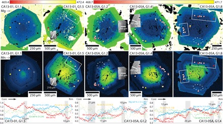
Color-enhanced wavelength-dispersive spectrometer (WDS) x-ray count maps for Mg (top row) and Mn (middle row) in garnets from Ring Mountain. Decreasing Mg and Mn content are indicated by yellow-green-blue-black color progression. Spots give location of Raman quartz analyses and are colored according to peak position (±1.5 cm−1 about 470.9 cm−1 for CA13-01; ±1.5 cm−1 about 470.2 cm−1 for CA13-05A; see data in Fig. 3D). Transparent spots correspond to partially transparent analyses in Fig. 3D. Spot size indicated is larger than the actual ~1-μm spot. White rectangles show notable zones with varying Raman response associated with compositional zoning boundaries (blowups provided in the Supplementary Materials). Dashed white ellipses highlight examples of zoning embayments/incursions. Gray backscattered electron (BSE) images indicate locations of quantitative (WDS) electron microprobe traverses at the bottom of the figure. Regions with steep compositional gradients in the traverses are indicated by vertical gray bands (with length scale indicated).
Fig. 2. Model for garnet zoning in response to pressure pulses.
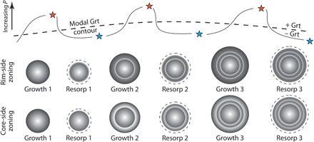
Conceptual models for development of rim-side (top row of garnets with Mn back diffusion during resorption) and core-side zoning (bottom row of garnets with no Mn back diffusion during resorption) by fluctuating garnet stability (growth-resorption cycles) in response to pressure pulses. Blue stars mark the onset of undrained conditions at the start of a phase of fluid overpressure development and garnet growth. Orange stars mark full overpressure conditions at the end of a growth phase (immediately before an earthquake event and fracturing due to dynamic stresses and relief of overpressure). Grt, garnet.
Franciscan garnet growth-dissolution cycles due to pressure(-temperature) fluctuations
The modal garnet contours for both rocks (see phase assemblage diagrams in Materials and Methods) suggest that modest changes in T or, more so, P might have triggered significant garnet production/loss and therefore caused the garnet zoning (that is 5 to 15 volume % growth/dissolution, relative to starting garnet content, for a ΔT of 40°C or a ΔP of 100 MPa). The gentle slopes in P-T space for both Mg- and Mn-in-garnet isopleths (see phase assemblage diagrams in Materials and Methods) are also consistent with rhythmic zoning in Mg and Mn having developed primarily in response to changes in P. Garnet zoning is complemented by irregular major-element zoning in glaucophane, omphacite, and zoisite in CA13-01, and glaucophane, hornblende, and zoisite in CA13-05A. Up-pressure dehydration reactions likely involved breakdown of glaucophane and zoisite to form garnet, jadeite, quartz, and H2O in CA13-01 and breakdown of hornblende and zoisite to form garnet, glaucophane, and H2O in CA13-05A (see Materials and Methods). To test the role of pressure fluctuations in driving garnet growth-dissolution cycles, we performed high-resolution barometry by Raman microspectroscopy on quartz inclusions in garnet and novel synchrotron Fourier transform infrared (FTIR) microspectroscopy on garnet.
Interrogating pressure pulses by spectroscopic methods
Raman barometry uses the pressure dependence of the position of peaks in the Raman spectra for an included mineral to calculate a residual pressure in the inclusion (38). This residual pressure results from differential expansion/contraction of host and inclusion following cooling and exhumation and, knowing the thermoelastic properties of the two minerals, can be used to calculate the P-T conditions of inclusion overgrowth by host. The properties of quartz and garnet yield an inclusion-host system far more sensitive to overgrowth pressure than temperature, meaning that the quartz-in-garnet system makes a good barometer (39). The position of (nominal 464 cm−1) quartz Raman A1g-mode peaks was measured in triplicate for 80 quartz-in-garnet inclusions across samples CA13-01 and CA13-05A. Images of quartz inclusions are provided in Fig. 3 (A to C), and detailed methods are provided in Materials and Methods. Repeat analyses produced consistent results, yielding a 2σ reproducibility of ±0.3 cm−1 (Fig. 3D). With exposed, cracked, or compound inclusions (n = 12) excluded, analyses from the two rocks show a significant spread in A1g-mode peak position (4.2 to 7.2 cm−1 above atmospheric for CA13-01 and 3.5 to 6.5 cm−1 above atmospheric for CA13-05A; Fig. 3D). Variable Raman A1g-mode peak position reflects variable residual pressure resulting from different overgrowth pressure. Using the calibration of Schmidt and Ziemann (40), the elastic model of Guiraud and Powell (41), and mineral properties recommended by Holland and Powell (42), the ranges of Raman A1g-mode peak shifts suggest P = 1.4 to 2.1 GPa (median, 1.9 GPa) for CA13-01 and P = 1.25 to 1.95 GPa (median, 1.5 GPa) for CA13-05A. Spatially, Raman A1g-mode peak position varies by 0.5 to 1.5 cm−1 over garnet radial distances of 10 to 50 μm and in association with the major-element growth zoning (Fig. 1); we infer that garnet growth-dissolution cycles were associated with fluctuations in metamorphic pressure of magnitude 100–350 MPa.
Fig. 3. Quartz-in-garnet Raman microspectroscopy results.
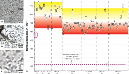
(A to C) Transmitted-light photomicrographs of some analyzed quartz inclusions in garnet. (D) Positions for the nominally 464 cm−1 A1g Raman peak for quartz inclusions in garnet from Ring Mountain, analyzed in triplicate. Inclusion circled in pink displays an abnormally low peak position despite lack of evidence that it was exposed during polishing or associated with visible cracks in garnet. Yellow-to-red background coloring is shown for reference to spot colors on garnet x-ray count maps in Fig. 1.
Raman barometry can only be applied where suitable inclusions occur. To obtain more complete records of pressure fluctuations during garnet growth/resorption, we also applied exploratory FTIR absorption spectroscopy for simultaneous garnet barometry and hygrometry. Experimental work has demonstrated a positive relationship between growth pressure and the OH content of pyrope garnet [at mantle pressures; (43, 44)], and the presence of nonstructural (molecular) H2O in garnet (an impurity) may also act as a proxy for the activity (partial pressure) of H2O (a hygrometer) during garnet growth. Structural OH and molecular H2O in garnet are associated with independent FTIR spectral peaks within the mid-infrared (IR) range. Interrogation of the fine major-element zoning in the Franciscan HP/LT garnets by FTIR spectroscopy requires high–spatial resolution and high-sensitivity measurement of a component that occurs in extremely low concentrations (<100 parts per million; garnet is nominally anhydrous), meaning that the technique requires an IR source with brightness only offered by synchrotron light. Transmission synchrotron FTIR microspectroscopy was performed on three garnets from CA13-01 and two from CA13-05A at the Australian Synchrotron and Diamond Light Source, UK. Detailed methods and results are provided in Materials and Methods.
Our new, qualitative synchrotron FTIR barometry and hygrometry technique identified regions with steep OH (and, to a lesser extent, H2O) concentration gradients in garnet (Fig. 4). These breaks in OH match the locations of the steep compositional gradients that mark garnet growth unconformities (Fig. 4). The correlation is with zoning in Mg and Mn but not in Ca, suggesting that fluctuations in OH do not simply reflect changes in proportion of the garnet end members that are most commonly hydrated (that is, grossular/andradite). Regardless of the polarity of Mn change across the unconformities in rim-side (CA13-01) and core-side (CA13-05A) zoning, OH content uniformly shows a sharp rim-side decrease (Fig. 4). This result is consistent with garnet growth (following dissolution) resuming at lower pressure than recorded in the garnet immediately core-side of the growth unconformity—that is, the synchrotron FTIR results indicate that dissolution was associated with a pressure drop. The synchrotron FTIR results demonstrate the linkage between pressure fluctuations and growth-dissolution cycles proposed in the model for development of the fine-scale, rhythmic compositional zoning in the garnets (Fig. 2). The relationship between OH and H2O in the garnets is complex, displaying covariance or anticorrelation in some regions and apparent independence in others. For these rocks, if systematic mechanistic linkages between pressure and fluid activity exist, then the synchrotron FTIR approach was unable to resolve them.
Fig. 4. Synchrotron FTIR microspectroscopy results.
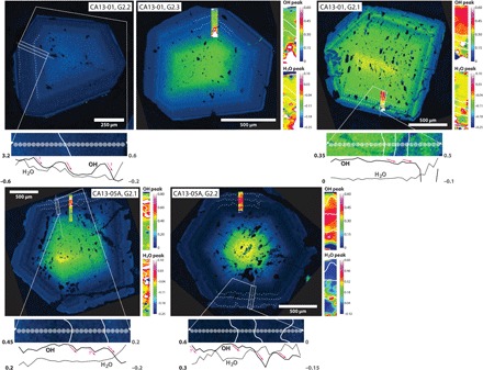
X-ray count maps for Mn in garnet and synchrotron FTIR analysis for one-dimensional (1D) traverses (bottom of each Mn map) and two-dimensional (2D) maps (right of each Mn map) for OH and H2O. Dashed white lines on Mn maps indicate the position of garnet growth unconformities and correspond to the white lines on the 1D FTIR traverses and 2D FTIR maps. Vertical axis on 1D traverse plots and colors of the 2D FTIR maps correspond to integrated intensity in the 3520 to 3620 cm−1 region for OH (a dimensionless measure of OH abundance) and in the 3350 to 3450 cm−1 region for H2O (a dimensionless measure of H2O abundance). Pink arrows on 1D traverse plots indicate regions of decreasing OH in the vicinity of garnet growth unconformities.
Short time scales for multiple pressure pulses
The presence of rutile and Ca-Na amphibole in CA13-05A constrains peak T = 535° to 595°C at P = 1.2 to 1.8 GPa, and the presence of glaucophane and the absence of chlorite in CA13-01 constrain peak T = 555° to 585°C for P = 1.4 to 1.95 GPa (see metamorphic assemblage diagrams in Materials and Methods). The presence of rutile inclusions at the edge of garnet rims is consistent with garnet zoning having developed during the peak-T phase of the metamorphism (T > 550°C), in agreement with other work on Ring Mountain HP/LT garnets (33) but in contrast to Franciscan HP/LT garnets with retrograde rims from Healdsburg (34, 45) and Jenner (46). The absence of lawsonite in both rocks suggests slightly lower maximum pressure than calculated from Raman barometry (<1.8 to 2.0 GPa versus 1.9 to 2.1 GPa). The counterclockwise P-T path experienced by these rocks involved relatively isobaric refrigeration after peak P-T, followed by monotonic cooling during exhumation (33, 34, 47). Preservation of fine-scale diffusion textures in garnet that developed during the peak-T, garnet rim-growth phase of the metamorphism (≤10 μm, Fig. 1) means that there was negligible modification during exhumation, despite evidence for a protracted exhumation (cooling) history (45, 48).
We use an error function approximation to 1D (radial) diffusion [equation of the form c(x,t) ∝ erf (x/√4Dt), where c, x, t, and D are concentration, position, time, and diffusivity, respectively] to reproduce Mn diffusion length scales associated with the garnet growth unconformities (≤10 μm, Fig. 1) and thereby constrain time scales for formation of the garnet zoning at near-peak metamorphic conditions. This approach assumes an initial step change in Mn concentration associated with the overgrowth/dissolution surface and no cutting effects or spatial homogenization due to spot size; the calculated estimates are therefore significantly biased toward longer durations. The Mn-in-garnet Arrhenius parameters of Chu and Ague (49) yield time scales of <300 thousand years (ky) for T > 550°C and P < 2.0 GPa; all the pressure-driven growth-dissolution cycles must have occurred within this time frame. Varying the overgrowth P-T from 550° to 590°C and from 1.2 to 2.0 GPa expands the estimate range to <30 to 300 ky. Allowing for Mn-in-garnet Arrhenius parameter values between the most diffusive (50) and the most retentive (51) published expands the range to <0.04 to 1.2 million years (My) (for mean P-T conditions; T = 570°C, P = 1.6 GPa). This range is compatible with global subduction speeds (52), indicating that most subducting crust passes through blueschist-facies conditions in ~1 My.
DISCUSSION
The diffusion length scale data indicate that at least four cycles of pressure fluctuation (of magnitude 100–350 MPa) occurred during HP/LT metamorphism, with net garnet growth length scales of <100 to 300 μm and metamorphic time scales very likely <<1 My. On the basis of major-element zoning in HP/LT garnets from Cuba, García-Casco et al. (31) hypothesized pressure fluctuations of similar magnitude during subduction, relating them to small-scale (physical) exhumation events during prograde metamorphism. The <<1 My time scales for garnet zoning development are significantly shorter than hypothesized time scales for multiple burial-exhumation cycles in HP/LT rocks that have been related to (i) “yo-yo tectonics,” involving (local or regional) switches between shortening and extension (35, 53–55); (ii) exhumation-erosion-deposition-subduction cycles (56); and (iii) numerically simulated “chaotic” mixing as a result of convection within a putative subduction channel (57–60). These “lithostatic pressure” models are limited by maximum rates of burial/exhumation; for example, four 7.5-km (mean magnitude, 225 MPa) burial-exhumation cycles at vertical movement rates approximating tectonic plate velocities—that is, 30 mm year−1, as in the subduction convection model of Gerya et al. (58)—require at least 2 My. It should be noted, however, that modeling of convection within the subduction channel also predicts periodic stalling of particles, more likely yielding time scales of 5 to 10 My per 200- to 500-MPa physical burial-exhumation cycle [for example, see figures 4 and 7 of the study by Gerya et al. (58)].
Garnet zoning marks seismic cycles during subduction
Figure 5 compares time scales for (i) megathrust earthquake recurrence, (ii) individual P pulses recorded in the HP/LT garnets of this study, and (iii) small-scale physical burial-exhumation cycles, showing that these constraints are most consistent with a seismic cycle origin for the garnet P pulse records. We relate rhythmic major-element zoning in the Franciscan garnets to cycles of pore fluid overpressure development and dissipation, linked to a seismically induced switch from locally undrained to drained conditions [that is, dynamic cracking and permeability changes following large earthquakes; see the works of Brenguier et al. (61) and Chaves and Schwartz (62)].
Fig. 5. Time scale comparisons.
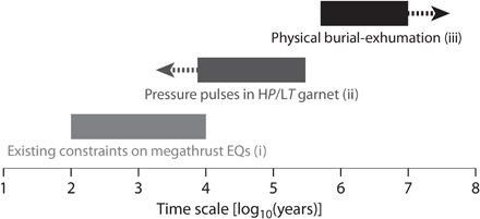
Time scales for individual cycles relating to (i) megathrust earthquake (EQ) events from tsunami and turbidite deposits and subsidence/uplift histories, (ii) garnet dissolution-growth cycles and associated P pulses as recorded in the garnets of this study, and (iii) physical burial-exhumation cycles relating to “yo-yo” tectonics, exhumation-erosion-deposition-subduction cycles, or convection within a putative subduction channel. Note that time scales for (ii) are maximum values due to bias toward overestimation in the approach used and the possibility that P pulses were significantly more numerous than recorded in the garnets (see text).
Although rocks do not typically have unconfined strength greater than 100 to 200 MPa, the effects of confinement and undrained conditions may allow pore fluid pressure excess (overpressure) to attain more significant values (63–65). Transient pressure pulses may mark porosity waves that transit the rock volume or cycles of overpressure development-dissipation linked to subduction zone seismicity. Porosity waves are only theorized to exist, whereas subduction zone seismicity is an observed phenomenon that has been associated with pore fluid overpressure (6, 9–12) and occurs at the depth and location of HP/LT metamorphism.
Our model for Franciscan garnet zoning invokes widespread rock fracture with passing seismic waves as the trigger for overpressure relief; changes in seismic velocity following megathrust earthquakes have been related to this mechanism of pore fluid pressure relief (61, 62). Rock fracture in response to dynamic stresses induced by a large earthquake may also produce a widespread permeability spike. Once fault healing and sealing are complete and conditions again become undrained, feedback between dehydration reactions and increasing metamorphic pressure—due to metamorphic pore fluid production—can lead to overpressure development and recommenced garnet growth, until dynamic stresses associated with a subsequent large earthquake again cause rock fracture and permeability increase. Not all earthquake events need be recorded, and those that were need not have been proximal; dynamic stresses and therefore likelihood of local fracturing and overpressure relief would be greatest for the most energetic events (for example, megathrusts). The model is consistent with our FTIR observation of a decrease in OH across the growth unconformity (Fig. 4). Intimate garnet growth zone δ18O variation in the Franciscan garnets [cf. Page et al. (45)] may also be expected for cycles of pore fluid purging with rock fracture, pressure drop, and permeability increase, followed by metamorphic dehydration and overpressure development under undrained conditions. Such work may offer a means to test the fluid overpressure relief (following large earthquakes) model presented here.
Multiple, rapid pressure fluctuations of 100 to 350 MPa, as observed here in association with garnet zoning, can be achieved by seismic cycles in subduction zones. Large earthquakes have recurrence intervals of 100 to 10,000 years (16–21). Although there is some overlap, the HP/LT garnets studied record frequencies mostly outside of this recurrence range (see Fig. 5). Not every earthquake will cause fracturing and overpressure dissipation; records are only made for those that produced sufficient seismic energy, directed at the rock volume. This, partnered with the potential for garnet dissolution to have removed evidence of earlier garnet growth-dissolution cycles (cannibalization of records of P pulses), can explain much of the potential discrepancy between time scales for HP/LT garnet records of P pulses and recurrence intervals for large subduction zone earthquakes (Fig. 5).
We conclude that major-element zoning in the HP/LT garnets records seismic cycles within the Franciscan subduction zone. These metamorphic features are common in HP/LT metamorphic rocks worldwide. In time, geochemical features of HP/LT garnets (and other minerals) may offer important new insights into the nature of subduction zone (paleo)seismicity and associations among stress evolution, earthquake magnitude and recurrence, and fluid transport/mobility in the crust.
MATERIALS AND METHODS
Sample descriptions
The article focuses on two rocks from Ring Mountain, San Francisco Bay, California. Sample CA13-01 [Global Positioning System (GPS): 37°55.073′ N, 122°29.583′ W] is an eclogite-blueschist with 11 volume % garnet. Sample CA13-05A (GPS: 37°54.729′ N, 122°29.122′ W) is a garnet-rich (32 volume %) amphibolite-blueschist. Garnet populations in both rocks are not uniform, with grain size in each varying from <0.5 mm to approximately 2 mm across. Both rocks are heterogeneous on a scale of 1 to 10 mm; CA13-01 has blueschist and eclogite layers, and CA13-05A has interfingered amphibolite and blueschist domains. Garnet in CA13-01 is commonly surrounded by phengite, whereas many garnets in CA13-05A are surrounded by clots of chlorite. Mineralogy for each sample is given below. Cross-polarized light microphotographs are provided in Fig. 6.
Fig. 6. Cross-polarized-light photomicrographs of samples CA13-01 and CA13-05A.
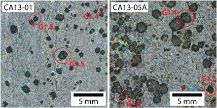
For each rock, some of the garnets mapped for major elements are circled in red.
(CA13-01) omphacite + garnet + glaucophane + phengite + titanite + zoisite + quartz + rutile
(CA13-05A) hornblende + garnet + glaucophane + zoisite + chlorite + rutile + phengite + omphacite + titanite
Tsujimori et al. (33) provided detailed descriptions of three HP/LT rocks from Ring Mountain. Their sample A is incredibly similar in mineralogy and appearance to our CA13-05A. For that sample, Tsujimori et al. (33) used petrography, garnet-omphacite-phengite thermobarometry, and equilibrium phase assemblage modeling to constrain garnet rim growth to the peak metamorphic phase, at T = 550° to 620°C and P = 2.2 to 2.5 GPa. Titanite overgrew rutile in both CA13-01 and CA13-05A, and chlorite overgrew garnet in CA13-05A. The Ring Mountain HP/LT rocks record a counterclockwise P-T path, with a metamorphic overprint relating to refrigeration before exhumation (33, 47). Chlorite and titanite in CA13-01 and titanite in CA13-05A are texturally post-peak metamorphic and grew during the exhumation (and refrigeration) phase of the rock history.
Electron microprobe methods, mineral chemistry and zoning, and Mn sources
Electron microprobe mapping method and results
WDS x-ray count maps for Al, Ca, Fe, Mg, and Mn were produced for seven garnets from CA13-01 and six garnets from CA13-05A. Measurements were made simultaneously on the five spectrometers of the Cameca SX 100 electron microprobe in the Department of Earth Science, University of California, Santa Barbara; Al, Ca, Fe, Mg, and Mn measurements were made on TAP, LPET, LLIF, LTAP, and LLIFF crystals, respectively. Run conditions were 15 keV and 200 nA, and pixel dwell times were 50 ms for all maps except for CA13-01 garnet 2.3, which used 100 ms. Mapping resolution was 2 to 5 μm, depending on the size of the garnet, and uniform in x and y. Color overlay maps for all garnets are provided in the Supplementary Materials.
Electron microprobe quantitative analysis method and results
Quantitative WDS analyses were produced for three radial garnet traverses (n = 670), a cluster of zoned zoisites from each of CA13-01 and CA13-05A (n = 24), and selected additional minerals from each sample (n = 36). All analyses were performed on the JEOL JXA 8600 electron microprobe in the Department of Earth & Planetary Sciences, Johns Hopkins University. Calibration used multiple natural reference materials. Garnet traverses were run at 15 keV and 40 nA and used a 1-μm spot size at 1-μm centers and 30-s on-peak and 10-s background counting times. All other analyses were run at 15 keV and 20 nA and used a 5-μm spot size and 30-s on-peak and 15-s background counting times.
Results for the radial garnet traverses are provided in Fig. 1. Compositions of typical examples of minerals from CA13-01 and CA13-05A are provided in Tables 1 and 2. The subsequent section discusses mineral chemical variability and zoning. All quantitative electron microprobe results are provided in the Supplementary Materials.
Table 1. Stoichiometric mineral compositions for CA13-01, based on assumed number of oxygen atoms shown.
Note that Fe in zoisite was assumed to be 100% Fe3+ and Fe in all other minerals was assumed to be 100% Fe2+. Gln, glaucophane; Omph, omphacite; Phe, phengite; Zo, zoisite.
| CA13-01 | |||||
| Gln | Omph | Phe | Zo 1 | Zo 2 | |
| Si | 7.99 | 2.02 | 3.49 | 2.91 | 2.92 |
| Ti | — | — | 0.01 | 0.01 | — |
| Al | 1.69 | 0.36 | 1.97 | 2.41 | 2.25 |
| Cr | — | — | — | — | — |
| Fe | 1.15 | 0.22 | 0.19 | 0.50 | 0.62 |
| Mn | — | — | — | — | 0.01 |
| Mg | 2.21 | 0.44 | 0.39 | 0.01 | — |
| Ca | 0.15 | 0.51 | — | 1.79 | 1.72 |
| Na | 1.87 | 0.46 | 0.03 | — | — |
| K | — | — | 0.87 | — | — |
| O | 23 | 6 | 11 | 12.5 | 12.5 |
| Al/(Al + Si) | 0.17 | 0.15 | 0.36 | 0.45 | 0.44 |
| Fe/(Fe + Al) | 0.41 | 0.38 | 0.09 | 0.17 | 0.21 |
| Fe/(Fe + Mg) | 0.34 | 0.34 | 0.33 | — | — |
| Na/(Na + Ca) | 0.93 | 0.48 | — | — | — |
| Na/(Na + K) | — | — | 0.04 | — | — |
| Total oxides (wt %) | 97.5 | 100.6 | 94.5 | 96.3 | 97.6 |
Table 2. Stoichiometric mineral compositions for CA13-05A, based on assumed number of oxygen atoms shown.
Note that Fe in zoisite was assumed to be 100% Fe3+ and Fe in all other minerals was assumed to be 100% Fe2+. Hbl, hornblende; Gln, glaucophane; Chl, chlorite; Aln, allanite.
| CA13-05A | |||||||
| Hbl 1 | Hbl 2 | Gln | Chl | Zo 1 | Zo 2 | Aln | |
| Si | 7.29 | 6.92 | 7.85 | 2.97 | 2.91 | 2.85 | 2.94 |
| Ti | 0.02 | 0.03 | 0.01 | 0.01 | 0.01 | 0.06 | 0.01 |
| Al | 1.24 | 1.91 | 1.77 | 2.32 | 2.41 | 2.09 | 2.01 |
| Cr | — | — | — | — | — | — | — |
| Fe | 1.56 | 1.57 | 1.30 | 1.84 | 0.51 | 0.73 | 0.87 |
| Mn | 0.02 | 0.01 | 0.01 | 0.02 | 0.01 | 0.02 | 0.02 |
| Mg | 3.00 | 2.67 | 2.21 | 2.68 | — | — | — |
| Ca | 1.50 | 1.47 | 0.12 | — | 1.77 | 1.70 | 1.40 |
| Na | 0.82 | 0.93 | 1.98 | — | — | — | — |
| K | 0.04 | 0.08 | — | 0.01 | — | — | — |
| O | 23 | 23 | 23 | 14 | 12.5 | 12.5 | 12.5 |
| Al/(Al + Si) | 0.15 | 0.22 | 0.18 | 0.44 | 0.45 | 0.42 | 0.41 |
| Fe/(Fe + Al) | 0.56 | 0.45 | 0.42 | 0.44 | 0.17 | 0.26 | 0.30 |
| Fe/(Fe + Mg) | 0.34 | 0.37 | 0.37 | 0.41 | — | — | — |
| Na/(Na + Ca) | 0.35 | 0.39 | 0.94 | — | — | — | — |
| Na/(Na + K) | 0.95 | 0.92 | — | — | — | — | — |
| Total oxides (wt %) | 97.1 | 98.9 | 97.1 | 86.1 | 96.7 | 97.2 | 81.0 |
Solid solution, mineral zoning, and Mn sources
Quantitative analyses demonstrate variability in composition for all minerals analyzed. Zoning in individual minerals can capture the full range of compositional variation observed for omphacite and zoisite in CA13-01 and hornblende and zoisite in CA13-05A. Amphiboles from CA13-01 are glaucophane, with Na/(Na + Ca) ≈ 94% (Fig. 7A). One population of amphibole in CA13-05A has similar glaucophane chemistry, whereas the other is a hornblende, with composition between actinolite and pargasite (Fig. 7A). Clinopyroxene in CA13-01 is omphacite, with 39 to 53 mole percent (mol %) jadeite (Fig. 7B). Chlorite in CA13-05A varies in composition, with Fe/(Fe + Mg) in the range 37 to 45% and Si increasing with Fe (Fig. 7C). White mica in CA13-01 also varies in composition, potentially defining two (phengitic) compositional arrays between celadonite and muscovite/paragonite (Fig. 7D). Phengite in CA13-01 has a Si content of 3.34 to 3.54 per formula unit.
Fig. 7. Mineral composition plots.
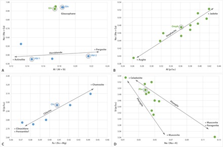
Plots showing solid solution for (A) amphibole, (B) clinopyroxene, (C) chlorite, and (D) white mica. Green data are for CA13-01, and blue data are for CA13-05A. Circled points are those whose geochemistry is given in Tables 1 and 2. p.f.u., per formula unit.
The compositional arrays and populations of Fig. 7 are reflected in BSE zoning in amphibole and omphacite. In addition, zoisite has fine-scale, oscillatory BSE zoning (Fig. 8). The zoning principally relates to Fe3+ and Al substitution in the epidote-zoisite solid solution (Fig. 8 and Tables 1 and 2) but also maps with Mn content, particularly in CA13-05A (Fig. 8). Complementary Mn zoning in zoisite to that observed in garnet (see Results) suggests that zoisite growth may have partially balanced garnet dissolution (and vice versa). This observation is consistent with parallelism of modal garnet and zoisite contours (Fig. 9). Garnet breakdown in the presence of H2O produces zoisite only if other reactants are able to balance the remaining chemistry. Garnet growth-dissolution reactions that mark P pulses (seismic loading-rupture cycles) may have involved garnet, jadeite, quartz, and H2O to yield Na amphibole and zoisite (and vice versa) in CA13-05C, and garnet, Na amphibole, and H2O to yield Na-Ca amphibole and zoisite (and vice versa) in CA13-01.
Fig. 8. Zoisite zoning and composition.
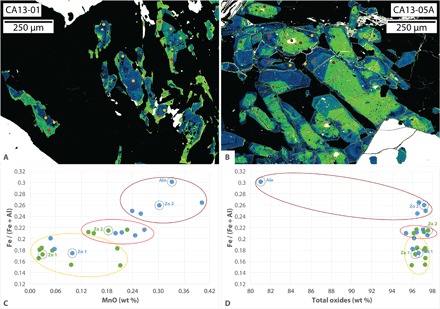
(A and B) Color overlay BSE maps for zoisite in CA13-01 and CA13-05A, highlighting fine-scale oscillatory zoning. (C) Discrimination plot showing positive correlation between Fe/(Fe + Al) and MnO in zoisite. (D) Discrimination plot showing one analysis distinct from others on the basis of lower oxide totals: an allanite that contains a high proportion of trace elements not analyzed for. Green data are for CA13-01, and blue data are for CA13-05A. Yellow, red, and maroon groupings match the color of analysis spots and low to high BSE response, respectively, in the maps. Spots on maps are three times larger than actual analyses.
Fig. 9. Results of thermodynamic modeling.
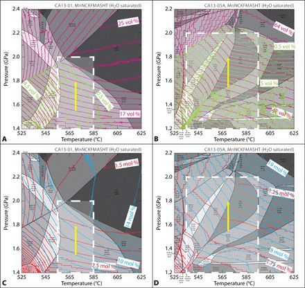
MnNCKFMASHT metamorphic assemblage diagrams for (A and C) CA13-01 and (B and D) CA13-05A. Phase assemblage fields and labels redrafted from the Perple_X output. White dashed box indicates approximate equilibrium P-T range of the peak-metamorphic assemblage in each rock. Green and pink contours in (A) and (B) are for modal zoisite and garnet, respectively; contour intervals of 0.5 volume % are used for zoisite, and contour intervals of 1 and 2 volume % are used for garnet in CA13-01 and CA13-05A, respectively. Note the significant congruence between zoisite and garnet contours, suggesting a metamorphic relationship between the phases (that is, the dissolution of one partially accommodates the growth of the other). The yellow arrow shows a 250-MPa isothermal P increase (starting at T = 570°C and P = 1.55 GPa), associated with an absolute gain of 5 volume % garnet and loss of 1.25 volume % zoisite for CA13-01 and an absolute gain of 9 volume % garnet and loss of 4 volume % zoisite for CA13-05A. Blue and red contours in (C) and (D) are for proportion of Mg garnet (pyrope) and Mn garnet (spessartine), respectively; contour intervals of 1 mol % are used for pyrope, and contour intervals of 0.25 mol % are used for spessartine.
The predicted garnet growth–to–zoisite dissolution ratio for an isothermal, 250-MPa P pulse starting at T = 570°C and P = 1.55 GPa is greater for CA13-01 (5:1.25) than for CA13-05A (9:4) (Fig. 9). The Mn-in-garnet–to–Mn-in-zoisite ratio is also greater for CA13-01 (approximately 1.0:0.1) than for CA13-05A (approximately 0.5:0.2). Hornblende, a product of garnet breakdown in CA13-05A, has up to 0.11 weight % (wt %) Mn. More accommodating hosts for Mn liberated during breakdown of garnet in CA13-05A may account for limited Mn back diffusion during garnet dissolution in CA13-05A (relative to CA13-01) and thus development of core-side rather than rim-side zoning in those garnets (see Fig. 2).
Whole-rock geochemistry and thermodynamic modeling method and results
Whole-rock geochemistry
Approximately 20 g of fresh and representative material from each of CA13-01 and CA13-05A was ground to powder in a tungsten carbide ring-and-puck mill. Lithium borate–fused glass beads were prepared for analysis with a sample (powder)-to-flux ratio of 1:5. Whole-rock geochemical analyses for major elements were performed on the PANalytical Axios Advanced x-ray fluorescence (XRF) spectrometer in the Department of Geology, University of Leicester. Results from the XRF work are provided in Table 3.
Table 3. Whole-rock geochemistry determined by XRF (values in wt %).
LOI, loss on ignition.
| SiO2 | TiO2 | Al2O3 | Fe2O3 | MnO | MgO | CaO | Na2O | K2O | P2O5 | SO3 | LOI | Total | |
| CA13-01 | 48.09 | 1.53 | 15.78 | 9.90 | 0.19 | 5.38 | 10.74 | 3.49 | 1.54 | 0.12 | <0.002 | 1.26 | 98.02 |
| CA13-05A | 40.85 | 2.84 | 16.43 | 17.23 | 0.35 | 6.36 | 12.53 | 1.11 | 0.13 | 0.10 | 0.112 | 1.28 | 99.32 |
Perple_X thermodynamic modeling of metamorphic phase equilibria
Thermodynamic modeling of metamorphic phase equilibria was performed in the MnNCKFMASHT system using the whole-rock compositions in Table 3. Whole-rock compositions were considered in preference to porphyroblast-subtracted compositions as the modeling was performed to interrogate sensitivity of garnet content (and growth or dissolution) to changes in P-T; a highly responsive, porphyroblast-involved system was assumed. For each model, the rock was considered saturated in H2O (set to 10 wt %), and Fe determined from XRF was recalculated to 100% ferrous (FeO); the only mineral in the rock with significant Fe3+ is epidote and that accounts for only a limited proportion of the zoisite (see Fig. 8). Modeling used version 6.6.6 of the Perple_X program (66, 67) and the 2004 update of the thermodynamic data set of Holland and Powell (68). Solution models used for the modeling (and references) are provided in the Supplementary Materials. Modeling was performed in the following ranges: T = 525° to 625°C and P = 1.4 to 2.4 GPa for CA13-01 and T = 525° to 625°C and P = 1.2 to 2.2 GPa for CA13-05A. Figure 9 shows metamorphic assemblage diagrams for both samples, with modal abundance contours for both garnet and zoisite (Fig. 9, A and B) and Mg garnet (pyrope) and Mn garnet (spessartine) (Fig. 9, C and D) overlain.
Raman thermobarometry method and results
Raman spectroscopy was performed on quartz inclusions in garnet using a HORIBA Jobin Yvon LabRAM HR Raman microscope in the Department of Chemistry, Durham University. All analyses used an 1800 grid (1800 grooves mm−1 grating), centered at 470 cm−1. For each quartz inclusion analysis, a 100× confocal objective lens was used to focus the 632.8-nm (red) HeNe laser to a spot size of 1 μm, centered in the inclusion. Quartz inclusions were inspected for exposure and/or cross-cutting cracks and analyzed in triplicate (three measurements in separate sessions). Precise peak positions were obtained by fitting Lorentzian curves to the nominal 464 cm−1 A1g-mode quartz Raman peak (for unknowns) using the software package Igor Pro. Curve fitting to the nominal 464 cm−1 A1g-mode peak in quartz used the portion of the spectrum 12 grid points on either side of the maximum intensity position, representing a total range of ~7.6 cm−1.
Instrument drift due to internal temperature equilibration was corrected for by quasi-hourly (duplicate) measurement of a pure sulfur Raman spectroscopy standard. Analyses on sulfur standards used a 50× objective and the same 632.8-nm (red) HeNe laser, focused to the position within the homogeneous standard that produced maximum spectrum intensity. For the sulfur standard spectra, curve fitting to the nominal 384.1 cm−1 mode peak used the portion of the spectra eight grid points on either side of the maximum intensity position (total range of ~5.1 cm−1), a Lorentzian form, and the software package Igor Pro. Drift in the position of the nominal 384.1 cm−1 sulfur peak during each Raman session was characterized by an equation of the form [y = –A exp(–t/b) + c], where y is the measured peak position minus the nominal 384.1 cm−1 value, t is the time since the first measurement of the session, A and b are shape constants, and c is a vertical shift constant. Values of A, b, and c for each session were calculated from a fit to the sulfur data for that session. Following correction for instrument drift at the time of measurement, unknowns were found to have an overall 2σ reproducibility of ±0.3 cm−1 for the peak position of the nominal 464 cm−1 A1g-mode quartz Raman peak, despite measurements being made on separate days (see Results and Fig. 3). Uncorrected spectra (with time stamps) for both quartz inclusions (unknowns) and sulfur standards are available at https://doi.org/10.7281/T1/SP6KO3.
Calculation of overgrowth P and T used the MATLAB QuiB Calc program of Ashley et al. (69), the quartz Raman calibration of Schmidt and Ziemann (40), mineral properties compiled in Holland and Powell (42), and the elastic model of Guiraud and Powell (41). Properties of garnet were calculated by linear interpolation between almandine, grossular, pyrope, and spessartine using respective molar ratios of 59:26:11:4 for CA13-01 and 56:28:15:1 for CA13-05A. These garnet compositions match microprobe-determined rim compositions for the garnets (see Fig. 1). For the same Raman shifts, the elastic model of Zhang (70) yields P estimates that are systematically higher by 100 to 200 MPa.
Synchrotron FTIR method and results
Microanalyses made at the Diamond Light Source were performed in transmission mode, with a spectral resolution of 16 cm−1 and a phase resolution of 128, using a 700 to 3200 cm−1 window. Analyses used a 36× objective and a 10 μm × 10 μm square slit size defining the detected area at the sample. Each spectrum was produced from 3000 scans, and backgrounds were collected before each traverse. Four traverses were run radially across the zoning at the garnet edge, using 10-μm centers (n = 39 for CA13-01 G1T2, n = 35 for CA13-01 G2T1, n = 76 for CA13-05A G1T1, and n = 46 for CA13-05A G2T1). Microanalyses made at the Australian Synchrotron were also performed in transmission mode, with a spectral resolution of 16 cm−1 and a phase resolution of 128, using a 700 to 3200 cm−1 window. Analyses again used a 36× objective but a circular spot size of 11.6 μm. Each spectrum was produced from 512 scans, and backgrounds were collected before each map. The 2D maps approximated 1D radial traverses, forming elongate grids with the following dimensions: 6 × 44 (n = 264) for CA13-01 G1T3, 5 × 39 (n = 195) for CA13-01 G2T2, 6 × 45 (n = 270) for CA13-01 G3T1, 6 × 81 (n = 486) for CA13-05A G1T2, and 6 × 48 (n = 288) for CA13-05A G2T2. All line maps used 12-μm centers in the “circumferential” direction and 6-μm centers (approximate 2× oversampling) in the radial direction. CA13-01 garnet 2, map G2T2, collected at the Australian Synchrotron, experienced irrecoverable issues with background correction.
In total, analyses were performed in transmission mode on three garnets from a clean, ~225-μm-thick wafer of sample CA13-01 and two garnets from a clean, ~280-μm-thick wafer of sample CA13-05A. Relative OH and H2O contents were calculated from the integrated intensity of the background-subtracted spectra in the 3520 to 3620 cm−1 and 3350 to 3450 cm−1 mid-IR regions, respectively. Integrated intensity calculations were performed with the OPUS software package using a linear integration baseline. Processed data for the integrated intensities of OH and H2O in the five garnets analyzed by synchrotron FTIR spectroscopy are provided in Fig. 4. Raw data (as OPUS files) for all synchrotron FTIR analyses are available at https://doi.org/10.7281/T1/SP6KO3.
Supplementary Material
Acknowledgments
K. Livi provided technical assistance for electron microprobe work at Johns Hopkins University. K. Chamberlain, G. Cooper, and E. Finch helped with the synchrotron FTIR work. A. Beeby provided access to the Raman Spectroscopy Laboratory at Durham University. Discussions with M. Brown helped hone the proposed model for seismicity-driven metamorphism. The paper benefitted greatly from the comments of five anonymous reviewers. Funding: Funding for the work was awarded by the Australian-American Fulbright Commission (Fulbright Victoria Fellowship to D.R.V.) and Durham University and Marie Skłodowska-Curie actions of the European Union (International Junior Research Fellowship to D.R.V.). Microprobe mapping work was supported by the University of California, Santa Barbara and NSF EAR-1219942. Synchrotron FTIR work was supported by a Rapid Access beamtime award from the Diamond Light Source (#SM14590) and a Merit Access beamtime award from the Australian Synchrotron (#11167). Author contributions: D.R.V. initiated and led the study, with input from B.R.H. and M.B.A.; D.R.V. wrote the paper, also with significant input from B.R.H. and M.B.A.; G.G.E.S. designed and performed the garnet electron microprobe mapping work; M.J.T., C.S.K., and G.C. facilitated (and helped interpret results from) the synchrotron FTIR work; and A.R.D. helped collect and analyze the Raman data. Competing interests: The authors declare that they have no competing interests. Data and materials availability: All data needed to evaluate the conclusions in the paper are present in the paper, in the Supplementary Materials, and/or in the online data repository at https://doi.org/10.7281/T1/SP6KO3. Data may also be requested from D.R.V.
SUPPLEMENTARY MATERIALS
Supplementary material for this article is available at http://advances.sciencemag.org/cgi/content/full/4/3/eaaq0234/DC1
fig. S1. Color overlay x-ray count maps for major divalent elements in Garnet 1.1 from CA13-01.
fig. S2. Color overlay x-ray count maps for major divalent elements in Garnet 1.4 from CA13-01.
fig. S3. Color overlay x-ray count maps for major divalent elements in Garnet 1.5 from CA13-01.
fig. S4. Color overlay x-ray count maps for major divalent elements in Garnet 1.6 from CA13-01.
fig. S5. Color overlay x-ray count maps for major divalent elements in Garnet 2.1 from CA13-01.
fig. S6. Color overlay x-ray count maps for major divalent elements in Garnet 2.2 from CA13-01.
fig. S7. Color overlay x-ray count maps for major divalent elements in Garnet 2.3 from CA13-01.
fig. S8. Color overlay x-ray count maps for major divalent elements in Garnet 1.2 from CA13-05A.
fig. S9. Color overlay x-ray count maps for major divalent elements in Garnet 1.4 from CA13-05A.
fig. S10. Color overlay x-ray count maps for major divalent elements in Garnet 1.7 from CA13-05A.
fig. S11. Color overlay x-ray count maps for major divalent elements in Garnet 1.8 from CA13-05A.
fig. S12. Color overlay x-ray count maps for major divalent elements in Garnet 2.1 from CA13-05A.
fig. S13. Color overlay x-ray count maps for major divalent elements in Garnet 2.2 from CA13-05A.
fig. S14. Color overlay x-ray count maps for Mg in garnets from CA13-01 and CA13-05A.
fig. S15. Color overlay x-ray count maps for Mn in garnets from CA13-01 and CA13-05A.
fig. S16. Blowups of regions indicated by white rectangles in (top row) fig. S14 and (bottom row) fig. S15.
Excel file
REFERENCES AND NOTES
- 1.Nakajima J., Hasegawa A., Kita S., Seismic evidence for reactivation of a buried hydrated fault in the Pacific slab by the 2011 M9.0 Tohoku earthquake. Geophys. Res. Lett. 38, L00G06 (2011). [Google Scholar]
- 2.Spandler C., Pettke T., Rubatto D., Internal and external fluid sources for eclogite-facies veins in the Monviso meta-ophiolite, western Alps: Implications for fluid flow in subduction zones. J. Petrol. 52, 1207–1236 (2011). [Google Scholar]
- 3.Davies J. H., The role of hydraulic fractures and intermediate-depth earthquakes in generating subduction-zone magmatism. Nature 398, 142–145 (1999). [Google Scholar]
- 4.John T., Gussone N., Podladchikov Y. Y., Bebout G. E., Dohmen R., Halama R., Klemd R., Magna T., Seitz H.-M., Volcanic arcs fed by rapid pulsed fluid flow through subducting slabs. Nat. Geosci. 5, 489–492 (2012). [Google Scholar]
- 5.Frohlich C., The nature of deep-focus earthquakes. Annu. Rev. Earth Planet. Sci. 17, 227–254 (1989). [Google Scholar]
- 6.S. Kirby, E. R. Engdahl, R. Denlinger, Intermediate-depth intraslab earthquakes and arc volcanism as physical expressions of crustal and uppermost mantle metamorphism in subducting slabs, in Subduction: Top to Bottom, G. E. Bebout, D. W. Scholl, S. H. Kirby, J. P. Platt, Eds. (Geophysical Monograph Series, American Geophysical Union, 1996), vol. 96, pp. 195–214. [Google Scholar]
- 7.Abers G. A., van Keken P. E., Kneller E. A., Ferris A., Stachnik J. C., The thermal structure of subduction zones constrained by seismic imaging: Implications for slab dehydration and wedge flow. Earth Planet. Sci. Lett. 241, 387–397 (2006). [Google Scholar]
- 8.Green H. W. II, Chen W.-P., Brudzinski M. R., Seismic evidence of negligible water carried below 400-km depth in subducting lithosphere. Nature 467, 828–831 (2010). [DOI] [PubMed] [Google Scholar]
- 9.Green H. W. II, Houston H., The mechanics of deep earthquakes. Annu. Rev. Earth Planet. Sci. 23, 169–213 (1995). [Google Scholar]
- 10.Hacker B. R., Peacock S. M., Abers G. A., Holloway S. D., Subduction factory 2. Are intermediate-depth earthquakes in subducting slabs linked to metamorphic dehydration reactions? J. Geophys. Res. 108, 2030 (2003). [Google Scholar]
- 11.Preston L. A., Creager K. C., Crosson R. S., Brocher T. M., Trehu A. M., Intraslab earthquakes: Dehydration of the cascadia slab. Science 302, 1197–1200 (2003). [DOI] [PubMed] [Google Scholar]
- 12.Jung H., Green H. W. II, Dobrzhinetskaya L. F., Intermediate-depth earthquake faulting by dehydration embrittlement with negative volume change. Nature 428, 545–549 (2004). [DOI] [PubMed] [Google Scholar]
- 13.Nakajima J., Uchida N., Shiina T., Hasegawa A., Hacker B. R., Kirby S. H., Intermediate-depth earthquakes facilitated by eclogitization-related stresses. Geology 41, 659–662 (2013). [Google Scholar]
- 14.Schubnel A., Brunet F., Hilairet N., Gasc J., Wang Y., Green H. W. II, Deep-focus earthquake analogs recorded at high pressure and temperature in the laboratory. Science 341, 1377–1380 (2013). [DOI] [PubMed] [Google Scholar]
- 15.Adams J., Paleoseismicity of the Cascadia subduction zone: Evidence from turbidites off the Oregon–Washington margin. Tectonics 9, 569–583 (1990). [Google Scholar]
- 16.Goldfinger C., Nelson C. H., Johnson J. E.; The Shipboard Scientific Party , Holocene earthquake records from the Cascadia subduction zone and northern San Andreas fault based on precise dating of offshore turbidites. Annu. Rev. Earth Planet. Sci. 31, 555–577 (2003). [Google Scholar]
- 17.Nanayama F., Satake K., Furukawa R., Shimokawa K., Atwater B. F., Shigeno K., Yamaki S., Unusually large earthquakes inferred from tsunami deposits along the Kuril trench. Nature 424, 660–663 (2003). [DOI] [PubMed] [Google Scholar]
- 18.Cisternas M., Atwater B. F., Torrejón F., Sawai Y., Machuca G., Lagos M., Eipert A., Youlton C., Salgado I., Kamataki T., Shishikura M., Rajendran C. P., Malik J. K., Rizal Y., Husni M., Predecessors of the giant 1960 Chile earthquake. Nature 437, 404–407 (2005). [DOI] [PubMed] [Google Scholar]
- 19.Monecke K., Finger W., Klarer D., Kongko W., McAdoo B. G., Moore A. L., Sudrajat S. U., A 1,000-year sediment record of tsunami recurrence in northern Sumatra. Nature 455, 1232–1234 (2008). [Google Scholar]
- 20.Gràcia E., Vizcaino A., Escutia C., Asioli A., Rodés Á., Pallàs R., Garcia-Orellana J., Lebreiro S., Goldfinger C., Holocene earthquake record offshore Portugal (SW Iberia): Testing turbidite paleoseismology in a slow-convergence margin. Quat. Sci. Rev. 29, 1156–1172 (2010). [Google Scholar]
- 21.Taylor F. W., Edwards R. L., Wasserburg G. J., Frohlich C., Seismic recurrence intervals and timing of aseismic subduction inferred from emerged corals and reefs of the Central Vanuatu (New Hebrides) frontal arc. J. Geophys. Res. 95, 393–408 (1990). [Google Scholar]
- 22.Nelson A. R., Shennan I., Long A. J., Identifying coseismic subsidence in tidal-wetland stratigraphic sequences at the Cascadia subduction zone of western North America. J. Geophys. Res. 101, 6115–6135 (1996). [Google Scholar]
- 23.Austrheim H., Boundy T. M., Pseudotachylytes generated during seismic faulting and eclogitization of the deep crust. Science 265, 82–83 (1994). [DOI] [PubMed] [Google Scholar]
- 24.John T., Schenk V., Interrelations between intermediate-depth earthquakes and fluid flow within subducting oceanic plates: Constraints from eclogite facies pseudotachylytes. Geology 34, 557–560 (2006). [Google Scholar]
- 25.Angiboust S., Agard P., Yamato P., Raimbourg H., Eclogite breccias in a subducted ophiolite: A record of intermediate-depth earthquakes? Geology 40, 707–710 (2012). [Google Scholar]
- 26.Bjørnerud M. G., Austrheim H., Lund M. G., Processes leading to eclogitization (densification) of subducted and tectonically buried crust. J. Geophys. Res. 107, 2252 (2002). [Google Scholar]
- 27.Raimbourg H., Goffé B., Jolivet L., Garnet reequilibration and growth in the eclogite facies and geodynamical evolution near peak metamorphic conditions. Contrib. Mineral. Petrol. 153, 1–28 (2007). [Google Scholar]
- 28.Camacho A., Lee J. K. W., Hensen B. J., Braun J., Short-lived orogenic cycles and the eclogitization of cold crust by spasmodic hot fluids. Nature 435, 1191–1196 (2005). [DOI] [PubMed] [Google Scholar]
- 29.Dragovic B., Samanta L. M., Baxter E. F., Selverstone J., Using garnet to constrain the duration and rate of water-releasing metamorphic reactions during subduction: An example from Sifnos, Greece. Chem. Geol. 314–317, 9–22 (2012). [Google Scholar]
- 30.Dudley P. P., Electron microprobe analyses of garnet in glaucophane schists and associated eclogites. Am. Mineral. 54, 1139–1150 (1969). [Google Scholar]
- 31.García-Casco A., Torres-Roldán R. L., Millán G., Monié P., Schneider J., Oscillatory zoning in eclogitic garnet and amphibole, Northern Serpentinite Melange, Cuba: A record of tectonic instability during subduction? J. Metam. Geol. 20, 581–598 (2002). [Google Scholar]
- 32.Davis P. B., Whitney D. L., Petrogenesis of lawsonite and epidote eclogite and blueschist, Sivrihisar Massif, Turkey. J. Metam. Geol. 24, 823–849 (2006). [Google Scholar]
- 33.Tsujimori T., Matsumoto K., Wakabayashi J., Liou J. G., Franciscan eclogite revisited: Reevaluation of the P–T evolution of tectonic blocks from Tiburon Peninsula, California, U.S.A. Mineral. Petrol. 88, 243–267 (2006). [Google Scholar]
- 34.Page F. Z., Armstrong L. S., Essene E. J., Mukasa S. B., Prograde and retrograde history of the Junction school eclogite, California, and an evaluation of garnet–phengite–clinopyroxene thermobarometry. Contrib. Mineral. Petrol. 153, 533–555 (2007). [Google Scholar]
- 35.Kabir M. F., Takasu A., Evidence for multiple burial–partial exhumation cycles from the Onodani eclogites in the Sambagawa metamorphic belt, central Shikoku, Japan. J. Metam. Geol. 28, 873–893 (2010). [Google Scholar]
- 36.Meyer M., Klemd R., John T., Gao J., Menneken M., An (in-)coherent metamorphic evolution of high-P eclogites and their host rocks in the Chinese southwest Tianshan? J. Metam. Geol. 34, 121–146 (2016). [Google Scholar]
- 37.T. Tsujimori, V. B. Sisson, J. G. Liou, G. E. Harlow, S. S. Sorensen, Petrologic characterization of Guatemalan lawsonite eclogite: Eclogitization of subducted oceanic crust in a cold subduction zone, in Ultrahigh Pressure Metamorphism: Deep Continental Subduction, B. R. Hacker, W. C. McClelland, J. G. Liou, Eds. (Geological Society of America Special Papers, 2006), vol. 403, pp. 147–168. [Google Scholar]
- 38.Kohn M. J., “Thermoba-Raman-try”: Calibration of spectroscopic barometers and thermometers for mineral inclusions. Earth Planet. Sci. Lett. 388, 187–196 (2014). [Google Scholar]
- 39.Ashley K. T., Steele-MacInnis M., Bodnar R. J., Darling R. S., Quartz-in-garnet inclusion barometry under fire: Reducing uncertainty from model estimates. Geology 44, 699–702 (2016). [Google Scholar]
- 40.Schmidt C., Ziemann M. A., In-situ Raman spectroscopy of quartz: A pressure sensor for hydrothermal diamond-anvil cell experiments at elevated temperatures. Am. Mineral. 85, 1725–1734 (2000). [Google Scholar]
- 41.Guiraud M., Powell R., P–V–T relationships and mineral equilibria in inclusions in minerals. Earth Planet. Sci. Lett. 244, 683–694 (2006). [Google Scholar]
- 42.Holland T. J. B., Powell R., An improved and extended internally consistent thermodynamic dataset for phases of petrological interest, involving a new equation of state for solids. J. Metam. Geol. 29, 333–383 (2011). [Google Scholar]
- 43.Lu R., Keppler H., Water solubility in pyrope to 100 kbar. Contrib. Mineral. Petrol. 129, 35–42 (1997). [Google Scholar]
- 44.Withers A. C., Wood B. J., Carroll M. R., The OH content of pyrope at high pressure. Chem. Geol. 147, 161–171 (1998). [Google Scholar]
- 45.Page F. Z., Essene E. J., Mukasa S. B., Valley J. W., A garnet–zircon oxygen isotope record of subduction and exhumation fluids from the Franciscan Complex, California. J. Petrol. 55, 103–131 (2014). [Google Scholar]
- 46.Krogh E. J., Oh C. W., Liou J. G., Polyphase and anticlockwise P-T evolution for Franciscan eclogites and blueschists from Jenner, California, USA. J. Metam. Geol. 12, 121–134 (1994). [Google Scholar]
- 47.Wakabayashi J., Counterclockwise P-T-t paths from amphibolites, Franciscan Complex, California: Relics from the early stages of subduction zone metamorphism. J. Geol. 98, 657–680 (1990). [Google Scholar]
- 48.Anczkiewicz R., Platt J. P., Thirlwall M. F., Wakabayashi J., Franciscan subduction off to a slow start: Evidence from high-precision Lu–Hf garnet ages on high-grade blocks. Earth Planet. Sci. Lett. 225, 147– 161 (2004). [Google Scholar]
- 49.Chu X., Ague J. J., Analysis of experimental data on divalent cation diffusion kinetics in aluminosilicate garnets with application to timescales of peak Barrovian metamorphism, Scotland. Contrib. Mineral. Petrol. 170, 25 (2015). [Google Scholar]
- 50.Chakraborty S., Ganguly J., Cation diffusion in aluminosilicate garnets: Experimental determination in spessartine–almandine diffusion couples, evaluation of effective binary diffusion coefficients, and applications. Contrib. Mineral. Petrol. 111, 74–86 (1992). [Google Scholar]
- 51.Carlson W. D., Rates of Fe, Mg, Mn, and Ca diffusion in garnet. Am. Mineral. 91, 1–11 (2006). [Google Scholar]
- 52.Syracuse E. M., Abers G. A., Global compilation of variations in slab depth beneath arc volcanoes and implications. Geochem. Geophys. Geosyst. 7, Q05017 (2006). [Google Scholar]
- 53.Beltrando M., Hermann J., Lister G. S., Compagnoni R., On the evolution of orogens: Pressure cycles and deformation mode switches. Earth Planet. Sci. Lett. 256, 372–388 (2007). [Google Scholar]
- 54.Rubatto D., Regis D., Hermann J., Boston K., Engi M., Beltrando M., McAlpine S. R. B., Yo-yo subduction recorded by accessory minerals in the Italian Western Alps. Nat. Geosci. 4, 338–342 (2011). [Google Scholar]
- 55.Lister G. S., Forster M. A., White mica 40Ar/39Ar age spectra and the timing of multiple episodes of high-P metamorphic mineral growth in the Cycladic eclogite–blueschist belt, Syros, Aegean Sea, Greece. J. Metam. Geol. 34, 401–421 (2016). [Google Scholar]
- 56.Wakabayashi J., Subducted sedimentary serpentinite mélanges: Record of multiple burial–exhumation cycles and subduction erosion. Tectonophysics 568–569, 230–247 (2012). [Google Scholar]
- 57.Gerya T., Stöckhert B., Two-dimensional numerical modelling of tectonic and metamorphic histories at active continental margins. Int. J. Earth Sci. 95, 250–274 (2006). [Google Scholar]
- 58.Gerya T. V., Stöckhert B., Perchuk A. L., Exhumation of high-pressure metamorphic rocks in a subduction channel: A numerical simulation. Tectonics 21, 1056 (2002). [Google Scholar]
- 59.Blanco-Quintero I. F., García-Casco A., Gerya T. V., Tectonic blocks in serpentinite mélange (eastern Cuba) reveal large-scale convective flow of the subduction channel. Geology 39, 79–82 (2011). [Google Scholar]
- 60.Li J.-L., Klemd R., Gao J., John T., Poly-cyclic metamorphic evolution of eclogite: Evidence for multistage burial–exhumation cycling in a subduction channel. J. Petrol. 57, 119–146 (2016). [Google Scholar]
- 61.Brenguier F., Campillo M., Takeda T., Aoki Y., Shapiro N. M., Briand X., Emoto K., Miyake H., Mapping pressurized volcanic fluids from induced crustal seismic velocity drops. Science 345, 80–82 (2014). [DOI] [PubMed] [Google Scholar]
- 62.Chaves E. J., Schwartz S. Y., Monitoring transient changes within overpressured regions of subduction zones using ambient seismic noise. Sci. Adv. 2, e1501289 (2016). [DOI] [PMC free article] [PubMed] [Google Scholar]
- 63.Vrijmoed J. C., Podladchikov Y. Y., Andersen T. B., Hartz E. H., An alternative model for ultra-high pressure in the Svartberget Fe–Ti garnet-peridotite, Western Gneiss Region, Norway. Eur. J. Mineral. 21, 1119–1133 (2009). [Google Scholar]
- 64.Padrón-Navarta J. A., Sánchez-Vizcaíno V. L., Garrido C. J., Gómez-Pugnaire M. T., Metamorphic record of high-pressure dehydration of antigorite serpentinite to chlorite harzburgite in a subduction setting (Cerro del Almirez, Nevado–Filábride complex, southern Spain). J. Petrol. 52, 2047–2078 (2011). [Google Scholar]
- 65.P. I. Nabelek, Hydrofracturing of sediment and hydrated MORB’s during subduction of ocean crust, American Geophysical Union Fall Meeting, Moscone South, 16 December 2015.
- 66.Connolly J. A. D., Computation of phase equilibria by linear programming: A tool for geodynamic modeling and its application to subduction zone decarbonation. Earth Planet. Sci. Lett. 236, 524–541 (2005). [Google Scholar]
- 67.Connolly J. A. D., The geodynamic equation of state: What and how. Geochem. Geophys. Geosyst. 10, Q10014 (2009). [Google Scholar]
- 68.Holland T. J. B., Powell R., An internally consistent thermodynamic data set for phases of petrological interest. J. Metam. Geol. 16, 309–343 (1998). [Google Scholar]
- 69.Ashley K. T., Steele-MacInnis M., Caddick M. J., QuIB Calc: A MATLAB® script for geobarometry based on Raman spectroscopy and elastic modeling of quartz inclusions in garnet. Comput. Geosci. 66, 155–157 (2014). [Google Scholar]
- 70.Zhang Y., Mechanical and phase equilibria in inclusion–host systems. Earth Planet. Sci. Lett. 157, 209–222 (1998). [Google Scholar]
- 71.Wei C. J., Powell R., Phase relations in high-pressure metapelites in the system KFMASH (K2O–FeO–MgO–Al2O3–SiO2–H2O) with application to natural rocks. Contrib. Mineral. Petrol. 145, 301–315 (2003). [Google Scholar]
- 72.White R. W., Powell R., Phillips G. N., A mineral equilibria study of the hydrothermal alteration in mafic greenschist facies rocks at Kalgoorlie, Western Australia. J. Metam. Geol. 21, 455–468 (2003). [Google Scholar]
- 73.Powell R., Holland T. J. B., Relating formulations of the thermodynamics of mineral solid solutions: Activity modeling of pyroxenes, amphiboles, and micas. Am. Mineral. 84, 1–14 (1999). [Google Scholar]
- 74.White R. W., Powell R., Holland T. J. B., Worley B. A., The effect of TiO2 and Fe2O3 on metapelitic assemblages at greenschist and amphibolite facies conditions: Mineral equilibria calculations in the system K2O–FeO–MgO–Al2O3–SiO2–H2O–TiO2–Fe2O3. J. Metam. Geol. 18, 497–511 (2000). [Google Scholar]
- 75.Holland T. J. B., Baker J., Powell R., Mixing properties and activity–composition relationships of chlorites in the system MgO–FeO–Al2O3–SiO2–H2O. Eur. J. Mineral. 10, 395–406 (1998). [Google Scholar]
- 76.Green E., Holland T. J. B., Powell R., An order-disorder model for omphacitic pyroxenes in the system jadeite-diopside-hedenbergite-acmite, with application to eclogitic rocks. Am. Mineral. 92, 1181–1189 (2007). [Google Scholar]
- 77.Diener J. F. A., Powell R., Revised activity–composition models for clinopyroxene and amphibole. J. Metam. Geol. 30, 131–142 (2012). [Google Scholar]
- 78.Fuhrman M. L., Lindsley D. H., Ternary-feldspar modeling and thermometry. Am. Mineral. 73, 201–215 (1998). [Google Scholar]
- 79.Connolly J. A. D., Trommsdorff V., Petrogenetic grids for metacarbonate rocks: Pressure-temperature phase-diagram projection for mixed-volatile systems. Contrib. Mineral. Petrol. 108, 93–105 (1991). [Google Scholar]
- 80.Holland T. J. B., Powell R., Calculation of phase relations involving haplogranitic melts using an internally consistent thermodynamic dataset. J. Petrol. 42, 673–683 (2001). [Google Scholar]
- 81.White R. W., Powell R., Holland T. J. B., Calculation of partial melting equilibria in the system Na2O–CaO–K2O–FeO–MgO–Al2O3–SiO2–H2O (NCKFMASH). J. Metam. Geol. 19, 139–153 (2001). [Google Scholar]
- 82.Coggon R., Holland T. J. B., Mixing properties of phengitic micas and revised garnet–phengite thermobarometers. J. Metam. Geol. 20, 683–696 (2002). [Google Scholar]
- 83.Auzanneau E., Schmidt M. W., Vielzeuf D., Connolly J. A. D., Titanium in phengite: A geobarometer for high temperature eclogites. Contrib. Mineral. Petrol. 159, 1–24 (2010). [Google Scholar]
Associated Data
This section collects any data citations, data availability statements, or supplementary materials included in this article.
Supplementary Materials
Supplementary material for this article is available at http://advances.sciencemag.org/cgi/content/full/4/3/eaaq0234/DC1
fig. S1. Color overlay x-ray count maps for major divalent elements in Garnet 1.1 from CA13-01.
fig. S2. Color overlay x-ray count maps for major divalent elements in Garnet 1.4 from CA13-01.
fig. S3. Color overlay x-ray count maps for major divalent elements in Garnet 1.5 from CA13-01.
fig. S4. Color overlay x-ray count maps for major divalent elements in Garnet 1.6 from CA13-01.
fig. S5. Color overlay x-ray count maps for major divalent elements in Garnet 2.1 from CA13-01.
fig. S6. Color overlay x-ray count maps for major divalent elements in Garnet 2.2 from CA13-01.
fig. S7. Color overlay x-ray count maps for major divalent elements in Garnet 2.3 from CA13-01.
fig. S8. Color overlay x-ray count maps for major divalent elements in Garnet 1.2 from CA13-05A.
fig. S9. Color overlay x-ray count maps for major divalent elements in Garnet 1.4 from CA13-05A.
fig. S10. Color overlay x-ray count maps for major divalent elements in Garnet 1.7 from CA13-05A.
fig. S11. Color overlay x-ray count maps for major divalent elements in Garnet 1.8 from CA13-05A.
fig. S12. Color overlay x-ray count maps for major divalent elements in Garnet 2.1 from CA13-05A.
fig. S13. Color overlay x-ray count maps for major divalent elements in Garnet 2.2 from CA13-05A.
fig. S14. Color overlay x-ray count maps for Mg in garnets from CA13-01 and CA13-05A.
fig. S15. Color overlay x-ray count maps for Mn in garnets from CA13-01 and CA13-05A.
fig. S16. Blowups of regions indicated by white rectangles in (top row) fig. S14 and (bottom row) fig. S15.
Excel file


