



Mayaro, chikungunya, and Venezuelan equine encephalitis viruses are closely related alphaviruses that are spread by mosquitos, causing diseases that produce similar influenza-like symptoms or more severe illnesses. Moreover, alphavirus infection symptoms can be similar to those of dengue or Zika disease, leading to underreporting of cases and potential misdiagnoses. New methods that can be used to detect antibody responses to multiple alphaviruses within the same assay would greatly aid disease surveillance efforts. However, possible antibody cross-reactivity between viruses can reduce the quality of laboratory results. Our results demonstrate that antibody responses to multiple alphaviruses can be specifically quantified within the same assay by using selected recombinant protein antigens and further show that Mayaro virus infections result in unique responses to viral envelope proteins.
KEYWORDS: alphavirus, humoral immunity, protein microarray, viral antigen
ABSTRACT
Mayaro virus (MAYV), Venezuelan equine encephalitis virus (VEEV), and chikungunya virus (CHIKV) are vector-borne alphaviruses that cocirculate in South America. Human infections by these viruses are frequently underdiagnosed or misdiagnosed, especially in areas with high dengue virus endemicity. Disease may progress to debilitating arthralgia (MAYV, CHIKV), encephalitis (VEEV), and death. Few standardized serological assays exist for specific human alphavirus infection detection, and antigen cross-reactivity can be problematic. Therefore, serological platforms that aid in the specific detection of multiple alphavirus infections will greatly expand disease surveillance for these emerging infections. In this study, serum samples from South American patients with PCR- and/or isolation-confirmed infections caused by MAYV, VEEV, and CHIKV were examined by using a protein microarray assembled with recombinant capsid, envelope protein 1 (E1), and E2 from nine New and Old World alphaviruses. Notably, specific antibody recognition of E1 was observed only with MAYV infections, whereas E2 was specifically targeted by antibodies from all of the alphavirus infections investigated, with evidence of cross-reactivity to E2 of o’nyong-nyong virus only in CHIKV-infected patient serum samples. Our findings suggest that alphavirus structural protein microarrays can distinguish infections caused by MAYV, VEEV, and CHIKV and that this multiplexed serological platform could be useful for high-throughput disease surveillance.
IMPORTANCE Mayaro, chikungunya, and Venezuelan equine encephalitis viruses are closely related alphaviruses that are spread by mosquitos, causing diseases that produce similar influenza-like symptoms or more severe illnesses. Moreover, alphavirus infection symptoms can be similar to those of dengue or Zika disease, leading to underreporting of cases and potential misdiagnoses. New methods that can be used to detect antibody responses to multiple alphaviruses within the same assay would greatly aid disease surveillance efforts. However, possible antibody cross-reactivity between viruses can reduce the quality of laboratory results. Our results demonstrate that antibody responses to multiple alphaviruses can be specifically quantified within the same assay by using selected recombinant protein antigens and further show that Mayaro virus infections result in unique responses to viral envelope proteins.
INTRODUCTION
Alphaviruses are members of the Togaviridae family of single-stranded, positive-sense RNA viruses that make up a major group of medically important, arthropod-borne viruses (arboviruses). Human infections range from asymptomatic, mild, undifferentiated, febrile illness to debilitating polyarthralgia, encephalitis, and death. Alphaviruses impact human health around the world, often in areas with heavy disease burdens from other arboviruses that cause infections that present similar early clinical symptoms. Countries in the Americas and the Caribbean are experiencing a waning epidemic encompassing over 2 million suspected infections from the new arrival in 2013 of chikungunya virus (CHIKV), an alphavirus that is associated with chronic and debilitating polyarthralgias (1). CHIKV is an Old World alphavirus that was isolated in 1952 during an outbreak in Tanzania (2–4). Since then, local transmission has been reported around the world, including 45 countries or territories throughout the Americas (5). The three distinct lineages of CHIKV (Asian, East Central South African, and West African) (6) are global disease threats that form part of the Semliki Forest serocomplex (7, 8). In addition to CHIKV, the current alphavirus disease burden in South America includes a diverse range of medically important species that are limited in geographic distribution to the New World. These viruses include the zoonotic Mayaro virus (MAYV), another member of the Semliki Forest virus complex, as well as species of the Venezuelan equine encephalitis, eastern equine encephalitis, and western equine encephalitis serocomplexes (7). There is growing concern that MAYV is poised to become the next emerging pathogen (9). Further, no licensed vaccines are available for any of these alphaviruses and their close phylogenic relationships may complicate specific immune responses. For example, immune antibody interference was documented in human vaccine trials involving the sequential administration of heterologous live attenuated alphaviruses that were not in the same serocomplex (10). Administration of an experimental CHIKV vaccine was sufficient to block human challenges with an attenuated Venezuelan equine encephalitis (VEEV), and VEEV infection similarly blocked human responses to a CHIKV vaccine challenge, likely because of CHIKV and VEEV serological cross-reactivity (10).
MAYV is a reemerging arthritogenic alphavirus (7, 11–14) with three lineages (D, L, and N) (15) that are found primarily in northern South America. However, the first MAYV infection in Haiti was recently reported (16), possibly indicating an expanded disease risk. The equine encephalitis viruses primarily infect equids but are also involved in sporadic outbreaks of human disease, including encephalitis, caused by strains in the eastern, western, and Venezuelan equine encephalitis serocomplexes (7, 17–19). Moreover, Madariaga viruses (7, 20), which are the South American members of the eastern equine encephalitis serocomplex, were recently linked to human disease (19), while reports of western equine encephalitis virus (WEEV) infections are declining (21, 22). In contrast to the other equine encephalitis viruses, species of the Venezuelan equine encephalitis serocomplex are associated primarily with larger disease outbreaks that occur in many South American countries (7, 17, 23), extending into Central America and Mexico (7). Specifically, disease outbreaks caused by epizootic/epidemic VEEV subtypes IAB and IC can rapidly surge to several thousand cases (18, 24, 25). The enzootic VEEV subtypes (ID and IE) have also been associated with clinical infections (18). Although New World alphavirus human infection cycles tend to be relatively small and contained, adaptation to new vector or host species by selective genetic mutations could conceivably drive expansion into new geographic regions. Despite the importance of monitoring infection dynamics, alphavirus disease surveillance remains problematic. The majority of cases in the recent CHIKV epidemic in the Americas were not laboratory confirmed (http://www.paho.org/hq/) but were instead classified by standard clinical criteria, including acute-phase fever onset (>38°C) with severe arthritis. Yet, many laboratory-confirmed cases of CHIKV infection do not meet the standard criteria of clinical symptoms (26–28). Misdiagnosis and underdiagnosis of infections are also likely because CHIKV, MAYV, and VEEV cocirculate with other arboviruses, including dengue virus (DENV), Zika virus, and yellow fever virus, which often cause similar, undifferentiated clinical features.
New high-throughput methods are needed to track the changing disease landscape of emerging infectious agents. While nucleic acid testing and virus isolation are important to identify disease etiologies, results are dependent on viremia levels that are only sufficient for detection during the first few days of infection. In the absence of viremia, enzyme-linked immunosorbent assays (ELISAs) and plaque reduction neutralization tests can be used to detect virus-specific antibodies, but these methods are not amenable to high-throughput analysis, and live-virus assays may require biosafety level 3 laboratory containment. Alternatively, serological assays that employ individually expressed viral proteins often yield species-specific signals with fewer cross-reactive antibody interactions than those that use whole viruses (29, 30). Further, assays based on protein microarrays are easily scaled to process large numbers of specimens against expandable antigen panels (29–31). An examination of the alphavirus proteome suggests several possible antigens that could be included in a microarray for analysis of antibody responses. The single-stranded RNA genome (~11.5 kb) consists of an open reading frame (ORF) encoding four nonstructural proteins (nsp1 to nsp4) that are involved in viral RNA transcription and replication and a second ORF that encodes envelope glycoproteins E1 and E2, capsid (C), and small polypeptides E3 and 6K (32). The nucleocapsid core, structured by the RNA genome and 240 C molecules, is surrounded by 240 E1-E2 heterodimers that are imbedded in a bilayered envelope of host-derived lipid and tiled into 80 trimeric spikes protruding from the surface of the virus. The E1 component of the spike protein is involved primarily in cell fusion, while E2 binds receptors for cell entry (32–34). For most alphaviruses, surface-exposed E1 and E2 are important targets of neutralizing antibodies (32). In addition, the C protein was demonstrated to be a target of antibodies from humans and lab animals (35–37), whereas the contribution of the remaining viral proteome to immune responses is not well established. To examine antibody responses to infection, we assembled a microarray with recombinant C, E1, and E2 antigens from four alphaviruses that cocirculate in South America (MAYV, VEEV, CHIKV, and WEEV) and from eastern equine encephalitis virus (EEEV), Ross River virus (RRV), and o’nyong-nyong virus (ONNV), which are geographically limited to North America and the Caribbean, Oceania, and Africa, respectively (7, 8, 20). Our study focused on MAYV, VEEV, and CHIKV infections that recently occurred in South America to explore the utility of this multiplexed platform for high-throughput disease surveillance.
RESULTS
Alphavirus antigen microarray.
Genes encoding the structural proteins (C, E1, and E2) of nine medically important alphaviruses were cloned and expressed (Table 1 and Fig. 1; see Table S1 and Fig. S1 in the supplemental material) for inclusion in the microarray by previously described methods (29). Arthritogenic CHIKV (three genotypes: Asian, ECSA, and WAf), MAYV, ONNV, and RRV and encephalitogenic VEEV, EEEV, and WEEV were all represented in the microarray. All recombinant protein antigens were deposited by Inkjet printing on nitrocellulose-coated glass surfaces. Antibody recognition of printed antigens was assessed with reference mouse antisera (Fig. 2). Antigen signal-to-cutoff ratios (SCRs) were calculated, and antigens with SCRs of >1 were considered positive. The mouse antibodies recognized C, E1, and/or E2 in a virus-specific manner, with the highest IgG binding to alphavirus antigens from the same species against which the antibodies were generated. In general, the antibody recognition pattern of the arthritogenic viruses was against E1 and E2, whereas C and E2 recognition was observed with encephalitis virus antibodies. Antibody cross-reactivity correlated with antigens derived from closely related species within the same antigen serocomplex (Table 1; Fig. S1D and E). With mouse CHIKV antiserum, for example, the highest E2 SCRs were obtained with the three CHIKV genotypes (Fig. 2A), which have sequence identities of 93 to 96% (Fig. S1), while the closely related ONNV and MAYV E2 proteins (sequence identities to the CHIKV E2 proteins of 90 to 93% and 67 to 68%, respectively) were also positive but with lower signal levels than the CHIKV E2 proteins. The mouse anti-CHIKV polyclonal antibody also recognized all three CHIKV E1 antigens (sequence identities of 97 to 97.5%) and ONNV E1, while all other E1 signal levels were 39 to 89% lower. Similarly, CHIKV rabbit antisera (Fig. S2) recognized all three CHIKV E2 proteins with the highest signal levels, and detectable cross-recognition was observed only with E2 proteins from ONNV and MAYV. The CHIKV rabbit antibodies also recognized Asian and WAf CHIKV E1 proteins, ONNV E1, Asian and ECSA CHIKV C proteins, and ONNV C protein, but all other antigens were not detected. Further, an anti-WEEV E2 rabbit polyclonal antibody recognized WEEV E2 specifically with no cross-reactivity to E2 proteins from the other alphaviruses (Fig. S2). Taken together, the reference antisera demonstrated that specific alphavirus antigen-binding antibodies were detectable with the protein microarray platform and supported further analysis with patient serum samples from natural infections.
TABLE 1 .
Alphaviruses represented in protein microarrays
| Virus | Lineage/subtype (strain) | Abbreviation | Serocomplexa |
|---|---|---|---|
| Chikungunya | Asian (181/25) | Asian CHIKV | Semliki Forest |
| Chikungunya | East/Central-South African (LR2006_OPY1) |
ECSA CHIKV | Semliki Forest |
| Chikungunya | West African (SH 3013) | WAf CHIKV | Semliki Forest |
| Mayaro | D (TRVL 4675) | MAYV | Semliki Forest |
| O’nyong-nyong | ONN (SG650) | ONNV | Semliki Forest |
| Ross River | Lineage I (T48) | RRV | Semliki Forest |
| Venezuelan equine encephalitis |
IAB (TC-83) | VEEV | Venezuelan equine encephalitis |
| Eastern equine encephalitis |
Lineage I (Florida91-4697) | EEEV | Eastern equine encephalitis |
| Western equine encephalitis |
Group B2 (71V1658) | WEEV | Western equine encephalitis |
Serocomplexes are according to reference 7.
FIG 1 .

Schematic representations of structural proteins included in the alphavirus protein microarray. (A) Schematics of ECSA CHIKV structural polyprotein (top) and domains that were cloned and expressed (bottom) with residue numbers indicated. The expressed domains are full-length C (purple) and truncated E1 (light blue) and E2 (royal blue). (B) Structural models of chikungunya virus trimeric spike and C molecules from cryoelectron microscopy (PDB code 3J2W). The E1-E2 heterodimer (colored as in panel A) is represented by a combination of space fill and ribbon structures to show domains that were (space fill) or were not (ribbon) included in the expression construct. The other two heterodimers (gray) are represented as ribbon structures and show the orientation in the trimeric spike. The full-length C protein (purple) was expressed for the protein microarrays, but only a partial C structure is available and is shown in the model.
FIG 2 .

Mouse polyclonal antibody recognition of alphavirus microarray proteins. Mouse polyclonal antibodies raised against whole alphaviruses of the Semliki Forest complex (A: CHIKV, MAYV, ONNV, and RRV, top to bottom) or equine encephalitis viruses (B: VEEV, EEEV, and WEEV, top to bottom). The mouse polyclonal antibody used is indicated in the upper left corner of each graph. The ratio of the antigen signal to the cutoff value was determined for each replicate spot. Cutoff values were determined as the mean signal of the control proteins plus 3 standard deviations. The average ratio for replicate antigen spots was determined, and ratios >1 are shown, with error bars representing the standard deviation. Vertical lines separate antigens into C, E1, and E2 groups.
Analyses of amino acid sequences of alphavirus proteins used in microarrays. Optimal multiple-sequence alignments were generated with the translated sequences of the cloned alphavirus genes and used for molecular phylogeny tree construction as described in Materials and Methods. Alignments of C (A), truncated E1 (B), and truncated E2 (C) were imported into BioEdit (v7.2.5) for shading and sequence identity and similarity analysis with the BLOSUM62 matrix. For shading, the threshold was set to 50% for identical (black) and similar (gray) residues. Residue numbering is indicated to the left and right of each sequence. Gaps are represented by dashes. Identity and similarity matrices (D) were constructed by using pairwise comparisons of the aligned sequences for C (top), truncated E1 (middle), and truncated E2 (bottom). The percent identity is unshaded, and the percent similarity is shaded in gray. Molecular phylogeny trees (E) were constructed for C (top left), truncated E1 (top right), and truncated E2 (bottom). Accession numbers for whole genomes most closely related to cloned sequences: Asian CHIKV, L37661; ECSA CHIKV, KT449801; WAf CHIKV, HM045816; MAYV, AF339482; ONNV, AF079456; RRV, DQ226993; VEEV, L01443; EEEV, AY705241; WEEV, GQ287645. Download FIG S1, PDF file, 0.2 MB (219.9KB, pdf) .
This is a work of the U.S. Government and is not subject to copyright protection in the United States. Foreign copyrights may apply.
Rabbit polyclonal antibody binding to alphavirus microarray antigens. Rabbit anti-CHIKV polyclonal antibody raised against whole inactivated CHIKV strain 181/25 (A) or rabbit anti-WEEV E2 peptide polyclonal antibody (B) was used to probe alphavirus protein microarrays. The ratio of the antigen signal to the cutoff value was determined for each replicate spot. Cutoff values were determined as the mean signal of the control proteins plus 3 standard deviations. The average ratio for replicate antigen spots was determined, and ratios >1 are shown with error bars representing standard deviations. Vertical lines separate antigens into C, E1, and E2 groups. Download FIG S2, PDF file, 0.03 MB (35.4KB, pdf) .
This is a work of the U.S. Government and is not subject to copyright protection in the United States. Foreign copyrights may apply.
Primer pairs used for cloning of alphavirus genes (5′-to-3′ direction). Download TABLE S1, PDF file, 0.1 MB (73.4KB, pdf) .
This is a work of the U.S. Government and is not subject to copyright protection in the United States. Foreign copyrights may apply.
Alphavirus antigen recognition by human antibodies.
Alphavirus infections were identified through an established clinic-based study initiated by United States Naval Medical Research Unit No. 6 (NAMRU-6) to ascertain the etiologic agents associated with undifferentiated febrile illness in South America. Alphavirus infections for acute-phase serum samples were identified by reverse transcription (RT)-PCR, virus isolation, and indirect immunofluorescence (Table 2). Anti-alphavirus antibodies were detected in all convalescent-phase serum samples by enzyme immunoassays (EIAs), demonstrating substantial cross-recognition of whole viruses, especially in the MAYV cohort (Table 2). Serum samples collected from patients with confirmed MAYV infections in Peru, including acute- and convalescent-phase and follow-up specimens, were examined in more detail with the protein microarray (Table 3). An increase in MAYV antigen recognition by IgG from the acute phase to the convalescent phase for most patients was observed in results from the protein microarray, and antibody levels decreased during the follow-up period (Fig. 3). MAYV E1 had the greatest increase in antibody recognition, but increases in MAYV C and E2 were also noted. In contrast, antibody recognition of VEEV antigens remained low or at the background level over the collection period (Fig. 3), with the exception of serum samples obtained from one individual. On the basis of the results of the MAYV infections, convalescent-phase serum samples from the MAYV, VEEV, and CHIKV infection groups were used to compare specific and cross-reactive antigen recognition patterns (Table 4). Because specimens were not collected prior to infection, serum samples from RT-PCR-confirmed influenza A virus infections in Peru were used for controls (Table 2). For each alphavirus-infected patient, the ratio of the antigen signal level to the mean antigen signal level of the influenza A virus group was determined to examine patterns of antibody recognition in each infection group (Fig. 4A). Across all of the alphavirus infection groups, specific and statistically significant E2 recognition was noted (Fig. 4). Patients in the MAYV-infected group presented statistically significant antibody binding to recombinant MAYV E1 (P < 0.0005) and E2 (P < 0.005) compared to that of noninfected controls, while in two cases antibodies recognized only E1 or E2 (Fig. 4B). The nearest neighbors of MAYV are RRV, CHIKV, and ONNV (Fig. S1E), all of which are members of the Semliki Forest serocomplex. However, cross-recognition of MAYV antibodies with the antigens of these viruses was not significant in the microarray assays (Fig. 4B). For four of the MAYV serum samples, low levels of antibody recognition of EEEV E1 were also detected. In the confirmed VEEV infections (Fig. 4B), the E2 antigen provided the best specificity (P < 0.0005) and there were no significant antibody interactions with any other alphavirus antigen. These data contrast with the mouse antiserum results (Fig. 2), which also demonstrated recognition of C and E1. Finally, CHIKV antibody interactions with E2 from all three CHIKV genotypes were significant (P < 0.05). On the basis of the E2 results, the overall abundance of human antibodies resulting from CHIKV infection was lower than that of human antibodies resulting from either MAYV or VEEV infection, perhaps explaining why the level of antibody binding to E1 was not as high as that demonstrated with the mouse and rabbit sera (Fig. 2; Fig. S2).
TABLE 2 .
Standard laboratory assays used for infection diagnosis and confirmation
| Infection | No. of samples positive/no. tested in: |
||||||
|---|---|---|---|---|---|---|---|
| Acute-phase assay |
Convalescent-phase EIA |
||||||
| RT-PCR | Virus culturea | MAYV IgM |
MAYV IgG |
VEEV IgM |
VEEV IgG |
CHIK IgM |
|
| MAYV | 10/10 | 9/10 | 10/10 | 7/8 | 3/10 | 6/8 | NDb |
| VEEV | 10/10 | 10/10 | 0/10 | 3/10 | 9/10 | 9/10 | ND |
| CHIKV | 10/10 | 7/9 | 0/10 | ND | 0/10 | ND | 9/9 |
| Influenza A virus |
10/10 | 10/10 | 0/10 | 0/1 | 0/10 | 1/1 | ND |
Viruses isolated in cultures were determined by immunofluorescence assay.
ND, not determined for any of the samples in that infection group.
TABLE 3 .
Human MAYV infection serum samples analyzed by protein microarrays
| Infection phase | No. of serum samples |
Time frame | Range of no. of days after acute- phase diagnosis (mean ± SD) |
|---|---|---|---|
| Acute | 10 | Jan 2011–May 2014 | NAa |
| Convalescent | 9 | Jan 2011–Jun 2014 | 13−22 (17 ± 3) |
| Follow-up | 10 | Apr 2011–Sept 2014 | 86−133 (100 ± 15) |
NA, not applicable.
FIG 3 .
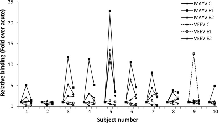
Recognition of MAYV antigens by antibodies from human MAYV infections. Serum samples from patients (n = 10) with PCR-confirmed MAYV infections were used to probe protein microarrays consisting of MAYV and VEEV antigens to detect IgG binding. Acute-phase, convalescent-phase, and follow-up serum samples were analyzed for subjects 1 to 9, while acute-phase and follow-up serum samples were analyzed for subject 10. For each individual, antigen signal levels were plotted relative to the acute-phase signal level.
TABLE 4 .
Human infection serum samples analyzed by protein microarrays
| Infection | Lineage | No. of serum samples |
Country(ies) | Time frame | Days after acute- phase diagnosis (mean ± SD) |
|---|---|---|---|---|---|
| VEEV | ID | 10 | Peru | Apr 2013–Jul 2013 |
10−30 (18 ± 8) |
| CHIKV | NDa | 10 | Colombia, Venezuela |
Jul 2014–Dec 2014 |
11−27 (19 ± 5) |
| Influenza virus | A | 10 | Peru | Aug 2012–Sept 2014 |
12−28 (18 ± 5) |
ND, not determined.
FIG 4 .

Antibody specificity of human alphavirus infections to alphavirus structural antigens. Serum samples from patients with alphavirus or influenza A virus (Flu A) infections were used to probe alphavirus protein microarrays to detect specific antigen-IgG binding. All serum samples, except one MAYV follow-up sample, were collected within 10 to 30 days of infection confirmation by RT-PCR. The signal levels of the alphavirus-infected subjects were compared to the mean signal level of each antigen in the influenza A virus group. Signal ratios were calculated as described in Materials and Methods. (A) Graphs showing individual and mean signal ratios for 10 patients per alphavirus infection group for MAYV (top), VEEV (middle), and CHIKV (bottom) with error bars representing the standard error of the mean. Vertical lines separate antigens into C, E1, and E2 groups. Horizontal dotted lines indicate an alphavirus infection signal to influenza A virus signal ratio of 1. (B) M statistics were used to identify antigens with significant recognition by antibodies in the alphavirus versus influenza A virus infection groups. Antigens with significant signal levels in the alphavirus infection groups, prevalence, and statistical significance are shown.
DISCUSSION
This report describes an analysis of serological immune responses to human MAYV, VEEV, and CHIKV infections that occurred in Peru, Colombia, and Venezuela from 2011 to 2014. The results were obtained with a high-throughput microarray that incorporated structural proteins from nine medically important alphaviruses, including antigens derived from alphaviruses that are not currently endemic to South America. One objective was to determine if sufficient assay specificity could be obtained with the viral structural proteins selected for inclusion in the microarray. Our results indicated that antibody cross-reactivity with reference mouse and rabbit antisera correlated with antigens derived from closely related species within the same antigen serocomplex. Because previous reports suggested that human MAYV serum samples cross-react with CHIKV and RRV antigens in ELISAs (38–40), another goal of the study presented here was to provide a more detailed analysis of antigen cross-reactivity. In contrast to the results obtained with animal reference antisera, we observed a high degree of specificity for the three human infection groups examined that was primarily based on independently printed E1 and E2 antigens.
Generally, outbreaks of alphavirus infections in the Americas have been sporadic and contained. However, cocirculation of closely related MAYV, VEEV, and CHIKV presents the potential for an increased public health concern. Before CHIKV spread throughout South America, one study of febrile illness etiologies encompassing multiple sites in Bolivia, Ecuador, Paraguay, and Peru found evidence of MAYV and VEEV infections in up to 8.2 or 7.0% of the subjects tested, respectively (17). Additional reports found MAYV exposure in at least 34% of the subjects at certain study sites (41, 42), while VEEV exposure was reported in up to 84% of the populations studied (18). After the recent spread of CHIKV throughout the New World, a study of acute febrile illness in Brazil identified CHIKV antibodies in 5 (6.7%) out of 75 DENV-negative samples and MAYV antibodies were detected in 15 (55.6%) out of 27 DENV- and CHIKV-negative samples (43). Misdiagnosis, especially in countries where DENV is endemic, can be problematic. For example, a study of acute febrile illness patients in Brazil with a clinical diagnosis of DENV found that infections in 13% of 46 arbovirus nucleic acid-positive samples were caused by MAYV (44). Misdiagnosis or immune interference caused by cocirculating viruses is more likely with the arrival and widespread occurrence of CHIKV, as recently noted for MAYV infections occurring in Brazil between December 2014 and January 2016 that were misreported as CHIKV infections (45). Further, arthritogenic CHIKV and MAYV should remain on surveillance watch lists for potential involvement in disease outbreaks, and improved surveillance tools are needed for this purpose. Interestingly, human serum samples from natural infections used in our study demonstrated highly specific antibody recognition with recombinant antigen probes, while we found a greater level of cross-reactivity with the reference mouse polyclonal antibodies. For MAYV infections, responses to E1 appeared to dominate the serological immune response, and 90% of the serum samples from MAYV infections recognized at least one of the MAYV envelope proteins, while antibodies from two individuals recognized only E1 or E2. The mouse data also suggested that the MAYV and RRV antigens share a low level of antibody recognition, yet we did not observe significant antigen cross-reactivity with serum samples from human MAYV infections or those of any other Semliki Forest complex members. Further, we observed specific antibody responses to VEEV E2 from the epizootic IAB lineage with 80% of the infections that were caused by enzootic VEEV subtype ID, and neither significant cross-reactivity to other E2 antigens nor human serum recognition of VEEV E1 or C was observed. Although differences in VEEV IAB and ID antigen sequences may account for reduced C and E1 recognition, further studies are needed to determine this conclusively. We noted that antibodies of some subjects in the MAYV disease group recognized the more distantly related EEEV E1 or VEEV E2 protein. Evaluations of EEEV infection patients, which were not included in the study described here, may help to determine if EEEV E1 antigen recognition by the MAYV serum is due to a low level of antibody cross-reactivity or prior alphavirus exposure. Recognition of VEEV E2 by convalescent-phase serum from one MAYV infection subject, without an apparent rise in levels of IgG for VEEV E2 compared to the acute-phase serum, indicated a possible history of prior exposure to VEEV. VEEV and MAYV are known to cocirculate, and one study showed that approximately 10% of VEEV infection serum samples also had MAYV IgM (17). These results suggest that the protein microarray can be used to detect possible prior exposures to alphaviruses, as well as increased antibody-antigen recognition from recent infections. Only E2 antigens of all three CHIKV lineages and ONNV were recognized by CHIKV human serum, in contrast to the reference mouse and rabbit antisera that also recognized E1 and C. As ONNV is the nearest neighbor of CHIKV, some degree of antibody cross-reactivity was not unexpected. Some reports have detected CHIKV C, E1, and/or E2 antibody recognition by serum samples from human CHIKV patients (36, 46–48), while the recognition of each antigen may be dependent on the sampling time postinfection. A possible change in specific antibody levels over time may explain why we observed antibody recognition of CHIKV E2 but not C or E1. Taken together, these data show that a side-by-side comparisons of antigen recognition in the multiplexed platform can be used to detect infection-specific antigen recognition patterns and to distinguish diseases caused by related viruses.
The emergence and spread of alphaviruses occur through genetic changes generated during viral replication that can lead to adaptation to new vector or host species (49–53). The global expansion of CHIKV, for example, was facilitated in part by adaptive genetic changes that produced increased vector competency in the urban mosquitos Aedes aegypti and A. albopictus (49–51, 54), which feed on human hosts. Interestingly, laboratory studies concluded that MAYV and VEEV could be transmitted by A. aegypti and A. albopictus (55–60), though neither virus has been confirmed to be naturally spread by these mosquitoes. Multiplexed surveillance assays that include both endemic and nonendemic viruses will be important for monitoring disease outbreaks and spread to new geographic regions. Further, the ability to expand this multiplex assay by the addition of new antigens that are synthesized from sequence data will provide a means to rapidly incorporate assays for new pathogens into disease surveillance matrices (29–31, 61).
MATERIALS AND METHODS
Ethics statement.
Research on human subjects was conducted in compliance with Department of Defense, Federal, and State statutes and regulations relating to the protection of human subjects and adhered to principles identified in the Belmont report (62). All of the human samples and associated data used in this study were gathered under an institutional review board-approved protocol, no. NAMRU6.2010.0010, and were authorized by study volunteers for future use. Use of the samples for retrospective analysis in this study was determined as not human subject research by the NAMRU-6 Research Administration Program (NAMRU6.2016.0001) and the USAMRIID Office of Human Use and Ethics (FY16-07).
Study cohort and sample collection.
Samples were collected and archived as part of a study to determine the etiology of febrile illnesses in Latin America. The criteria for inclusion in this study required subjects to present to selected health facilities with an elevated temperature (≥38°C oral, tympanic, or rectal; ≥37.5°C axillary) that had lasted no more than 5 days. Patients who presented with readily diagnosable sources of infection or who were not 5 years old or older were excluded. The sample subset used in this study was collected from patients in Peru (MAYV, VEEV, and influenza A virus) or Colombia and Venezuela (CHIKV). Acute-phase samples were tested for arboviral infections by isolation and/or RT-PCR (Table 2) as previously described (11, 17, 23). For positive samples, convalescent-phase samples were collected within 10 to 30 days postdiagnosis and EIAs for IgM and IgG were performed as previously described (Table 2) (11, 17). Follow-up samples were collected from MAYV-infected patients approximately 3 months after the acute infection phase.
Alphavirus gene cloning.
Synthetic genes or alphavirus cDNAs were used for PCR amplification and cloning of alphavirus genes for the expression of C, E1, and E2 proteins from nine alphavirus species (Table 1; Table S1). Cloning was designed such that all C-encoding genes expressed full-length proteins, while all E1- and E2-encoding genes were truncated to express the following regions of the ectodomains: E1 domains I and II (starting with the first proline at the N terminus) and E2 domains A, B, and C (Fig. 1; Fig. S1). To clone E1 and E2 from Asian CHIK, VEEV, MAYV, and RRV, cDNA templates for PCR were produced by RT with viral RNAs and the SuperScript III First-Strand Synthesis System (Invitrogen, Carlsbad, CA). Asian CHIKV (strain185/25) and VEEV (strain TC-83) RNAs were obtained from Integrated BioTherapeutics, Inc. (Rockville, MD). For MAYV RNA, Phase Lock Gel-Heavy tubes (Eppendorf, Hauppauge, NY) were used with a MAYV TRIzol preparation (strain TRVL 4675) obtained from the World Reference Center for Emerging Viruses and Arboviruses/National Institute of Allergy and Infectious Diseases (WRCEVA/NIAID; Robert Tesh). RRV RNA was extracted from freeze-dried infected suckling mouse brain (strain T48) purchased from the American Type Culture Collection (Manassas, VA) with the QIAamp Viral RNA Minikit (Qiagen, Germantown, MD). The PCR amplification product of the EEEV C-encoding gene was kindly provided by Pamela Glass (USAMRIID). Gene synthesis was used to produce templates for the following targets (Table 1): C (Asian CHIKV, ECSA CHIKV, WAf CHIKV, MAYV, RRV, ONNV, VEEV, and WEEV), E1, and E2 (ECSA CHIKV, WAf CHIKV, ONNV, EEEV, and WEEV). Most synthesized genes were provided as gene fragments produced by Integrated DNA Technologies, Inc. (Coralville, IA), or Life Technologies, Inc. (Carlsbad, CA), except the EEEV and WEEV E2-encoding genes, which were synthesized and cloned into pUC57 (GenScript, Piscataway, NJ). All synthesized genes were codon optimized for expression in Escherichia coli, except those for E1 and E2 of ECSA CHIK, WAf CHIK, EEEV, and WEEV. PCR amplification of genes was performed with Phusion High-Fidelity PCR master mix (New England Biolabs, Ipswich, MA), and purified PCR products were inserted into the pENTR/TEV/D-TOPO plasmid (Life Technologies, Inc.) by TOPO cloning. Colony PCR and sequencing were performed to identify entry clones with the appropriate insert. Sequence-verified entry clones were shuttled into pDEST17 (Life Technologies, Inc.), an N-terminally 6×His-tagged expression plasmid, with LR Clonase II (Life Technologies, Inc.).
Alphavirus protein expression and purification.
Sequence-verified alphavirus constructs were expressed in E. coli BL21-AI by using Luria broth (300 ml) supplemented with 100 μg/ml ampicillin and 0.1% glucose. Proteins were induced at mid-log phase with the addition of 0.2% arabinose. Optimal induction conditions were determined for each protein, and bacteria were grown at either 30°C (3 to 4 h) or 18°C (18 to 20 h) (CHIK E1tr, 25°C for 18 h) prior to harvesting by centrifugation. Cell pellets were stored at −80°C prior to lysis with 10 ml of Bacterial Protein Extraction Reagent (Thermo, Fisher Scientific, Waltham, MA) containing 0.2 mg/ml lysozyme (Sigma-Aldrich, St. Louis, MO), EDTA-free 2× Halt protease inhibitor cocktail (Thermo, Fisher Scientific), 2 mM phenylmethylsulfonyl fluoride, and 100 U of DNase I (Pierce). Soluble and insoluble proteins were separated by centrifugation (15,000 × g, 10 min) and analyzed by SDS-PAGE (Bio-Rad, Hercules, CA), followed by Coomassie staining and Western blot analysis with a mouse anti-polyhistidine monoclonal antibody (clone HIS-1; Sigma-Aldrich) detected with horseradish peroxidase-conjugated goat anti-mouse antibody (Bio-Rad). All expressed proteins were present in inclusion bodies and purified as previously described, with some modifications (29, 63). Briefly, insoluble protein pellets were washed twice with a buffer consisting of 50 mM Tris-HCl (pH 7.4), 1 M urea, and 1% Triton X-100 and then washed once with 50 mM Tris-HCl (pH 7.4) with centrifugation at 15,000 × g for 10 min between washing steps. Pellets were resuspended in 50 mM Tris-HCl (pH 7.4) prior to storage at −80°C. Purified inclusion body suspensions were thawed and centrifuged at 17,000 × g for 10 min. Pellets were resuspended in solubilization buffer containing 50 mM HEPES (pH 7.3), 140 mM NaCl, 2 mM dl-dithiothreitol (DTT), and 1% sodium dodecyl sulfate (SDS) and heated to 99°C for 5 to 15 min. Solubilized protein was centrifuged at 17,000 × g for 10 min, and glycerol (25%) was added to the supernatant prior to storage at −80°C. Solubilized proteins were analyzed by SDS-PAGE, and protein concentration and purity were determined with the Agilent protein 230 kit and a Bioanalyzer 2100 instrument (Agilent Technologies, Santa Clara, CA).
Alphavirus protein microarrays.
Prior to printing of purified recombinant antigens, samples were diluted to 200 ng/μl in protein microarray printing buffer (50 mM HEPES, 140 mM NaCl, 2 mM DTT, pH 7.3) with glycerol (40%). Replicate spots (n = 6) of alphavirus proteins and controls were printed onto microporous-nitrocellulose-coated slides (ONCYTE SuperNOVA; Grace Bio-Labs, Inc., Bend, OR) with an Inkjet microarray printer (ArrayJet, Roslin, United Kingdom) at 65% humidity. Control proteins included bovine serum albumin (BSA), E. coli-expressed Rift Valley fever virus glycoprotein, and antigens from DENV and influenza virus hemagglutinin proteins (Immune Technology Corp., New York, NY), along with human, mouse, monkey, goat, and rabbit IgG (Rockland Immunochemicals Inc., Limerick, PA). The printed microarray slides were desiccated (12 h) and stored frozen (−20°C) until use. Spot deposition and the quality of spotted proteins were determined with SYPRO Ruby protein stain and mouse anti-polyhistidine monoclonal antibody (clone HIS-1; Sigma-Aldrich).
Protein microarray assays.
Mouse polyclonal antibodies were provided by the WRCEVA/NIAID and were produced as mouse hyperimmune ascitic fluid with intraperitoneal injections of alphavirus-infected newborn mouse brain homogenate (R. Tesh, personal communication). The strains used to generate mouse polyclonal antibodies were CHIKV Ross (ECSA lineage), ONNV MP-30, VEEV TC-83 (IAB lineage), EEEV Alabama, and others not specified. Rabbit polyclonal antibodies that were raised against sucrose-purified CHIKV 181/25 or a WEEV E2 peptide were obtained from Integrated BioTherapeutics, Inc. (Rockville, MD). Assay steps were performed at 22°C protected from light. Slides were blocked for 1 h with Super G blocking buffer (Grace Bio-Labs, Inc.) and washed three times (5 min each time) with wash buffer (1× PBS [pH 7.4], 0.2% Tween 20, 1% BSA) prior to the addition of samples. Anti-alphavirus mouse serum (1:50), anti-CHIKV and -WEEV E2 polyclonal antibodies (1:500), and human serum samples (1:150) were diluted in probe buffer (1× PBS [pH 7.4], 0.1% Tween 20, 1% BSA) and incubated with E. coli lysate (1 mg/ml; Promega, Madison, WI) for 1 h with gentle mixing to preclear the samples of nonspecific antibodies. Samples were pelleted at 17,000 × g for 5 min, and the cleared supernatant was added to the microarrays. Samples were incubated for 1.5 h with gentle rocking and then washed five times (5 min each time) with wash buffer. Alexa Fluor 647-conjugated secondary antibodies were diluted in probe buffer and used to detect antibodies bound to the microarrayed antigens. Goat anti-mouse IgG (1:2,000; Invitrogen), goat anti-rabbit IgG (1:2,000; Invitrogen), or goat anti-human γ-specific IgG (1:1,000; Southern Biotechnologies, Birmingham, AL) was incubated with the arrays for 1 h. Arrays were washed with wash buffer (3×, 5 min each time), rinsed with ultrapure water (2×, 2 min each time), and dried (16 h, 22°C).
Microarray data acquisition and analysis.
Microarray slides were imaged with a GenePix 4400A confocal laser scanner (Molecular Devices, Sunnyvale, CA) at a wavelength of 635 nm. GenePix Pro 7 software was used to obtain local background-subtracted median fluorescence intensity for all data analyses. For mouse or rabbit polyclonal antibodies, scanner settings (power and gain) were adjusted for an optimal signal without saturation (>65,000 relative fluorescence units [RFU]) of antigen spots. Outliers among the data replicates that were identified with a modified Z score (median absolute deviation, >3.5) were excluded from the final analyses. Cutoff values for each assay were determined from the fluorescence signals of the antigen control spots and were calculated as the mean plus 3 standard deviations. Antigen SCRs were calculated, and an SCR of >1 was considered positive. For human serum samples, the scanner power was set to 100 and the gain setting was adjusted for each infection group to achieve an optimal signal without antigen signal saturation for any of the samples. Intra-array normalization of data from the acute-phase, convalescent-phase, or follow-up MAYV samples was done by using the negative-control spots. Subsequently, for each individual, the acute-phase antigen signal levels were set to 1 and the antigen signal levels from the convalescent-phase and follow-up time points were adjusted relative to the acute-phase antigen signal levels. For comparisons of convalescent-phase human alphavirus infection sample groups to the influenza A virus-infected group, ProtoArray Prospector v5.1 (Invitrogen) was used to quantile normalize the data. An M statistics analysis, with a minimal signal of 500 RFU and a minimal signal gap of 200 RFU, was used for serum group comparisons to identify antigens with significant reactivity. Microarrays probed with serum samples from the influenza A virus group or an alphavirus-naive study volunteer cohort from the United States were compared to confirm that the influenza A virus group did not have significant recognition of the printed alphavirus proteins (data not shown). Further, antigens that were significantly recognized by antibodies among the alphavirus infection group compared to the influenza A virus control group were identified on the basis of the M statistics test (ProtoArray Prospector v5.1). Signal ratios were determined after data outliers were identified and removed, as described above, to compare the antibody-binding signal levels of the alphavirus infection groups and the influenza A virus group. The antibody-binding signal levels in each array probed with serum samples from the alphavirus infection groups were divided by the mean signal levels from the influenza A virus group.
Sequence alignments and molecular phylogeny.
Multiple-sequence alignments of the C, E1, and E2 amino acid sequences of the nine alphaviruses represented on the protein microarray were generated. Three alignments generated by Clustal W2 (64) by using BLOSUM62 as the substitution matrix and gap opening penalties of 5, 10, and 25 with all other settings as default were applied in T-Coffee Combine (65, 66) to obtain a single optimized alignment. Sequence identities and similarities in the optimized alignment were determined by using the sequence identity matrix and pairwise alignment features of BioEdit software (v7.2.5) (67). Phylogenetic trees were generated by using the PhyML interface (v3.0) for maximum-likelihood trees (68) with the BLOSUM62 matrix, and the number of substitution rate categories was set to four. The starting tree was BioNJ with optimization by using tree topology and branch length and tree improvement using subtree pruning and regrafting. Phylogram images were generated by using the Newick format tree output in TreeView v1.6.6 (69).
ACKNOWLEDGMENTS
We thank Robert Tesh of WRCEVA/NIAID for kindly providing the mouse polyclonal antibodies and MAYV TRIzol preparation. We thank the DIRESA Loreto, the Instituto Nacional de Salud, and the Peruvian Ministry of Health for their support in establishing and maintaining the passive surveillance activities in Iquitos, Peru. We also thank Moo-Jin Suh for mass spectrometric analysis of proteins, Pamela J. Glass for providing the PCR product for EEEV C cloning, Cyra M. Ranji for technical assistance, and Sarah Jenkins for help with the management of the NAMRU-6 nonhuman subject protocol.
This research was supported in part by the National Academies of Sciences, Engineering, and Medicine through a National Research Council research associateship awarded to J.L.S. Grant support for this project was provided in part by the NIAID (R01AI096215 to R.G.U.). Collection of samples from patients in South America was performed with support from the Armed Forces Health Surveillance Center-Global Emerging Infections Surveillance (AFHSC-GEIS) (80000.82000.25GB.B0016 and 847705.82000.25GB.B0016).
The opinions, interpretations, and conclusions in this report are those of the authors and do not necessarily reflect the official policy or position of the United States Army, the Department of the Navy, the Department of Defense, or the United States Government.
REFERENCES
- 1.Pan American Health Organization 2017. Chikungunya: data, maps and statistics. Pan American Health Organization, Washington, DC: http://www.paho.org/hq/index.php?option=com_topics&view=readall&cid=5927&Itemid=40931&lang=en. Accessed: 5 September 2017. [Google Scholar]
- 2.Lumsden WH. 1955. An epidemic of virus disease in Southern Province, Tanganyika Territory, in 1952–53. II. General description and epidemiology. Trans R Soc Trop Med Hyg 49:33–57. [DOI] [PubMed] [Google Scholar]
- 3.Robinson MC. 1955. An epidemic of virus disease in Southern Province, Tanganyika Territory, in 1952–53. I. Clinical features. Trans R Soc Trop Med Hyg 49:28–32. [DOI] [PubMed] [Google Scholar]
- 4.Ross RW. 1956. The Newala epidemic. III. The virus: isolation, pathogenic properties and relationship to the epidemic. J Hyg (Lond) 54:177–191. doi: 10.1017/S0022172400044442. [DOI] [PMC free article] [PubMed] [Google Scholar]
- 5.Centers for Disease Control and Prevention 2016. Chikungunya virus: geographic distribution. Centers for Disease Control and Prevention, Atlanta, GA. https://www.cdc.gov/chikungunya/geo/index.html. Accessed: 5 September 2017. [Google Scholar]
- 6.Powers AM, Brault AC, Tesh RB, Weaver SC. 2000. Re-emergence of chikungunya and o’nyong-nyong viruses: evidence for distinct geographical lineages and distant evolutionary relationships. J Gen Virol 81:471–479. doi: 10.1099/0022-1317-81-2-471. [DOI] [PubMed] [Google Scholar]
- 7.Calisher CH, Karabatsos K. 1988. Arbovirus serogroups: definition and geographic distribution, p 19–57. In Monath TP (ed), The arboviruses: epidemiology and ecology, vol. 1 CRC Press, Boca Raton, FL. [Google Scholar]
- 8.Calisher CH, Shope RE, Brandt W, Casals J, Karabatsos N, Murphy FA, Tesh RB, Wiebe ME. 1980. Proposed antigenic classification of registered arboviruses I. Togaviridae, Alphavirus. Intervirology 14:229–232. doi: 10.1159/000149190. [DOI] [PubMed] [Google Scholar]
- 9.Hotez PJ, Murray KO. 2017. Dengue, West Nile virus, chikungunya, Zika—and now Mayaro? PLoS Negl Trop Dis 11:e0005462. doi: 10.1371/journal.pntd.0005462. [DOI] [PMC free article] [PubMed] [Google Scholar]
- 10.McClain DJ, Pittman PR, Ramsburg HH, Nelson GO, Rossi CA, Mangiafico JA, Schmaljohn AL, Malinoski FJ. 1998. Immunologic interference from sequential administration of live attenuated alphavirus vaccines. J Infect Dis 177:634–641. doi: 10.1086/514240. [DOI] [PubMed] [Google Scholar]
- 11.Halsey ES, Siles C, Guevara C, Vilcarromero S, Jhonston EJ, Ramal C, Aguilar PV, Ampuero JS. 2013. Mayaro virus infection, Amazon Basin region, Peru, 2010–2013. Emerg Infect Dis 19:1839–1842. doi: 10.3201/eid1911.130777. [DOI] [PMC free article] [PubMed] [Google Scholar]
- 12.Santiago FW, Halsey ES, Siles C, Vilcarromero S, Guevara C, Silvas JA, Ramal C, Ampuero JS, Aguilar PV. 2015. Long-term arthralgia after Mayaro virus infection correlates with sustained pro-inflammatory cytokine response. PLoS Negl Trop Dis 9:e0004104. doi: 10.1371/journal.pntd.0004104. [DOI] [PMC free article] [PubMed] [Google Scholar]
- 13.Torres JR, Russell KL, Vasquez C, Barrera R, Tesh RB, Salas R, Watts DM. 2004. Family cluster of Mayaro fever, Venezuela. Emerg Infect Dis 10:1304–1306. doi: 10.3201/eid1007.030860. [DOI] [PMC free article] [PubMed] [Google Scholar]
- 14.Muñoz M, Navarro JC. 2012. Mayaro: a re-emerging arbovirus in Venezuela and Latin America. Biomedica 32:286–302. (In Spanish.) doi: 10.1590/S0120-41572012000300017. [DOI] [PubMed] [Google Scholar]
- 15.Auguste AJ, Liria J, Forrester NL, Giambalvo D, Moncada M, Long KC, Morón D, de Manzione N, Tesh RB, Halsey ES, Kochel TJ, Hernandez R, Navarro JC, Weaver SC. 2015. Evolutionary and ecological characterization of Mayaro virus strains isolated during an outbreak, Venezuela, 2010. Emerg Infect Dis 21:1742–1750. doi: 10.3201/eid2110.141660. [DOI] [PMC free article] [PubMed] [Google Scholar]
- 16.Lednicky J, De Rochars VM, Elbadry M, Loeb J, Telisma T, Chavannes S, Anilis G, Cella E, Ciccozzi M, Okech B, Salemi M, Morris JG Jr.. 2016. Mayaro virus in child with acute febrile illness, Haiti, 2015. Emerg Infect Dis 22:2000–2002. doi: 10.3201/eid2211.161015. [DOI] [PMC free article] [PubMed] [Google Scholar]
- 17.Forshey BM, Guevara C, Laguna-Torres VA, Cespedes M, Vargas J, Gianella A, Vallejo E, Madrid C, Aguayo N, Gotuzzo E, Suarez V, Morales AM, Beingolea L, Reyes N, Perez J, Negrete M, Rocha C, Morrison AC, Russell KL, Blair PJ, Olson JG, Kochel TJ, NMRCD Febrile Surveillance Working Group . 2010. Arboviral etiologies of acute febrile illnesses in western South America, 2000–2007. PLoS Negl Trop Dis 4:e787. doi: 10.1371/journal.pntd.0000787. [DOI] [PMC free article] [PubMed] [Google Scholar]
- 18.Aguilar PV, Estrada-Franco JG, Navarro-Lopez R, Ferro C, Haddow AD, Weaver SC. 2011. Endemic Venezuelan equine encephalitis in the Americas: hidden under the dengue umbrella. Future Virol 6:721–740. doi: 10.2217/FVL.11.5. [DOI] [PMC free article] [PubMed] [Google Scholar]
- 19.Carrera JP, Forrester N, Wang E, Vittor AY, Haddow AD, López-Vergès S, Abadía I, Castaño E, Sosa N, Báez C, Estripeaut D, Díaz Y, Beltrán D, Cisneros J, Cedeño HG, Travassos da Rosa AP, Hernandez H, Martínez-Torres AO, Tesh RB, Weaver SC. 2013. Eastern equine encephalitis in Latin America. N Engl J Med 369:732–744. doi: 10.1056/NEJMoa1212628. [DOI] [PMC free article] [PubMed] [Google Scholar]
- 20.Arrigo NC, Adams AP, Weaver SC. 2010. Evolutionary patterns of eastern equine encephalitis virus in North versus South America suggest ecological differences and taxonomic revision. J Virol 84:1014–1025. doi: 10.1128/JVI.01586-09. [DOI] [PMC free article] [PubMed] [Google Scholar]
- 21.Bergren NA, Auguste AJ, Forrester NL, Negi SS, Braun WA, Weaver SC. 2014. Western equine encephalitis virus: evolutionary analysis of a declining alphavirus based on complete genome sequences. J Virol 88:9260–9267. doi: 10.1128/JVI.01463-14. [DOI] [PMC free article] [PubMed] [Google Scholar]
- 22.Forrester NL, Kenney JL, Deardorff E, Wang E, Weaver SC. 2008. Western equine encephalitis submergence: lack of evidence for a decline in virus virulence. Virology 380:170–172. doi: 10.1016/j.virol.2008.08.012. [DOI] [PMC free article] [PubMed] [Google Scholar]
- 23.Morrison AC, Forshey BM, Notyce D, Astete H, Lopez V, Rocha C, Carrion R, Carey C, Eza D, Montgomery JM, Kochel TJ. 2008. Venezuelan equine encephalitis virus in Iquitos, Peru: urban transmission of a sylvatic strain. PLoS Negl Trop Dis 2:e349. doi: 10.1371/journal.pntd.0000349. [DOI] [PMC free article] [PubMed] [Google Scholar]
- 24.Rivas F, Diaz LA, Cardenas VM, Daza E, Bruzon L, Alcala A, De la Hoz O, Caceres FM, Aristizabal G, Martinez JW, Revelo D, De la Hoz F, Boshell J, Camacho T, Calderon L, Olano VA, Villarreal LI, Roselli D, Alvarez G, Ludwig G, Tsai T. 1997. Epidemic Venezuelan equine encephalitis in la Guajira, Colombia, 1995. J Infect Dis 175:828–832. doi: 10.1086/513978. [DOI] [PubMed] [Google Scholar]
- 25.Weaver SC, Salas R, Rico-Hesse R, Ludwig GV, Oberste MS, Boshell J, Tesh RB. 1996. Re-emergence of epidemic Venezuelan equine encephalomyelitis in South America. VEE Study Group. Lancet 348:436–440. doi: 10.1016/S0140-6736(96)02275-1. [DOI] [PubMed] [Google Scholar]
- 26.Cunha RV, Trinta KS, Montalbano CA, Sucupira MVF, de Lima MM, Marques E, Romanholi IH, Croda J. 2017. Seroprevalence of chikungunya virus in a rural community in Brazil. PLoS Negl Trop Dis 11:e0005319. doi: 10.1371/journal.pntd.0005319. [DOI] [PMC free article] [PubMed] [Google Scholar]
- 27.Gay N, Rousset D, Huc P, Matheus S, Ledrans M, Rosine J, Cassadou S, Noël H. 2016. Seroprevalence of Asian lineage chikungunya virus infection on Saint Martin Island, 7 months after the 2013 emergence. Am J Trop Med Hyg 94:393–396. doi: 10.4269/ajtmh.15-0308. [DOI] [PMC free article] [PubMed] [Google Scholar]
- 28.Kuan G, Ramirez S, Gresh L, Ojeda S, Melendez M, Sanchez N, Collado D, Garcia N, Mercado JC, Gordon A, Balmaseda A, Harris E. 2016. Seroprevalence of anti-chikungunya virus antibodies in children and adults in Managua, Nicaragua, after the first chikungunya epidemic, 2014–2015. PLoS Negl Trop Dis 10:e0004773. doi: 10.1371/journal.pntd.0004773. [DOI] [PMC free article] [PubMed] [Google Scholar]
- 29.Keasey SL, Pugh CL, Jensen SM, Smith JL, Hontz RD, Durbin AP, Dudley DM, O’Connor DH, Ulrich RG. 2017. Antibody responses to Zika virus infections in environments of flavivirus endemicity. Clin Vaccine Immunol 24:e00036-17. doi: 10.1128/CVI.00036-17. [DOI] [PMC free article] [PubMed] [Google Scholar]
- 30.Natesan M, Jensen SM, Keasey SL, Kamata T, Kuehne AI, Stonier SW, Lutwama JJ, Lobel L, Dye JM, Ulrich RG. 2016. Human survivors of disease outbreaks caused by Ebola or Marburg virus exhibit cross-reactive and long-lived antibody responses. Clin Vaccine Immunol 23:717–724. doi: 10.1128/CVI.00107-16. [DOI] [PMC free article] [PubMed] [Google Scholar]
- 31.Kamata T, Natesan M, Warfield K, Aman MJ, Ulrich RG. 2014. Determination of specific antibody responses to the six species of Ebola and Marburg viruses by multiplexed protein microarrays. Clin Vaccine Immunol 21:1605–1612. doi: 10.1128/CVI.00484-14. [DOI] [PMC free article] [PubMed] [Google Scholar]
- 32.Strauss JH, Strauss EG. 1994. The alphaviruses: gene expression, replication, and evolution. Microbiol Rev 58:491–562. [DOI] [PMC free article] [PubMed] [Google Scholar]
- 33.Jose J, Snyder JE, Kuhn RJ. 2009. A structural and functional perspective of alphavirus replication and assembly. Future Microbiol 4:837–856. doi: 10.2217/fmb.09.59. [DOI] [PMC free article] [PubMed] [Google Scholar]
- 34.Mukhopadhyay S, Zhang W, Gabler S, Chipman PR, Strauss EG, Strauss JH, Baker TS, Kuhn RJ, Rossmann MG. 2006. Mapping the structure and function of the E1 and E2 glycoproteins in alphaviruses. Structure 14:63–73. doi: 10.1016/j.str.2005.07.025. [DOI] [PMC free article] [PubMed] [Google Scholar]
- 35.Dalrymple JM, Vogel SN, Teramoto AY, Russell PK. 1973. Antigenic components of group A arbovirus virions. J Virol 12:1034–1042. [DOI] [PMC free article] [PubMed] [Google Scholar]
- 36.Kam YW, Lee WW, Simarmata D, Harjanto S, Teng TS, Tolou H, Chow A, Lin RT, Leo YS, Rénia L, Ng LF. 2012. Longitudinal analysis of the human antibody response to chikungunya virus infection: implications for serodiagnosis and vaccine development. J Virol 86:13005–13015. doi: 10.1128/JVI.01780-12. [DOI] [PMC free article] [PubMed] [Google Scholar]
- 37.Kam YW, Simarmata D, Chow A, Her Z, Teng TS, Ong EK, Rénia L, Leo YS, Ng LF. 2012. Early appearance of neutralizing immunoglobulin G3 antibodies is associated with chikungunya virus clearance and long-term clinical protection. J Infect Dis 205:1147–1154. doi: 10.1093/infdis/jis033. [DOI] [PMC free article] [PubMed] [Google Scholar]
- 38.Calisher CH, el-Kafrawi AO, Al-Deen Mahmud MI, Travassos da Rosa AP, Bartz CR, Brummer-Korvenkontio M, Haksohusodo S, Suharyono W. 1986. Complex-specific immunoglobulin M antibody patterns in humans infected with alphaviruses. J Clin Microbiol 23:155–159. [DOI] [PMC free article] [PubMed] [Google Scholar]
- 39.Hassing RJ, Leparc-Goffart I, Tolou H, van Doornum G, van Genderen PJ. 2010. Cross-reactivity of antibodies to viruses belonging to the Semliki Forest serocomplex. Euro Surveill 15:19588 (Letter.) http://www.eurosurveillance.org/content/10.2807/ese.15.23.19588-en. [PubMed] [Google Scholar]
- 40.Prat CM, Flusin O, Panella A, Tenebray B, Lanciotti R, Leparc-Goffart I. 2014. Evaluation of commercially available serologic diagnostic tests for chikungunya virus. Emerg Infect Dis 20:2129–2132. doi: 10.3201/eid2012.141269. [DOI] [PMC free article] [PubMed] [Google Scholar]
- 41.Talarmin A, Chandler LJ, Kazanji M, de Thoisy B, Debon P, Lelarge J, Labeau B, Bourreau E, Vié JC, Shope RE, Sarthou JL. 1998. Mayaro virus fever in French Guiana: isolation, identification, and seroprevalence. Am J Trop Med Hyg 59:452–456. doi: 10.4269/ajtmh.1998.59.452. [DOI] [PubMed] [Google Scholar]
- 42.Azevedo RS, Silva EV, Carvalho VL, Rodrigues SG, Nunes-Neto JP, Monteiro H, Peixoto VS, Chiang JO, Nunes MR, Vasconcelos PF. 2009. Mayaro fever virus, Brazilian Amazon. Emerg Infect Dis 15:1830–1832. doi: 10.3201/eid1511.090461. [DOI] [PMC free article] [PubMed] [Google Scholar]
- 43.Brunini S, França DDS, Silva JB, Silva LN, Silva FPA, Spadoni M, Rezza G. 2017. High frequency of Mayaro virus IgM among febrile patients, central Brazil. Emerg Infect Dis 23:1025–1026. doi: 10.3201/eid2306.160929. [DOI] [PMC free article] [PubMed] [Google Scholar]
- 44.Vieira CJ, Silva DJ, Barreto ES, Siqueira CE, Colombo TE, Ozanic K, Schmidt DJ, Drumond BP, Mondini A, Nogueira ML, Bronzoni RV. 2015. Detection of Mayaro virus infections during a dengue outbreak in Mato Grosso, Brazil. Acta Trop 147:12–16. doi: 10.1016/j.actatropica.2015.03.020. [DOI] [PubMed] [Google Scholar]
- 45.Esposito DLA, Fonseca BALD. 2017. Will Mayaro virus be responsible for the next outbreak of an arthropod-borne virus in Brazil? Braz J Infect Dis 21:540–544. doi: 10.1016/j.bjid.2017.06.002. [DOI] [PMC free article] [PubMed] [Google Scholar]
- 46.Cho B, Jeon BY, Kim J, Noh J, Kim J, Park M, Park S. 2008. Expression and evaluation of chikungunya virus E1 and E2 envelope proteins for serodiagnosis of chikungunya virus infection. Yonsei Med J 49:828–835. doi: 10.3349/ymj.2008.49.5.828. [DOI] [PMC free article] [PubMed] [Google Scholar]
- 47.Kowalzik S, Xuan NV, Weissbrich B, Scheiner B, Schied T, Drosten C, Müller A, Stich A, Rethwilm A, Bodem J. 2008. Characterisation of a chikungunya virus from a German patient returning from Mauritius and development of a serological test. Med Microbiol Immunol 197:381–386. doi: 10.1007/s00430-008-0090-5. [DOI] [PubMed] [Google Scholar]
- 48.Cho B, Kim J, Cho JE, Jeon BY, Park S. 2008. Expression of the capsid protein of chikungunya virus in a baculovirus for serodiagnosis of chikungunya disease. J Virol Methods 154:154–159. doi: 10.1016/j.jviromet.2008.07.031. [DOI] [PubMed] [Google Scholar]
- 49.Schuffenecker I, Iteman I, Michault A, Murri S, Frangeul L, Vaney MC, Lavenir R, Pardigon N, Reynes JM, Pettinelli F, Biscornet L, Diancourt L, Michel S, Duquerroy S, Guigon G, Frenkiel MP, Bréhin AC, Cubito N, Desprès P, Kunst F, Rey FA, Zeller H, Brisse S. 2006. Genome microevolution of chikungunya viruses causing the Indian Ocean outbreak. PLoS Med 3:e263. doi: 10.1371/journal.pmed.0030263. [DOI] [PMC free article] [PubMed] [Google Scholar]
- 50.Tsetsarkin KA, Vanlandingham DL, McGee CE, Higgs S. 2007. A single mutation in chikungunya virus affects vector specificity and epidemic potential. PLoS Pathog 3:e201. doi: 10.1371/journal.ppat.0030201. [DOI] [PMC free article] [PubMed] [Google Scholar]
- 51.Vazeille M, Moutailler S, Coudrier D, Rousseaux C, Khun H, Huerre M, Thiria J, Dehecq JS, Fontenille D, Schuffenecker I, Despres P, Failloux AB. 2007. Two chikungunya isolates from the outbreak of la Reunion (Indian Ocean) exhibit different patterns of infection in the mosquito, Aedes albopictus. PLoS One 2:e1168. doi: 10.1371/journal.pone.0001168. [DOI] [PMC free article] [PubMed] [Google Scholar]
- 52.Brault AC, Powers AM, Holmes EC, Woelk CH, Weaver SC. 2002. Positively charged amino acid substitutions in the E2 envelope glycoprotein are associated with the emergence of Venezuelan equine encephalitis virus. J Virol 76:1718–1730. doi: 10.1128/JVI.76.4.1718-1730.2002. [DOI] [PMC free article] [PubMed] [Google Scholar]
- 53.Brault AC, Powers AM, Ortiz D, Estrada-Franco JG, Navarro-Lopez R, Weaver SC. 2004. Venezuelan equine encephalitis emergence: enhanced vector infection from a single amino acid substitution in the envelope glycoprotein. Proc Natl Acad Sci U S A 101:11344–11349. doi: 10.1073/pnas.0402905101. [DOI] [PMC free article] [PubMed] [Google Scholar]
- 54.Weaver SC, Reisen WK. 2010. Present and future arboviral threats. Antiviral Res 85:328–345. doi: 10.1016/j.antiviral.2009.10.008. [DOI] [PMC free article] [PubMed] [Google Scholar]
- 55.Smith GC, Francy DB. 1991. Laboratory studies of a Brazilian strain of Aedes albopictus as a potential vector of Mayaro and Oropouche viruses. J Am Mosq Control Assoc 7:89–93. [PubMed] [Google Scholar]
- 56.Long KC, Ziegler SA, Thangamani S, Hausser NL, Kochel TJ, Higgs S, Tesh RB. 2011. Experimental transmission of Mayaro virus by Aedes aegypti. Am J Trop Med Hyg 85:750–757. doi: 10.4269/ajtmh.2011.11-0359. [DOI] [PMC free article] [PubMed] [Google Scholar]
- 57.Ortiz DI, Kang W, Weaver SC. 2008. Susceptibility of Ae. aegypti (Diptera: Culicidae) to infection with epidemic (subtype IC) and enzootic (subtypes ID, IIIC, IIID) Venezuelan equine encephalitis complex alphaviruses. J Med Entomol 45:1117–1125. doi: 10.1093/jmedent/45.6.1117. [DOI] [PubMed] [Google Scholar]
- 58.Turell MJ, Beaman JR. 1992. Experimental transmission of Venezuelan equine encephalomyelitis virus by a strain of Aedes albopictus (Diptera: Culicidae) from New Orleans, Louisiana. J Med Entomol 29:802–805. doi: 10.1093/jmedent/29.5.802. [DOI] [PubMed] [Google Scholar]
- 59.Beaman JR, Turell MJ. 1991. Transmission of Venezuelan equine encephalomyelitis virus by strains of Aedes albopictus (Diptera: Culicidae) collected in North and South America. J Med Entomol 28:161–164. doi: 10.1093/jmedent/28.1.161. [DOI] [PubMed] [Google Scholar]
- 60.Fernández Z, Moncayo AC, Carrara AS, Forattini OP, Weaver SC. 2003. Vector competence of rural and urban strains of Aedes (Stegomyia) albopictus (Diptera: Culicidae) from Sao Paulo State, Brazil for IC, ID, and IF subtypes of Venezuelan equine encephalitis virus. J Med Entomol 40:522–527. doi: 10.1603/0022-2585-40.4.522. [DOI] [PubMed] [Google Scholar]
- 61.Fernandez S, Cisney ED, Tikhonov AP, Schweitzer B, Putnak RJ, Simmons M, Ulrich RG. 2011. Antibody recognition of the dengue virus proteome and implications for development of vaccines. Clin Vaccine Immunol 18:523–532. doi: 10.1128/CVI.00016-11. [DOI] [PMC free article] [PubMed] [Google Scholar]
- 62.National Commission for the Protection of Human Subjects of Biomedical and Behavioral Research 1979. The Belmont report: ethical principles and guidelines for the protection of human subjects of research. National Commission for the Protection of Human Subjects of Biomedical and Behavioral Research, Bethesda, MD: https://videocast.nih.gov/pdf/ohrp_belmont_report.pdf. [Google Scholar]
- 63.Palmer I, Wingfield PT. 2012. Preparation and extraction of insoluble (inclusion-body) proteins from Escherichia coli. Curr Protoc Protein Sci Chapter 6:Unit6.3. doi: 10.1002/0471140864.ps0603s70. [DOI] [PMC free article] [PubMed] [Google Scholar]
- 64.Larkin MA, Blackshields G, Brown NP, Chenna R, McGettigan PA, McWilliam H, Valentin F, Wallace IM, Wilm A, Lopez R, Thompson JD, Gibson TJ, Higgins DG. 2007. Clustal W and Clustal X version 2.0. Bioinformatics 23:2947–2948. doi: 10.1093/bioinformatics/btm404. [DOI] [PubMed] [Google Scholar]
- 65.Di Tommaso P, Moretti S, Xenarios I, Orobitg M, Montanyola A, Chang JM, Taly JF, Notredame C. 2011. T-Coffee: a web server for the multiple sequence alignment of protein and RNA sequences using structural information and homology extension. Nucleic Acids Res 39:W13–W17. doi: 10.1093/nar/gkr245. [DOI] [PMC free article] [PubMed] [Google Scholar]
- 66.Notredame C, Higgins DG, Heringa J. 2000. T-Coffee: a novel method for fast and accurate multiple sequence alignment. J Mol Biol 302:205–217. doi: 10.1006/jmbi.2000.4042. [DOI] [PubMed] [Google Scholar]
- 67.Hall TA. 1999. BioEdit: a user-friendly biological sequence alignment editor and analysis program for Windows 95/98/NT. Nucleic Acids Symp Ser 1999:95–98. [Google Scholar]
- 68.Guindon S, Dufayard JF, Lefort V, Anisimova M, Hordijk W, Gascuel O. 2010. New algorithms and methods to estimate maximum-likelihood phylogenies: assessing the performance of PhyML 3.0. Syst Biol 59:307–321. doi: 10.1093/sysbio/syq010. [DOI] [PubMed] [Google Scholar]
- 69.Page RD. 1996. TreeView: an application to display phylogenetic trees on personal computers. Comput Appl Biosci 12:357–358. [DOI] [PubMed] [Google Scholar]
Associated Data
This section collects any data citations, data availability statements, or supplementary materials included in this article.
Supplementary Materials
Analyses of amino acid sequences of alphavirus proteins used in microarrays. Optimal multiple-sequence alignments were generated with the translated sequences of the cloned alphavirus genes and used for molecular phylogeny tree construction as described in Materials and Methods. Alignments of C (A), truncated E1 (B), and truncated E2 (C) were imported into BioEdit (v7.2.5) for shading and sequence identity and similarity analysis with the BLOSUM62 matrix. For shading, the threshold was set to 50% for identical (black) and similar (gray) residues. Residue numbering is indicated to the left and right of each sequence. Gaps are represented by dashes. Identity and similarity matrices (D) were constructed by using pairwise comparisons of the aligned sequences for C (top), truncated E1 (middle), and truncated E2 (bottom). The percent identity is unshaded, and the percent similarity is shaded in gray. Molecular phylogeny trees (E) were constructed for C (top left), truncated E1 (top right), and truncated E2 (bottom). Accession numbers for whole genomes most closely related to cloned sequences: Asian CHIKV, L37661; ECSA CHIKV, KT449801; WAf CHIKV, HM045816; MAYV, AF339482; ONNV, AF079456; RRV, DQ226993; VEEV, L01443; EEEV, AY705241; WEEV, GQ287645. Download FIG S1, PDF file, 0.2 MB (219.9KB, pdf) .
This is a work of the U.S. Government and is not subject to copyright protection in the United States. Foreign copyrights may apply.
Rabbit polyclonal antibody binding to alphavirus microarray antigens. Rabbit anti-CHIKV polyclonal antibody raised against whole inactivated CHIKV strain 181/25 (A) or rabbit anti-WEEV E2 peptide polyclonal antibody (B) was used to probe alphavirus protein microarrays. The ratio of the antigen signal to the cutoff value was determined for each replicate spot. Cutoff values were determined as the mean signal of the control proteins plus 3 standard deviations. The average ratio for replicate antigen spots was determined, and ratios >1 are shown with error bars representing standard deviations. Vertical lines separate antigens into C, E1, and E2 groups. Download FIG S2, PDF file, 0.03 MB (35.4KB, pdf) .
This is a work of the U.S. Government and is not subject to copyright protection in the United States. Foreign copyrights may apply.
Primer pairs used for cloning of alphavirus genes (5′-to-3′ direction). Download TABLE S1, PDF file, 0.1 MB (73.4KB, pdf) .
This is a work of the U.S. Government and is not subject to copyright protection in the United States. Foreign copyrights may apply.