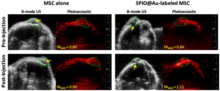
Figure 5.

In vivo photoacoustic (PA) imaging of mouse brain at 72 h after intra-carotid artery injection of SPIO@Au-labeled MSCs or MSCs alone. Yellow arrows on B-mode ultrasound (US) images of the brain indicate the bolt placement where the U87 cells were implanted into the brain. The PA images were taken at 810 nm, and the signal intensity was calculated within the green regions of interest. Average PA (PAAVE) values were obtained by averaging all PA intensity values above the signal-noise threshold within the regions of interest.
