Abstract
The present study is the first reported case of a patient undergoing esophagectomy with ectopic aortic arch secondary to a large esophageal cancer, which was pre-operatively misdiagnosed with a right-side aortic arch (RAA). The patient, a 54-year-old male, was first admitted to our hospital for esophagectomy owing to esophageal squamous cancer and had complained of progressive dysphasia for 3 months. Chest computed tomography (CT) revealed a mass in the middle thoracic esophagus. Furthermore, the three-dimensional CT of the thoracic great arteries showed a possible RAA and a curved descending aorta. After preoperative evaluation, the approach of using a left thoracotomy with cervical anastomosis was successfully performed and favorable short-term outcomes were achieved. According to previous reports, and the experience of the presented case, we emphasize clear recognition of the anatomical situation in the upper mediastinum and the importance of an optimal surgical approach for esophagectomy.
Keywords: Esophageal cancer, right-side aortic arch (RAA), left thoracotomy
Introduction
Several previous cases with deviation of the aortic arch in patients with esophageal carcinoma have been documented (1). Almost all previous cases were due to a scarce congenital anomaly: right-side aortic arch (RAA). However, we observed an acquired “RAA-like” patient resulting from a large esophageal cancer which was pre-operatively misdiagnosed as a RAA. Through the left thoracotomy and cervical anastomosis, complete resection of the tumor and pertinent mediastinal lymph nodes was performed and favorable short-term outcomes were achieved during the follow-up period.
Case presentation
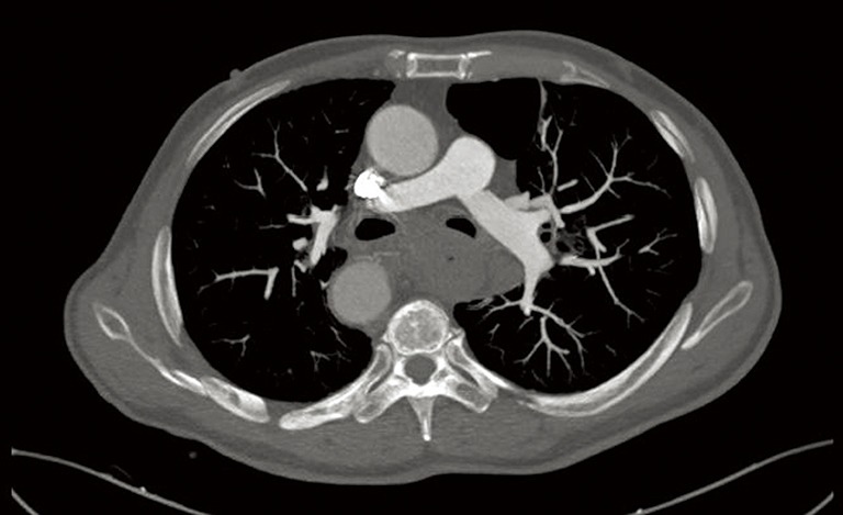
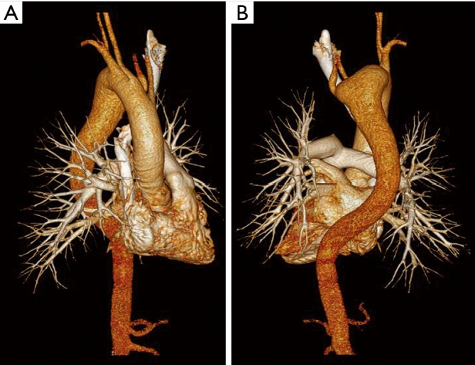
In March 2016, a 54-year-old male was admitted to our hospital with esophageal squamous cancer and the chief complaint of progressive dysphasia which had persisted for3 months. Before admission, esophageal endoscopy and concomitant biopsy were performed at a local hospital which revealed a squamous cancer in the middle third of the esophagus. In addition, his thoracic enhanced computed tomography (CT) on admission suggested a large esophageal cancer (Figure 1). The three-dimensional CT of the thoracic great arteries indicated a possible RAA and a curved descending aorta (Figure 2). Accordingly, the initial diagnosis of esophageal cancer associated with RAA was established.
Figure 1.

Contrast-enhanced CT scan of the chest showed an esophageal mass with a vague border. CT, computed tomography.
Figure 2.

Three-dimensional CT scan demonstrated the right transposition of the aortic arch and the curved descending aorta. (A) Frontal; (B) posterior. CT, computed tomography.
The patient received 3 cycles of neoadjuvant chemotherapy consisting of etoposide plus cisplatin, with an intention to proceed with resection if the tumor responded sufficiently. Unfortunately, there was a minor efficacy. After a third round of chemotherapy, the patient required surgical treatment in order to relieve obstruction of the digestive tract.
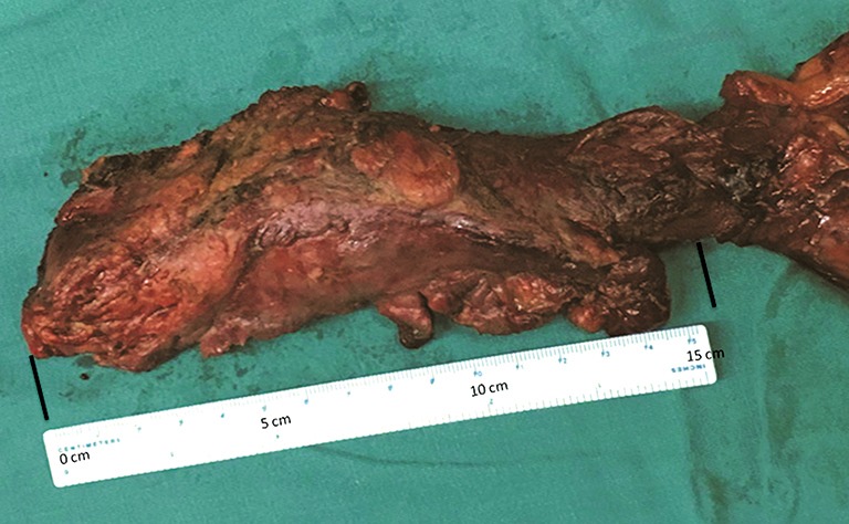
In consideration of the putative congenital RAA and the tumor size shown by chest CT, left thoracotomy was performed in the right-lateral decubitus position at an angle of 80° in order to dissect the esophagus. The left recurrent laryngeal nerve was exposed and thoracic duct was ligated. Unexpectedly, when mobilizing the thoracic esophagus, we found a very large exogenous esophageal neoplasm, up to 15 cm in length and 4 cm in diameter (Figure 3), involving its near tissues and compressing the aortic arch and descending aorta to the right. The pre-operative diagnosis of RAA was corrected to an acquired transposition of the aortic arch. No malformations of the branches originating from the aortic arch were found. Without incident, subtotal esophagectomy and dissection of the regional lymph nodes was performed using open surgery in thoracic phase.
Figure 3.

Macroscopic appearance of the post-operative esophageal specimens (15 cm × 4 cm).
Subsequently, a diaphragmatic incision was made to expose the abdominal cavity. Through the incision, the stomach was completely mobilized and then delivered into the thoracic cavity. Afterwards, a left cervical incision was performed and a gastric conduit was reconstructed and then sent to left side of the neck. This cervical anastomosis was fashioned with a circular mechanical stapler and reinforced by hand-sewn sutures in the neck phase (2). The post-operative course was uneventful with no sign of vocal cord paralysis. Via the post-operative histopathological examination, negative surgical margin was found and clinical stage IIIC (T4bN0M0G2) was determined. Nine months after the esophagectomy, the patient died due to local recurrence after two cycles of chemotherapy.
Informed consent was obtained from the patient for publication of this case report and any accompanying images.
Discussion
RAA is most commonly due to congenital anomaly which is about 0.14% prevalentand often associated with malformations of large arteries (3). However, acquired transposition of the aortic arch has been seldom reported in patients with esophageal cancer. In the present case report, we describe an extremely unusual ectopic aortic arch attributing to a large esophageal cancer.
We misdiagnosed this case as RAA associated with esophageal cancer before surgery, a complicating condition which has been documented repeatedly (4,5). Notably, with respect to surgical intervention, comprehensive identification of the mediastinal anatomical situation should be closely focused in esophageal cancer cases with congenital or acquired anomaly of the aortic arch. If esophageal cancer is associated with vascular malformations in the thorax, a surgical plan should be made predominately depending on the anatomic situation of the lesion and adjacent structures, particularly the recurrent laryngeal nerves. In such cases, minimally invasive surgery is difficult to carry out. Several surgical techniques of esophagectomy have been successfully performed in RAA patients with esophageal cancer including left and right thoracotomy plus sternotomy or laparotomy (3,6-8). In this case, we performed a left thoracotomy plus cervical anastomosis due to the large and advanced lesion. Fortunately, this approach reached a promising short-term outcome.
Accordingly, for optimal management during the peri-operative period of esophagectomy, it is very important to recognize the anatomical situation in the upper mediastinum to perform a safe and curative operation.
Acknowledgements
None.
Informed Consent: Informed consent was obtained from the patient for publication of this manuscript and any accompanying images.
Footnotes
Conflicts of Interest: The authors have no conflicts of interest to declare.
References
- 1.Kubo N, Ohira M, Lee T, et al. Successful resection of esophageal cancer with right aortic arch by video-assisted thoracoscopic surgery: a case report. Anticancer Res 2013;33:1635-40. [PubMed] [Google Scholar]
- 2.Zhang W, Chen X, Liu K, et al. Comparison of survival outcomes between transthoracic and transabdominal surgical approaches in patients with Siewert-II/III esophagogastric junction adenocarcinoma: a single-institution retrospective cohort study. Chin J Cancer Res 2016;28:413-22. 10.21147/j.issn.1000-9604.2016.04.04 [DOI] [PMC free article] [PubMed] [Google Scholar]
- 3.Kinoshita Y, Udagawa H, Kajiyama Y, et al. Esophageal cancer and right aortic arch associated with a vascular ring. Dis Esophagus 1999;12:216-8. 10.1046/j.1442-2050.1999.00025.x [DOI] [PubMed] [Google Scholar]
- 4.Kanaji S, Nakamura T, Otowa Y, et al. Thoracoscopic esophagectomy in the prone position for esophageal cancer with right aortic arch: case report. Anticancer Res 2013;33:4515-9. [PubMed] [Google Scholar]
- 5.Saito R, Kitamura M, Suzuki H, et al. Esophageal cancer associated with right aortic arch: report of two cases. Surg Today 1999;29:1164-7. 10.1007/BF02482266 [DOI] [PubMed] [Google Scholar]
- 6.Guillem P, Fontaine C, Triboulet JP. Esophageal cancer resection and right aortic arch: successful approach through left thoracotomy. Dis Esophagus 1999;12:212-5. 10.1046/j.1442-2050.1999.00024.x [DOI] [PubMed] [Google Scholar]
- 7.Krishnamurthy A. Esophageal cancer associated with right aortic arch. J Cancer Res Ther 2013;9:336-7. 10.4103/0973-1482.113429 [DOI] [PubMed] [Google Scholar]
- 8.Shimakawa T, Naritaka Y, Wagatuma Y, et al. Esophageal cancer associated with right aortic arch: a case study. Anticancer Res 2006;26:3733-8. [PubMed] [Google Scholar]


