The treatment of hemophilia consists of clotting factor replacement to prevent and control bleeding. The development of an inhibitor is one of the most serious complications of the disease, rendering patients refractory to factor replacement therapy. Hemostasis may be achieved in patients with high antibody titers by administering bypassing agents (BPA).1,2 The two major BPA are activated prothrombin complex concentrate (APCC) and recombinant activated factor VII (rFVIIa).
These agents have complex and multiple modes of action, but in all cases, the final product generated by BPA is thrombin. Our group and others have therefore suggested that the thrombin generation assay (TGA) may be a reasonable tool to assess the hemostatic properties of these agents.3–6
Emicizumab offers the potential of a novel treatment approach for hemophilia A, with or without inhibitors.7 In case of breakthrough bleeding while on emicizumab treatment, patients with inhibitors still require treatment with BPA. Given the mechanism of action of emicizumab, there may be some inherent risks associated when combining this drug with additional anti-hemophilic treatments, in particular with APCCs, which contain factor (F)IX/IXa and FX/Xa, major substrates of emicizumab.8 Clinical observations support this hypothesis, since several thrombotic complications, following treatment with BPA, were reported during the HAVEN-1 study evaluating the efficacy and the safety of emicizumab in the prophylaxis setting.9 Thus, it is recommended to avoid combining APCC with emicizumab.
Our group and others reported promising results showing a certain correlation between the clinical bleeding phenotype of patients with hemophilia and their thrombin generation (TG) capacity.10,11 Furthermore, it has been shown that a three-step protocol using TGA might be useful in order to individually tailor bypassing therapy.3–6 Thus, one can hypothesize that the use of TGA may help to limit adverse events that may occur when emicizumab is used in combination with other procoagulant molecules.
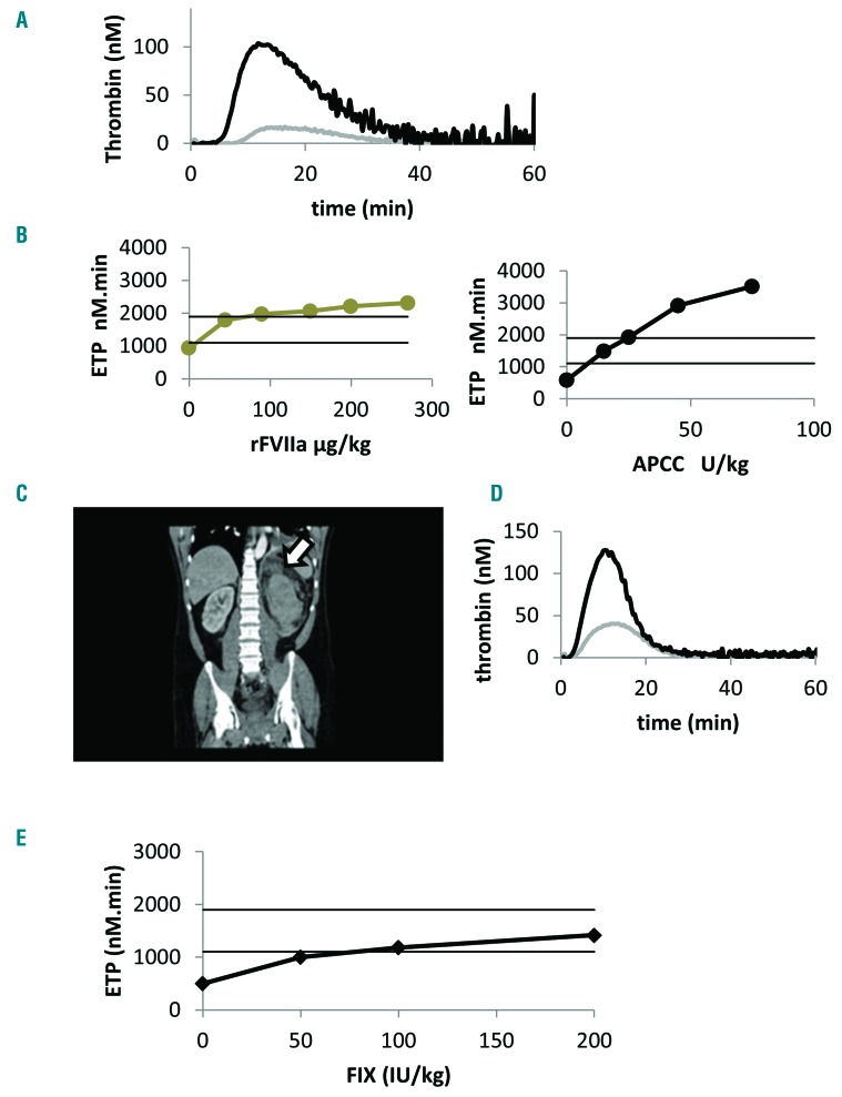
The patient was a 44-year-old with severe hemophilia A patient with anti-FVIII alloantibodies of 7 BU.mL−1. He underwent several surgeries for severe hemophilic arthropathy. For each surgery, the dosage of BPAs was adjusted using TGA. Platelet-poor and platelet-rich plasmas were prepared from venous blood collected into citrate tubes loaded with corn trypsin inhibitor 1.45μM (Haematologic Technologies, VT, USA). Calibrated automated TGA was used with a low tissue factor concentration of 1pM as previously described,3,10,11 and according to the recent standardization recommendations published by the International Society on Thrombosis and Hemostasis (ISTH) FVIII/FIX subcommittee.12 The results of the in vitro spiking experiments confirmed the higher efficacy of the APCC in this particular patient, who was a clinically poor responder to rFVIIa. After an infusion of APCC 75 U.kg−1, a complete correction of endogenous thrombin potential (ETP) was observed (mean ETP±SD = 1,334±212 nM.min) (Figure 1A). The patient underwent seven orthopedic procedures with APCC 75 U.kg−1 infused every eight hours with satisfactory clinical efficacy. No perioperative bleeding complications occurred with a blood loss ranging between 170 and 600 mL. For several years, the patient was on APCC 75 U.kg−1 treatment on demand.
Figure 1.
Personalized management of breakthrough bleeds in a patient on prophylaxis with emicizumab. (A) Representative ex vivo thrombin generation curve obtained before prophylaxis with emicizumab showing a full normalization of ETP one hour after the infusion of APCC 75 U.kg−1. Results were obtained in platelet-poor plasma using a final concentration of tissue factor at 1pM and phospholipids at 4μM. (B) Representative thrombin generation results obtained before and after the in vitro addition of BPAs during emicizumab prophylaxis of 1.5 mg.kg−1 weekly. Two horizontal black lines represent the normal range of thrombin generation (ETP= 1487±372 nM.min; mean±2SD). It is noteworthy that the baseline thrombin generation capacity induced by emicizumab 1.5 mg.kg−1 was higher in platelet-rich plasma compared to platelet-poor plasma (ETP= 940 vs. 585 nM.min respectively). The efficacy of APCC was tested in platelet-poor plasma using a final concentration of tissue factor at 1pM and phospholipids at 4μM. rFVIIa was tested in platelet-rich plasma in the presence of TF 1pM. (C) Contrast-enhanced CT scan of the abdomen and pelvis, showing a left perirenal hematoma (arrow). (D) Thrombin generation curves obtained during prophylaxis with emicizumab, before initiating APCC (gray curve) and at day three of APCC treatment with 15 U.kg−1 showing a full normalization of ETP with no hypercoagulability. In vivo thrombin generation capacity induced by emicizumab prophylaxis (gray curve Figure 1D, ETP=600 nM.min) was higher than that observed in the absence of procoagulant treatment as represented in Figure 1A (gray curve ETP=115 nM.min). In addition, it was interesting to observe a prolonged action of APCC when combined with emicizumab. This might be explained by the mechanism of action and the prolonged half-life of emicizumab that might influence the half-life of its targets FIX and FX. Results were obtained in platelet-poor plasma using a final concentration of tissue factor at 1pM and phospholipids at 4μM. (E) Thrombin generation results obtained before and after the in vitro addition of rFIX (Benefix) during emicizumab prophylaxis of 1.5 mg.kg−1 weekly. The efficacy of rFIX 0-25-50-100 IU.kg−1 was tested in platelet-poor plasma using a final concentration of tissue factor at 1pM and phospholipids at 4μM. ETP: endogenous thrombin potential; APCC: activated prothrombin complex concentrate: rFVIIa: recombinant activated factor VII; FIX: factor IX.
In 2016, the patient participated in the HAVEN-1 trial. On week six of the study, while receiving maintenance doses of emicizumab 1.5 mg.kg−1 weekly, with stable plasma concentrations, he gave his informed consent to test the combined in vitro effect of emicizumab and BPA using TGA. TGA was performed in the presence of five different dosages of APCC (0-15-25–45–75–100 U.kg−1) and rFVIIa (0-45–90–150-200–270 μg.kg−1). TG was measured in the same pre-analytical and analytical conditions as were the previous surgeries.12
A dose-dependent increase of TG was observed with both BPAs, in platelet-rich and platelet-poor plasma. However, very low dosages of APCC, i.e., 15 to 25 U.kg−1, were sufficient to fully normalize TG in our patient, and higher concentrations which were tested induced very high TG capacity, suggesting a potential risk of thrombosis (Figure 1B).
Six months later, the patient experienced an acute spontaneous arterial bleeding while on prophylaxis with emicizumab 1.5 mg.kg−1. He had a dissection of an aberrant renal artery, supplying the inferior pole of the left kidney, resulting in a perirenal hematoma of 16×10×7cm (Figure 1C). The patient was suffering from arterial dysplasia, high blood pressure and dyslipidemia. Based on his predetermined response to BPAs, the patient was able to benefit from an individually tailored bypassing therapy with APCC. He received a single dose of 25 U.kg−1 followed by 15 U.kg−1 every 12 hours for two weeks followed by 15 U.kg−1 every 24 hours for one additional week, making for a total treatment duration of three weeks. The frequency of APCC infusions was reduced from 12 hours to 24 hours on the basis of favorable clinical evolution, normal hemoglobin levels for several days and the ultrasound exam, which showed a significant reduction of the perirenal hematoma. During the treatment period, ETP was 2039nM.min with APCC 25 U.kg−1 and 1488nM.min with APCC 15U.kg−1 (Figure 1D). In our center, normal TG capacity is 1487±372nM.min (mean ETP±2SD), as determined in 100 healthy volunteers.3 The patient safely recovered with no thrombotic complications during the three weeks of APCC treatment. No thromboprophylaxis was prescribed during the treatment with APCC.
Given the mechanism of action of emicizumab, we hypothesized that during prophylaxis with emicizumab, recombinant FIX (rFIX) concentrates containing activated FIX (FIXa) might improve TG.13,14 We performed in vitro spiking experiments with increasing concentrations (0–25–50 and 100 IU/dL) of rFIX (Benefix®, Pfizer, France). The patient had a normal plasma FIX level of 98 IU/dL. We observed a dose-dependent correction of TG with no hypercoagulability (Figure 1E).
There is no routine laboratory assay to monitor the efficacy of emicizumab.8 APTT-based clotting assays determining FVIII activity or chromogenic FX activation assays were previously suggested to estimate clotting activity of emicizumab.13 However, these assays may not reflect the combined effect of emicizumab and BPAs. As the final product generated with both emicizumab and BPA is thrombin, TGA could be a good approach to monitor these therapies.
This case illustrates that TGA may help physicians to determine the individual profile of patients receiving combined treatment with BPA and emicizumab, and to personalize bypassing therapy when treating breakthrough bleeds.
The HAVEN-1 study reported 9% of thrombotic complications in patients who had received multiple infusions of APCC, no thrombotic events were reported with rFVIIa. Oldenburg et al.9 emphasized that the combined use of APCC and emicizumab prophylaxis was associated with a substantial risk of toxic effects. The manufacturers recommend not administering more than 50U.kg−1 of APCC (and no more than 100U.kg−1 daily) or 90μg.kg−1 of rFVIIa in patients receiving emicizumab prophylaxis.
The higher potential of thrombotic events observed with APCC may be explained by the major substrates of emicizumab (FIXa and FX) as well as the product generated (activated factor X [FXa]), which are all supplied by APCCs.8 In this patient, TG was substantially increased after the in vitro addition of APCC 50U.kg−1, suggesting a certain state of hypercoagulability and risk of thrombosis. Thus, the use of APCC 50U.kg−1 might not be entirely safe in all patients receiving emicizumab. In addition, despite the absence of thrombosis observed during the HAVEN-1 trial, no information is available on the coagulation capacity of the patients receiving emicizumab prophylaxis and multiple infusions of rFVIIa, given every 2–4 hours. In our patient, TG was above the normal range after the in vitro addition of rFVIIa at high dosages >150μg.kg−1. Therefore, it may be wise to determine the BPA dosages required to normalize TG for each patient receiving prophylaxis with emicizumab.
Another concern is the cost of these therapies. In this patient, a similar TG capacity was observed with APCC 15U.kg−1 and rFVIIa 90μg.kg−1, the latter being the most expensive treatment option requiring frequent infusions. The personalized approach, using global hemostasis assays, may help to limit the cost of BPA in this context and to choose the minimal dosage of BPA which is capable of normalizing the coagulation capacity of each single patient.
The individual coagulation capacity of patients receiving combined emicizumab and BPA may be very different. Therefore, required dosages of APCC or rFVIIa to achieve hemostasis may also differ from one individual to another. TGA may be a valuable surrogate marker for the combined use of procoagulant agents such as emicizumab and BPAs.
FVIII is one of the least abundant coagulation factors in plasma (0.001 μM). Given the higher levels of FIX (0.090 μM) and FX (0.135 μM), activated FVIII is therefore the limiting factor for FXa formation. During prophylaxis with emicizumab, the drug concentration in plasma is around 0.370 μM.8 As a result, the rate-limiting factor for FXa generation is no longer FVIIIa, but the concentration of FIXa generated. One can therefore hypothesize that the addition of rFIX concentrates containing FIXa, and a higher rate of FIX activation by FXIa,14,15 might improve FXa and TG in hemophilia A patients with emicizumab prophylaxis. Our in vitro results confirmed this hypothesis and showed that rFIX dose-dependently improved TG in the presence of emicizumab. In our patient, rFIX 100U.kg−1 normalized TG and induced a similar TG capacity to APCC 15 U.kg−1, which had effectively achieved hemostasis in the context of acute serious bleeding. Thus, in patients receiving prophylaxis with emicizumab, recombinant FIX concentrates may represent an alternative to BPA, with a lower potential risk of thrombotic complications.
Supplementary Material
Footnotes
Information on authorship, contributions, and financial & other disclosures was provided by the authors and is available with the online version of this article at www.haematologica.org.
References
- 1.Astermark J. Treatment of the bleeding in inhibitor patient. Semin Thromb Haemost. 2003;29(1):77–86 [DOI] [PubMed] [Google Scholar]
- 2.Ingerslev J. Strategies for the treatment of inhibitor patients. Haematologica. 2000;85(10):15–20 [PubMed] [Google Scholar]
- 3.Dargaud Y, Lienhart A, Negrier C. Prospective assessment of thrombin generation test for dose monitoring of bypassing therapy in hemophilia patients with inhibitors undergoing elective surgery. Blood. 2010;116(25):5734–5737 [DOI] [PubMed] [Google Scholar]
- 4.Van Veen JJ, Gatt A, Bowyer AE, Cooper PC, Kitchen S, Makris M. The effect of tissue factor concentration on calibrated automated thrombography in the presence of inhibitor bypass agents. Int J Lab Hematol. 2009;31(2):189–198 [DOI] [PubMed] [Google Scholar]
- 5.Luna-Záizar H, Beltrán-Miranda CP, Esparza-Flores MA, et al. Thrombin generation as objective parameter of treatment response in patients with severe haemophilia A and high-titre inhibitors. Haemophilia. 2014;20(1):e7–14 [DOI] [PubMed] [Google Scholar]
- 6.Turecek PL, Váradi K, Keil B, et al. Factor VIII inhibitor-bypassing agents act by inducing thrombin generation and can be monitored by a thrombin generation assay. Pathophysiol Haemost Thromb. 2003;33(1):16–22. [DOI] [PubMed] [Google Scholar]
- 7.Uchida N, Sambe T, Yoneyama K, et al. A first in human phase 1 study of ACE 910, a novel factor VIII-mimetic bispecific antibody, in healthy subjects. Blood. 2016;127(13):1633–1641 [DOI] [PMC free article] [PubMed] [Google Scholar]
- 8.Lenting PJ, Denis CV, Christophe OD. Emicizumab, a bispecific antibody recognizing coagulation factors IX and X: how does it actually compare to factor VIII¿ Blood. 2017;130(23):2463–2468 [DOI] [PubMed] [Google Scholar]
- 9.Oldenburg J, Mahlangu JN, Kim B, et al. Emicizumab prophylaxis in hemophilia A with inhibitors. N Eng J Med. 2017;377(9):809–818 [DOI] [PubMed] [Google Scholar]
- 10.Dargaud Y, Beguin S, Lienhart A, et al. Evaluation of thrombin generating capacity in plasma from patients with hemophilia A and B. Thromb Haemost. 2005;93(3):475–480 [DOI] [PubMed] [Google Scholar]
- 11.Dargaud Y, Lienhart A, Meunier S, et al. Major surgery in a severe haemophilia A patient with high titre inhibitor: use of the thrombin generation test in the therapeutic decision. Haemophilia. 2005;11(5):552–558 [DOI] [PubMed] [Google Scholar]
- 12.Dargaud Y, Wolberg AS, Gray E, Negrier C, Hemker HC. Subcommittee on Factor VIII, Factor IX, and rare coagulation disorders. Proposal for standardized preanalytical and analytical conditions for measuring thrombin generation in hemophilia: communication from the SSC of the ISTH. J Thromb Haemost. 2017;15(8):1704–1707 [DOI] [PMC free article] [PubMed] [Google Scholar]
- 13.Soeda T, Kitazawa T, Muto A, et al. In vitro characterization of ACE910, a humanized bispecific antibody to factors IXa and X. Haemophilia. 2014;20-(s3):77 [Google Scholar]
- 14.Yu Y, Millar M. Measurement of factor IX activity in plasma-derived and recombinant concentrates: insights from thrombin generation and activation-based assays. J Thromb Haemost. 2014;12(1):62–70 [DOI] [PubMed] [Google Scholar]
- 15.Barrowcliffe TW. Insights from factor IX activation studies with chromogenic assays: implications of disparate product results. Haemophilia. 2010;16(6):9–12. [DOI] [PubMed] [Google Scholar]
Associated Data
This section collects any data citations, data availability statements, or supplementary materials included in this article.


