Abstract
Background:
As the most viable method for investigating in vivo anterior cruciate ligament (ACL) rupture, video analysis is critical for understanding ACL injury mechanisms and advancing preventative training programs. Despite the limited number of published studies involving video analysis, much has been gained through evaluating actual injury scenarios.
Methods:
Studies meeting criteria for this systematic review were collected by performing a broad search of the ACL literature with use of variations and combinations of video recordings and ACL injuries. Both descriptive and analytical studies were included.
Results:
Descriptive studies have identified specific conditions that increase the likelihood of an ACL injury. These conditions include close proximity to opposing players or other perturbations, high shoe-surface friction, and landing on the heel or the flat portion of the foot. Analytical studies have identified high-risk joint angles on landing, such as a combination of decreased ankle plantar flexion, decreased knee flexion, and increased hip flexion.
Conclusions:
The high-risk landing position appears to influence the likelihood of ACL injury to a much greater extent than inherent risk factors. As such, on the basis of the results of video analysis, preventative training should be applied broadly. Kinematic data from video analysis have provided insights into the dominant forces that are responsible for the injury (i.e., axial compression with potential contributions from quadriceps contraction and valgus loading). With the advances in video technology currently underway, video analysis will likely lead to enhanced understanding of non-contact ACL injury.
Anterior cruciate ligament (ACL) rupture is a devastating injury for professional and recreational athletes. The short-term disability and long-term increased risk of osteoarthritis1, as well as the economic impact on the patient and health-care system, emphasize the importance of injury prevention. As the most practical means of investigating in vivo ACL disruption, analysis of video captured at the moment of injury is critical for understanding injury mechanisms and advancing preventative training programs.
The basic premise of video analysis is that video recorded during a sporting event often captures high-quality images of the athlete during an ACL injury. Analysis of these images can provide insights into the mechanisms of injury and can help to devise preventative strategies. The majority of video studies have focused on non-contact scenarios, defined as those involving no contact or a minor perturbation to the body without direct contact to the knee or tackling of the injured athlete. While originally qualitative in design, the field has evolved to include quantitative 2-dimensional (2D) and 3-dimensional (3D) techniques (Fig. 1).
Fig. 1.
Flow diagram illustrating the evolution of video analysis from qualitative to quasi-quantitative to quantitative study designs. Of note, the quasi-quantitative and quantitative study designs are subdivided into their respective subgroups; in addition, the dates and combined number of subjects analyzed for each study design are shown beneath each category.
Recent statements from the ACL Research Retreat have called for more video-based studies2. Prior to moving forward, it is crucial to understand what published studies have provided to our understanding of ACL injury and how video analyses can be improved. The aim of the present review is to summarize the contributions of video analysis to our understanding of the mechanisms of non-contact ACL injury (NC-ACLI) and potential preventative strategies, while highlighting gaps in the current literature.
Methods
Search Methods
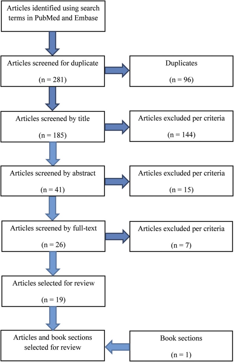
Studies meeting criteria for this review were identified by performing a broad search of the ACL literature through August 2015 (Table I). The electronic database search was performed in PubMed and Embase with use of the following search terms: (“videotape recording” or “videotape” or “video”) and (“anterior cruciate ligament” or [“anterior and cruciate”] or “acl”) and ([“wounds and injuries”] or “wounds” or “injuries”). Following removal of duplicates, 185 articles were screened in sequential steps by title, abstract, and full text (Fig. 2). We excluded studies involving non-human subjects, studies involving in vitro or cadaveric study designs, studies involving post-injury analysis, studies not written in the English language, letters, reviews, and abstracts. The bibliographies of the included papers were also reviewed to include pertinent book sections that were not present in the above databases. In total, 20 studies met the criteria for the review.
Fig. 2.
Flow diagram of the search method.
TABLE I.
Video Studies by Design*
| Study | Sport | Sample Size | Key Results† |
|---|---|---|---|
| Qualitative | |||
| Ettlinger et al.21 (1995) | Recreational alpine skiing | 10 subjects | A training program utilizing video recordings of actual injury scenarios reduced the prevalence of ACL injury by 62% in ski patrol and ski instructors |
| Ebstrup and Bojsen-Møller14 (2000) | Team sports | 15 subjects | The majority of NC-ACLIs occurred during jumping and landing actions, followed by immediate side-stepping maneuvers |
| Boden et al.7 (2000) | Team sports | 23 subjects | 65% (15 of 23) of ACLIs involved NC scenarios, and 35% (8 of 23) involved contact scenarios; all NC-ACLIs occurred with the knee close to full extension during landing or deceleration maneuvers; the majority of NC-ACLIs occurred with an opposing player in close proximity |
| Teitz18 (2001) | Team sports | Unreported | Most NC-ACLIs occurred while landing with the center of gravity located posterior to the knee |
| Lightfoot et al.33 (2005) | Collegiate wrestling | 6 subjects | All ACL injuries occurred near terminal knee extension; 83% (5 of 6) occurred with the foot planted firmly on the ground and involved rotational stress on the weight-bearing knee |
| Bere et al.65 (2011) | Professional alpine skiing | 20 subjects | Inconsistent piste (e.g., small bumps), ill-prepared jumps and spill zones, and icy conditions were cited as the most common factors predisposing to ACL injury |
| Bere et al.62 (2011) | Professional alpine skiing | 20 subjects; 19 controls | All injury scenarios demonstrated backward or inward loss of balance; the skiers’ bindings did not release during any ACL injury scenarios |
| Quasi-quantitative | |||
| Olsen et al.12 (2004) | Female team handball | 20 subjects | 63% (12 of 19) of NC-ACLIs involved a plant-and-cut maneuver with the knee close to full extension and the foot firmly fixed outside of the area directly beneath the COM; the average binned knee-flexion angle for subjects sustaining NC-ACLI was 15°; 75% (15 of 20) of NC-ACLIs occurred on artificial surfaces (higher shoe-surface friction), and 25% (5 of 20) occurred on wooden surfaces (lower shoe-surface friction); 95% (18 of 19) of NC-ACLIs occurred on offense, all while the subject was in possession of the ball; 63% (12 of 19) of NC-ACLIs involved some type of perturbation |
| Cochrane et al.8 (2007) | Australian football | 34 subjects | 56% (19 of 34) of ACLIs involved NC scenarios, and 44% (15 of 34) involved contact scenarios; 68% (13 of 19) of NC-ACLIs occurred during landing or side-stepping maneuvers |
| Krosshaug et al.9 (2007) | Basketball | 39 subjects | 74% (29 of 39) of NC-ACLIs occurred while on offense; 79% (22 of 28) of NC-ACLIs occurred with an opponent within 1 m; females sustaining an NC-ACLI landed with significantly higher knee (p = 0.034) and hip flexion (p = 0.043) at initial contact relative to males; females demonstrated valgus collapse 5.3 times more frequently than males |
| Brophy et al.11 (2015) | European football | 55 subjects | 73% (40 of 55) of NC-ACLIs occurred while defending, and females (20 of 23) were significantly (p = 0.045) more likely than males to be defending; 83% (20 of 24) of NC-ACLIs occurred with an opposing player within 1 or 2 yards |
| Waldén et al.13 (2015) | Male professional European football | 39 subjects | 64% (25 of 39) of NC-ACLIs occurred during side-stepping maneuvers; the average binned knee-flexion angle for subjects sustaining NC-ACLI was 6°; 95% (37 of 39) of NC-ACLIs occurred in dry weather conditions (higher shoe-surface friction), and 5% (2 of 37) occurred in wet weather conditions (lower shoe-surface friction); 77% (30 of 39) of NC-ACLIs occurred while defending |
| 2D quantitative | |||
| Boden et al.10 (2009) | Team and individual sports | 29 subjects; 27 controls | All subjects with NC-ACLIs first contacted the ground with the hindfoot or entire flat foot, attained the flat foot position 1.5 video frame sequences sooner than controls, and demonstrated 12° less plantar flexion of the ankle throughout the injury scenario; no significant differences in knee abduction or flexion angles were present between subjects sustaining NC-ACLIs and controls at initial contact (subjects sustaining NC-ACLIs demonstrated 18° of knee flexion at initial contact); NC-ACLIs were associated with a 19° increase in mean hip-flexion angle during the first 90 msec after initial contact; females sustaining NC-ACLI were found to be performing deceleration maneuvers in 78% (14 of 18) of injury scenarios, whereas males were found to be landing in 64% (7 of 11); all NC-ACLIs occurred while in possession of the ball or while guarding an opposing player in possession of the ball; 96% (26 of 27) of NC-ACLIs occurred with an opposing player within 1 m |
| Hewett et al.15 (2009) | Team and individual sports | 23 subjects; 6 controls | Females sustaining NC-ACLI demonstrated a 41° increase in knee abduction after initial contact, whereas males demonstrated a 15° increase; females sustaining NC-ACLI demonstrated an average 10° lateral trunk angle at initial contact, whereas males demonstrated an average angle of 3° |
| Sheehan et al.16 (2012) | Team sports | 20 subjects; 20 controls | Subjects with NC-ACLIs demonstrated a COM_BOS/femoral length ratio of 1.5, whereas healthy controls demonstrated a ratio of 0.7; the COM_BOS/femoral length ratio discriminated between injured and uninjured athletes with 80% accuracy |
| Sasaki et al.66 (2015) | Female European football | 60 subjects | The COM_BOS demonstrated significant inverse correlation (−0.6; p < 0.001) with trunk angle and positive correlation (0.9; p < 0.001) with limb angle |
| 3D quantitative | |||
| Koga et al.3 (2010) | Female team handball | 10 | All NC-ACLIs occurred while on offense; in all NC-ACLIs, the knee-flexion angle was <30° at initial contact; 70% (7 of 10) of NC-ACLIs occurred while cutting, and 30% (3 of 10) occurred on 1-leg landings; all NC-ACLIs demonstrated neutral abduction at initial contact with an average increase of 12° of valgus by 40 msec; the mean knee-flexion angle was 23° at initial contact and increased to 47° by 40 msec; sudden changes in the joint angular motion and peak vertical GRFs occurred within 40 msec after initial contact |
| Koga et al.4 (2011) | Male professional European football | 1 | Anterior tibial translation initiated 20 msec after initial contact; by 30 msec, approximately 9 mm of anterior translation had occurred |
| Bere et al.5 (2013) | Professional alpine skiing | 2 | NC-ACLI scenarios demonstrated an average increase of 34° of knee flexion and 11° of internal rotation immediately following initial contact |
| Dai et al.6 (2015) | Javelin throwing | 1 subject; 3 controls | Greater forward COM velocity and less vertical COM velocity in addition to decreased knee flexion and knee angular velocity occurred during the NC-ACLI series; anterior tibial translation beyond the anterior border of the patella occurred at 30% of the delivery phase, corresponding to 49.5 msec after initial contact |
Multiple video analyses employed >1 technique; for these studies, the primary technique was used for categorization.
COM = center of mass, BOS = base of support, COM_BOS = distance between center of mass and base of support, and GRF = ground-reaction force.
Study Designs
All types of video study designs were included in this review: qualitative analyses, quasi-quantitative 2D analyses, and quantitative analyses. Qualitative analyses rely on experts in biomechanics and sports medicine to describe and categorize injury scenarios without directly measuring body position at the time of injury. Quasi-quantitative analyses involve the visual evaluation of body position during NC-ACLIs in order to allow for the estimation of joint angles or to bin the data into general categories. Examples of binned data include the position of the knee on landing (e.g., extended or flexed) and the part of the foot that makes initial contact with the ground (e.g., heel or toes). Similar to qualitative analyses, no direct measures of body position are acquired. Quantitative analyses differ from qualitative and quasi-quantitative studies in that joint angles and body positions are directly measured in either 2 or 3 dimensions. There are 3 different types of quantitative designs: 2D quantitative, 3D modeling, and direct linear transformation. 2D quantitative analyses use images pulled from video to directly measure joint angles and distances (e.g., from the center of mass to the base of support) with use of various image-processing software packages. 3D modeling analyses superimpose a skeletal structure over the athlete in video frames captured from multiple cameras at various angles relative to the field of play to determine position3-5. Estimates of initial foot contact are used to temporally co-locate the multiple video feeds while common features in simultaneous video frames are used to spatially co-locate the images. 3D kinematic data also can be obtained with use of cameras calibrated for direct linear transformation analysis6. This technology can be used to follow an athlete’s movement during competition. It has the capacity to track body position in space to a high degree of accuracy with use of multiple, high-definition cameras placed at specific locations relative to the playing field. Yet, the position of the athlete relative to the cameras must be predictable a priori. Thus, sports such as track and field are well suited for this technology.
Results
Qualitative Analyses
Qualitative analyses have identified common features present during NC-ACLIs. Specifically, those studies have revealed a higher prevalence of non-contact, compared with contact, injury situations7,8 (Table I). They also have identified scenarios that predispose an athlete to an NC-ACLI, including close proximity to opposing players or minor perturbation7,9-12, increased shoe-surface friction12,13, high-risk maneuvers (e.g., decelerating, sidestepping3,8,12-14), and landing on the heel or the flat portion of the foot10. Analyses of sex-related differences with use of qualitative techniques have revealed a higher prevalence of NC-ACLI in females when decelerating as compared with a higher prevalence of rupture in males when performing jumping maneuvers10. Sport-specific trends have also been identified, including a higher prevalence of injury while on offense in team handball as compared with a higher prevalence of rupture while on defense in European football11-13. Descriptive analyses have revealed a higher prevalence of injury in team sports, particularly while athletes possess the ball or defend an opponent in possession of the ball10,12.
Quantitative Analyses
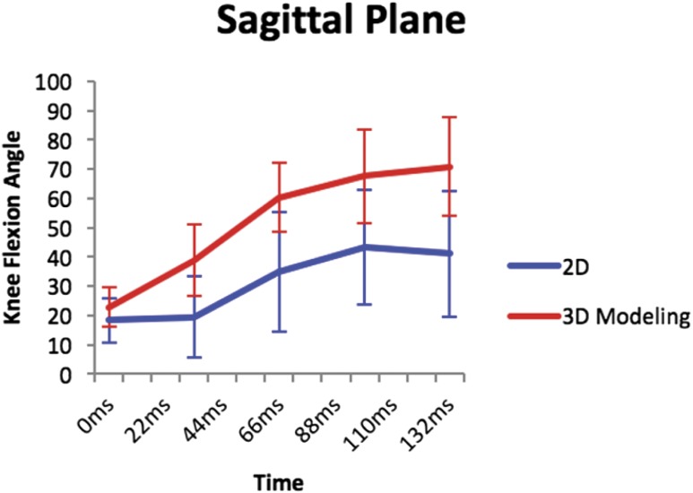
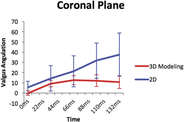
Quantitative studies have identified joint angles at the time of landing that likely increase the risk of rupture3-6,10,15,16. In addition, the cumulative results of those studies have supported new hypotheses regarding dominant forces involved in NC-ACLIs3,6,17 and have provided estimates of ACL rupture timing3,6,10. In sports that involve jumping and cutting, NC-ACLIs appear to occur with the knee flexed <30° in neutral varus-valgus angulation at initial contact10. The average knee-flexion angle (and standard deviation) obtained across all subjects in 4 separate studies3,10,12,13 was 16° ± 8.5°. A comparison of the 2D and 3D studies with the largest NC-ACLI cohorts3,10 showed that the differences in knee flexion and varus angles were smallest at initial contact and at 33 msec (difference in flexion, 4.5° and 19°, respectively; difference in varus, 5.5° and 5.13°, respectively) (Figs. 3 and 4). However, the 2D studies trended toward lower knee-flexion angles and higher valgus angles relative to the 3D studies at time intervals distant from landing (maximum difference in flexion, 29.79°; maximum difference in valgus, 26.96°).
Fig. 3.
Line graph showing sagittal data points from the quantitative 2D and 3D modeling techniques (individual data from the 2D analysis were made available by one of the authors [B.P.B.] from a previous study10, and individual data from the 3D analysis3 were obtained with use of WebPlotDigitizer/app). The error bars indicate 1 standard deviation.
Fig. 4.
Line graph showing coronal data points from the quantitative 2D and 3D modeling techniques (individual data from the 2D analysis were made available by one of the authors [B.P.B.] from a previous study10, and individual data from the 3D analysis3 were obtained with use of WebPlotDigitizer/app). The error bars indicate 1 standard deviation.
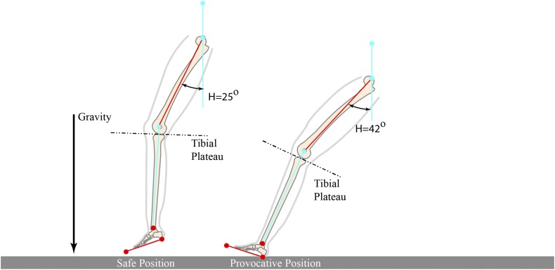
Boden et al.10, in a quantitative analysis, noted a trend toward less knee flexion on landing when subjects who sustained an NC-ACLI injury were compared with uninjured controls performing a similar movement, although the difference did not reach significance. That 2D study demonstrated that the subjects who experienced an ACL disruption landed with a less plantar-flexed ankle (landing flatfooted or on the heel) and a more flexed hip relative to controls. The authors concluded that landing with an extended knee alone is likely less of a risk than landing with this combined posture, which was defined as the provocative position (Fig. 5). Although only a case study, the report by Bere et al. demonstrated that the landing positions of 2 skiers who sustained an NC-ACLI mirrored the “provocative” position5.
Fig. 5.
Photographs and illustrations depicting provocative (L) and safe (R) landing position. These figures demonstrate the average joint angles at initial contact for athletes at risk of sustaining NC-ACLI and healthy controls. The average hip angles were obtained from the study by Sheehan et al.16. The average ankle and knee angles were obtained from the study by Boden et al.17. It should be noted that the images are still frames (not obtained from video) and were manipulated to place the athlete in the average provocative and safe positions. (Reprinted, with modification, from: Boden BP, Breit I, Sheehan FT. Tibiofemoral alignment: contributing factors to noncontact anterior cruciate ligament injury. J Bone Joint Surg Am. 2009 Oct;91[10]:2381-9.)
Quantitative studies also have suggested that the position of the base of support relative to the center of mass during a 1-legged landing maneuver is likely a factor in the occurrence of an NC-ACLI. Sheehan et al., in a study of patients who were matched for sex, sport, and maneuver just prior to injury, found that the distance between the center of mass and the base of support, normalized by the femoral length, discriminated between patients with ACL disruption and controls with an accuracy of 80%16. Although the study was not quantitative, Teitz also observed that most NC-ACLIs occurred in athletes who landed with the center of mass located posterior to the base of support18. Similarly, a laboratory-based study demonstrated that leaning forward while landing likely protected against NC-ACLI by bringing the center of mass closer to the base of support19.
Finally, quantitative studies have provided estimates of NC-ACLI timing. Koga et al.3 identified abrupt changes in joint angular positions between 20 and 50 msec after initial contact, which is the same time frame (33 msec) as the sudden change in joint kinematics documented in the work by Boden et al.10. Dai et al.6 observed anterior translation of the tibial plateau beyond the anterior border of the patella at 49.5 msec after initial contact.
Discussion
The primary goal of video analysis is to increase the understanding of NC-ACLI. The combined results of the studies to date suggest that increased demands placed on the neuromuscular system likely disrupt the motor control patterns that protect the knee during athletic activity, thus exposing the player to an NC-ACLI. By helping to identify high-risk scenarios, many aspects of which are inherently malleable, the clinical impact of video analysis has been evident in its contribution to the development of screening algorithms20 and preventative strategies21-26. This is in contrast to the plethora of studies evaluating inherent fixed (nonmalleable) risk factors (condylar notch width, tibial slope, etc.)27-31. The use of multiple inherent risk factors in a predictive model has only produced weak predictability of an NC-ACLI27,31. In contrast, combined body position at the time of landing has demonstrated a strong ability to discriminate between maneuvers that will and will not result in an NC-ACLI16. Thus, currently, it appears that the maneuver being performed at the time of injury has more influence on the likelihood of NC-ACLI than inherent fixed (nonmalleable) risk factors. As such, preventative training should be applied broadly, a conclusion recently supported in the economic analysis by Swart et al.32.
Application of Video Analysis in Understanding High-Risk Joint Positions
Video analysis highlights the importance of combined hip, knee, and ankle alignment during landing scenarios. As demonstrated in the study by Boden et al.10, knee-flexion angles alone were not found to significantly affect the risk of rupture when injured patients were compared with uninjured controls. However, in many studies, low flexion angles in combination with increased hip flexion and decreased ankle plantar flexion have appeared to predispose the athlete to injury3,6-8,10-13,33.
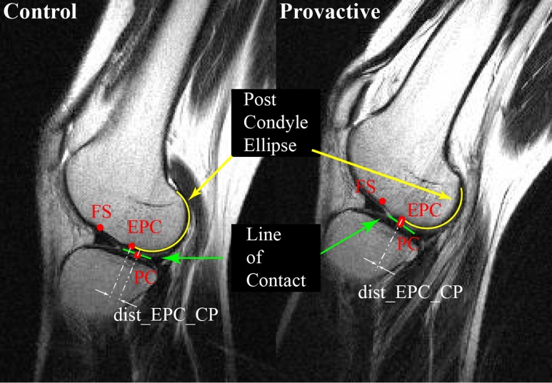
On the basis of the results of video analysis, the increased hip flexion (relative to vertical) seen in the provocative position may increase the risk of NC-ACLI by means of 3 synergistic mechanisms. First, hip flexion increases the slope of the posterior aspect of the tibial plateau relative to the gravitational vector (Fig. 6). On the basis of the combined results of 2 video-based studies16,17, the average difference in dynamic tibial plateau slope (the lateral tibial plateau relative to gravity at initial contact) between athletes who sustain an NC-ACLI and controls is approximately 21°. In contrast, a systematic review evaluating the difference in the inherent tibial plateau slope (the lateral tibial plateau relative to the long axis of the tibia) between injured subjects and healthy controls demonstrated an average difference of just 1.5°34. Thus, the difference between cohorts for the dynamic slope is 14 times greater than the difference between cohorts for the inherent slope. If the knee is subjected to substantial axial compression while in the provocative position, then the lateral femoral condyle is predisposed to posterior subluxation due to the increased slope of the tibial plateau. The resultant anterior tibial translation and internal rotation, the latter of which occurs because of the difference in slope of the medial and lateral tibial plateaus, place substantial stress on the ACL35. Next, combined knee extension with hip flexion shifts the contact point of the lateral femoral condyle to the more anterior flat portion of the condyle versus the rounded posterior portion (Fig. 7)17,35. This enhances the probability of the condyle sliding posteriorly on the tibial plateau instead of rolling as normally occurs during knee flexion. Finally, hip flexion brings the foot forward, which increases the distance between the center of mass and the base of support, thus predisposing to NC-ACLI16,18,19.
Fig. 6.
Illustrations showing the variation in tibial slope at low hip-flexion angles (safe position) and high hip-flexion angles (provocative position) relative to the gravitational vector. The average hip angles were obtained from the study by Sheehan et al.16. The average ankle and knee angles were obtained from the study by Boden et al.17. It should be noted that the inherent slope of the tibial plateau for both images was assumed to be 6°. (Reprinted, with modification, from: Boden BP, Breit I, Sheehan FT. Tibiofemoral alignment: contributing factors to noncontact anterior cruciate ligament injury. J Bone Joint Surg Am. 2009 Oct;91[10]:2381-9.)
Fig. 7.
Magnetic resonance images of the same knee in the control and provocative positions, showing the tibiofemoral joint contact (green), the elliptical outline of the posterior femoral condyle (EPC) (yellow), the distance from the midpoint of the tibiofemoral line of contact (PC) to the point at which the elliptical outline of the posterior femoral condyle diverges from the cortical bone (dist_EPC_CP) (white); and the femoral sulcus (FS) location. (Reprinted, with modification, from: Boden BP, Breit I, Sheehan FT. Tibiofemoral alignment: contributing factors to noncontact anterior cruciate ligament injury. J Bone Joint Surg Am. 2009 Oct;91[10]:2381-9.)
Landing with a less plantar-flexed ankle likely predisposes to NC-ACLI by limiting the absorptive capacity of the distal part of the lower extremity10. In athletes who land with a less plantar-flexed ankle, foot strike is likely to occur in a flat-footed position (or at the time of heel strike, just prior to a flat-footed position). In this posture, the ankle is effectively locked into a single position, and the ground-reaction forces are passed directly to the knee with minimal absorption by the calf muscles that normally takes places through eccentric muscle contraction. The subsequent increase in impulsive forces absorbed by the knee likely predisposes to NC-ACLI.
Contribution of Video Analysis to Understanding the Timing of ACL Rupture
Determining the timing of ACL rupture is crucial to understanding and preventing ACL injury as it directs the investigation of injury scenarios to key moments. On the basis of early qualitative and quasi-quantitative analysis, it was assumed that the NC-ACLI occurred “at or shortly after foot strike.”12 However, this assumption was based not on kinematics but rather on expert opinion. Newer quantitative analyses still cannot pinpoint the exact moment of rupture, but, as suggested by Koga et al., an abrupt change in kinematics likely indicates the moment of disruption3. Specifically, if the forces acting on the knee abruptly change (i.e., if the restraint of the ACL is lost), a sudden kinematic acceleration would follow. The time from initial contact to likely ACL rupture as reported by Boden et al.10 (33 msec) coincides exactly with peak ACL strain identified in a recent modeling study36 and is within the range suggested by the data of Koga et al.3. In addition, the anterior translation of the tibia observed at 49.5 msec after initial contact in the study by Dai et al.6 suggested that rupture occurred prior to this time point. Thus, the initial expert opinion has been substantiated with quantitative data, and ACL rupture likely occurs in the majority of cases between 30 and 40 msec, and certainly within 50 msec, after initial contact.
Contribution of Video Analysis to the Understanding of Forces Responsible for NC-ACLI
The direct kinematic evidence garnered from quantitative video analyses provides important insights into the long-standing debate in the literature pertaining to the dominant forces causing NC-ACLI. Multiple studies have supported excessive valgus load as the dominant factor37-40, whereas others have suggested that disruption is due to impingement41, quadriceps-hamstrings muscle imbalance42-44, and/or substantial axial compression44-46. Currently, the collective results of video analyses support axial compression as the dominant force causing NC-ACLI, with potential contributions from valgus loading and quadriceps muscle contraction.
The axial-compression theory was supported in cadaveric studies that identified substantial ACL strain capable of causing rupture during simulated axial loading46,47. The theory suggests that compressive impulses acting on the posterolateral tibial slope cause posterior translation of the lateral femoral condyle relative to the tibia. The resultant anterior tibial translation and internal rotation cause ACL rupture. Athletes who land flatfooted or close to this position are limited in their ability to dissipate ground-reaction forces at the ankle10. Thus, impulsive forces are passed directly to the knee. If the compressive force is above the injury threshold, the knee buckles (i.e., anterior tibial translation and internal rotation occur), and the ACL is ruptured48. In addition, if the athlete recruits the quadriceps in an attempt to bring the center of mass back over the base of support (and prevent a fall), the compressive force at the knee is amplified and an anterior shear force is placed on the tibia16,49. These explanations for NC-ACLI are specific to the axial-compression model and coincide with the provocative position identified on video analysis.
Video analyses also have clarified the potential role of valgus loading in NC-ACLI. Studies comparing injured subjects with uninjured controls have demonstrated no differences in valgus angles at initial contact10,15. In addition, to our knowledge, no quantitative video study has identified overt valgus collapse at initial contact. When observed, valgus collapse has been found to occur several hundred milliseconds after the presumed moment of rupture10. This finding suggests that the majority of valgus identified on video analysis occurs after NC-ACLI. Findings from a study on bone bruise patterns similarly suggested that valgus loading is a less-dominant force in NC-ACLIs as only 5° of valgus was identified at initial contact42. In addition, the prevalence of medial bone bruising recently was observed to be higher than earlier reported. Wittstein et al.50 found that 16 (57%) of 28 males and 27 (60%) of 45 females had medial and lateral bone bruising. Attempts to explain the etiology of medial bruising in the context of valgus loading have led to the concept of the contrecoup mechanism of NC-ACLI. This model suggests that valgus loading leads to ACL disruption followed by an abrupt varus rotation, resulting in impact on the medial aspect of the joint37,47,51-60. Findings from the overwhelming majority of video analyses oppose this theory, as the knee is in neutral or slight valgus angulation at initial contact and progresses into valgus thereafter3,7,9,10,12. It is more likely that, similar to the lateral knee bone bruises, the medial bone bruises are the result of an axial impaction injury, which occurs shortly after initial contact.
It should be noted, however, that in injured athletes, higher valgus angles have been identified in females compared with males15. When higher valgus positions are present at the knee, the resultant increased compressive force on the lateral aspect of the knee lowers the impulsive force necessary to reach the threshold for NC-ACLI61. This increased valgus may contribute to the increased rate of NC-ACLI in female athletes as compared with their male counterparts.
Future Research
Even with the key insights that video analysis has brought to the understanding of the mechanism of NC-ACLI, there are numerous areas for improvement. To our knowledge, only 5 studies have included controls6,10,15,16,62, of which only 3 matched for both sex and sport6,16,62. Without controls, support for the presumed risk factors is limited to observational evidence. Furthermore, unmatched studies cannot account for potential confounding factors specific to the sport, sex, and the maneuver being performed at the time of injury. Future analyses must include non-injured controls, ideally with the same athlete performing similar actions. Such analyses will allow for the identification of subtle differences that are present during rupture in addition to clarifying if an athlete can land in the same position as in the injury scenario and not sustain an NC-ACLI. An example of an ideally matched internal control was recently described by Dai et al.6. In that study, prior to ACL disruption, the athlete was recorded performing the same maneuver 3 times. Comparison of the non-injurious and injurious sequences revealed keen insights into the mechanism of injury involving horizontal and vertical center-of-mass velocities. This approach, using the same maneuver by the same athlete as a control, is unique and should be continued.
The study design that represents the best use of the researcher’s time and resources is currently a point of contention (Table II). It appears that the insights gained from qualitative and quasi-quantitative studies have been exhausted and that the field will benefit most by advancing quantitative techniques. Quantitative 2D and 3D designs both measure specific joint positions. Differences between the 2D and 3D measures (Figs. 3 and 4) potentially could be due to methodological differences but also may arise from the analysis of different sports or from the fact that angles in different cardinal planes were typically measured from the same subject in the 3D studies and from different subjects in the 2D studies.
TABLE II.
Video Analysis Study Designs
| Primary Aim | Advantages | Disadvantages | |
|---|---|---|---|
| Qualitative | Describe and categorize NC-ACLI scenarios | Identify environmental risk factors and gross motor patterns | Provide limited insights into mechanisms of NC-ACLI |
| Quasi-quantitative | Estimate joint angles and bin findings into categories | Determine general trends in body position during NC-ACLI | Lack the precision to determine high-risk joint positions |
| Quantitative | |||
| 2D | Directly measure joint angles and body positions during NC-ACLI | Ability to collect larger sample size due to public-domain videos and efficient analyses | Single plane fails to account for all 6 degrees of freedom; accuracy unassessed in validation studies; requires cardinal planes, which can be difficult to obtain |
| 3D modeling | Directly measure joint angles and body positions during NC-ACLI | Any perpendicular camera views are adequate | Extensive time required for the analysis, criticized for low accuracy |
| Direct linear translation | Directly measure joint angles and body positions during NC-ACLI | Closest approximation to controlled laboratory settings | Difficulty obtaining sufficient numbers for comparative studies |
The advantages of 2D study designs include larger sample sizes (due to the broad collection of public-domain videos featuring ACL injuries) and relatively quick analysis. The disadvantage of the 2D study design is that a single plane is used to measure joint angles, which fails to account for all 6 degrees of freedom. As a result, unaccounted internal or external rotation may distort sagittal and coronal measurements. Future 2D analyses must account for this potential risk of systematic error. In addition, studies assessing the validity of 2D techniques have not been performed against a gold standard such as motion analysis. This critical step is essential to guide the field and to enable researchers to design protocols based on defined accuracies. The validation study by Krosshaug and Bahr assessing 3D modeling serves as an example63.
Among 3D techniques, modeling has been criticized6 for low accuracy63. In addition, the technique requires 1 to 2 months per subject to complete3. In contrast, 3D direct linear transformation is the closest approximation to the controlled laboratory setting and is the best application of video analysis. However, because it captures NC-ACLIs so infrequently, and only in sports with predictable player positions, the application of this technique is currently limited to case reports.
Importantly, 2D and 3D measurements are most similar at early time intervals for knee flexion and valgus angulation (Figs. 3 and 4). These time intervals likely represent the critical frames during the injury scenario when ground-reaction forces are distributed to the ACL, resulting in rupture. Joint measurements at distant time intervals are of less importance as they likely occur after the ACL rupture. Therefore, prioritizing quantitative 2D techniques in the investigation of knee flexion and varus angulation can likely save time and resources. However, without vertical camera angles or prominent signposts (such as skis), both 2D and 3D techniques offer limited ability to assess internal or external rotation. This limitation, which is more prominent in 2D analyses, reflects the apparent symmetry of the femur and tibia about their central axes and the resultant difficulty in identifying unique landmarks for measurements of internal and external rotation.
Finally, there have been attempts to extend 3D modeling to estimate anterior tibial translation4. While innovative, the accuracy of this technique is inadequate for delineating the narrow difference between safe and stressed positions. An investigation of tibial translation in a controlled setting using skin surface markers identified that tracking the tibia was inherently associated with 3.2 mm of systemic error64. The additional error introduced by the femur at least doubles this value. Because 3D modeling-based video analysis is likely less accurate than motion capture, data obtained using modeling-based analyses are too crude for the investigation of tibial translation. This measurement should be limited to settings with cameras calibrated for direct linear transformation as this technique has demonstrated the accuracy necessary to apply the data in a clinically useful manner.
Summary
Despite the small number of published studies and the specific areas of potential improvement, video analysis has directly contributed to the understanding of ACL injuries in numerous ways. Key injury scenarios have been described, including close proximity to other players (often associated with minor perturbations), increased shoe-surface friction, and landing on the flat portion of the foot. A combination of decreased ankle plantar flexion, low knee flexion, and increased hip flexion has been defined as the provocative position. This landing position appears to influence the likelihood of NC-ACLI to a much greater extent than inherent fixed risk factors. On the basis of videotape identification of the provocative landing position for NC-ACLI, along with cadaveric studies, axial compression appears to be the primary mechanism of injury. With the improvements in video technology currently underway and the recommendations stated in this review, video analysis will likely lead to even better understanding of NC-ACLI.
Note:
This research was supported by the Intramural Research Program of the National Institutes of Health (NIH), Clinical Center, Functional and Applied Biomechanics Section. This research was also made possible through the NIH Medical Research Scholars Program, a public-private partnership supported jointly by the NIH and generous contributions to the Foundation for the NIH from Pfizer, the Leona M. and Harry B. Helmsley Charitable Trust, and the Howard Hughes Medical Institute, as well as other private donors. For a complete list, visit the website at http://fnih.org.The authors also thank informationist Judith Welsh, NIH Library, for editing this manuscript.
Footnotes
Investigation performed at the Clinical Center of the National Institutes of Health, Bethesda, Maryland
Disclosure: The authors indicated that no external funding was received for any aspect of this work. On the Disclosure of Potential Conflicts of Interest forms, which are provided with the online version of the article, one or more of the authors checked “yes” to indicate that the author had a relevant financial relationship in the biomedical arena outside the submitted work.
References
- 1.Luc B, Gribble PA, Pietrosimone BG. Osteoarthritis prevalence following anterior cruciate ligament reconstruction: a systematic review and numbers-needed-to-treat analysis. J Athl Train. 2014. Nov-Dec;49(6):806-19. [DOI] [PMC free article] [PubMed] [Google Scholar]
- 2.Shultz SJ, Schmitz RJ, Benjaminse A, Chaudhari AM, Collins M, Padua DA. ACL Research Retreat VI: an update on ACL injury risk and prevention. J Athl Train. 2012. Sep-Oct;47(5):591-603. [DOI] [PMC free article] [PubMed] [Google Scholar]
- 3.Koga H, Nakamae A, Shima Y, Iwasa J, Myklebust G, Engebretsen L, Bahr R, Krosshaug T. Mechanisms for noncontact anterior cruciate ligament injuries: knee joint kinematics in 10 injury situations from female team handball and basketball. Am J Sports Med. 2010. November;38(11):2218-25. Epub 2010 Jul 01. [DOI] [PubMed] [Google Scholar]
- 4.Koga H, Bahr R, Myklebust G, Engebretsen L, Grund T, Krosshaug T. Estimating anterior tibial translation from model-based image-matching of a noncontact anterior cruciate ligament injury in professional football: a case report. Clin J Sport Med. 2011. May;21(3):271-4. [DOI] [PubMed] [Google Scholar]
- 5.Bere T, Mok KM, Koga H, Krosshaug T, Nordsletten L, Bahr R. Kinematics of anterior cruciate ligament ruptures in World Cup alpine skiing: 2 case reports of the slip-catch mechanism. Am J Sports Med. 2013. May;41(5):1067-73. Epub 2013 Feb 28. [DOI] [PubMed] [Google Scholar]
- 6.Dai B, Mao M, Garrett WE, Yu B. Biomechanical characteristics of an anterior cruciate ligament injury in javelin throwing. J Sport Health Sci. 2015;4(4):333-40. [Google Scholar]
- 7.Boden BP, Dean GS, Feagin JA, Jr, Garrett WE., Jr Mechanisms of anterior cruciate ligament injury. Orthopedics. 2000. June;23(6):573-8. [DOI] [PubMed] [Google Scholar]
- 8.Cochrane JL, Lloyd DG, Buttfield A, Seward H, McGivern J. Characteristics of anterior cruciate ligament injuries in Australian football. J Sci Med Sport. 2007. April;10(2):96-104. Epub 2006 Jun 27. [DOI] [PubMed] [Google Scholar]
- 9.Krosshaug T, Nakamae A, Boden BP, Engebretsen L, Smith G, Slauterbeck JR, Hewett TE, Bahr R. Mechanisms of anterior cruciate ligament injury in basketball: video analysis of 39 cases. Am J Sports Med. 2007. March;35(3):359-67. Epub 2006 Nov 7. [DOI] [PubMed] [Google Scholar]
- 10.Boden BP, Torg JS, Knowles SB, Hewett TE. Video analysis of anterior cruciate ligament injury: abnormalities in hip and ankle kinematics. Am J Sports Med. 2009. February;37(2):252-9. [DOI] [PubMed] [Google Scholar]
- 11.Brophy RH, Stepan JG, Silvers HJ, Mandelbaum BR. Defending puts the anterior cruciate ligament at risk during soccer: a gender-based analysis. Sports Health. 2015. May;7(3):244-9. [DOI] [PMC free article] [PubMed] [Google Scholar]
- 12.Olsen OE, Myklebust G, Engebretsen L, Bahr R. Injury mechanisms for anterior cruciate ligament injuries in team handball: a systematic video analysis. Am J Sports Med. 2004. June;32(4):1002-12. [DOI] [PubMed] [Google Scholar]
- 13.Waldén M, Krosshaug T, Bjørneboe J, Andersen TE, Faul O, Hägglund M. Three distinct mechanisms predominate in non-contact anterior cruciate ligament injuries in male professional football players: a systematic video analysis of 39 cases. Br J Sports Med. 2015. November;49(22):1452-60. Epub 2015 Apr 23. [DOI] [PMC free article] [PubMed] [Google Scholar]
- 14.Ebstrup JF, Bojsen-Møller F. Anterior cruciate ligament injury in indoor ball games. Scand J Med Sci Sports. 2000. April;10(2):114-6. [DOI] [PubMed] [Google Scholar]
- 15.Hewett TE, Torg JS, Boden BP. Video analysis of trunk and knee motion during non-contact anterior cruciate ligament injury in female athletes: lateral trunk and knee abduction motion are combined components of the injury mechanism. Br J Sports Med. 2009. June;43(6):417-22. Epub 2009 Apr 15. [DOI] [PMC free article] [PubMed] [Google Scholar]
- 16.Sheehan FT, Sipprell WH, 3rd, Boden BP. Dynamic sagittal plane trunk control during anterior cruciate ligament injury. Am J Sports Med. 2012. May;40(5):1068-74. Epub 2012 Mar 1. [DOI] [PMC free article] [PubMed] [Google Scholar]
- 17.Boden BP, Sheehan FT, Torg JS, Hewett TE. Noncontact anterior cruciate ligament injuries: mechanisms and risk factors. J Am Acad Orthop Surg. 2010. September;18(9):520-7. [DOI] [PMC free article] [PubMed] [Google Scholar]
- 18.Teitz C. Video analysis of ACL injuries. In: Griffin LY, editor. Prevention of non-contact ACL injuries. Rosemont, IL: American Academy of Orthopaedic Surgeons; 2001. p 87-92. [Google Scholar]
- 19.Shimokochi Y, Ambegaonkar JP, Meyer EG, Lee SY, Shultz SJ. Changing sagittal plane body position during single-leg landings influences the risk of non-contact anterior cruciate ligament injury. Knee Surg Sports Traumatol Arthrosc. 2013. April;21(4):888-97. Epub 2012 Apr 28. [DOI] [PMC free article] [PubMed] [Google Scholar]
- 20.Myer GD, Ford KR, Brent JL, Hewett TE. An integrated approach to change the outcome part I: neuromuscular screening methods to identify high ACL injury risk athletes. J Strength Cond Res. 2012. August;26(8):2265-71. [DOI] [PMC free article] [PubMed] [Google Scholar]
- 21.Ettlinger CF, Johnson RJ, Shealy JE. A method to help reduce the risk of serious knee sprains incurred in alpine skiing. Am J Sports Med. 1995. Sep-Oct;23(5):531-7. [DOI] [PubMed] [Google Scholar]
- 22.Myklebust G, Engebretsen L, Braekken IH, Skjølberg A, Olsen OE, Bahr R. Prevention of anterior cruciate ligament injuries in female team handball players: a prospective intervention study over three seasons. Clin J Sport Med. 2003. March;13(2):71-8. [DOI] [PubMed] [Google Scholar]
- 23.Herman DC, Oñate JA, Weinhold PS, Guskiewicz KM, Garrett WE, Yu B, Padua DA. The effects of feedback with and without strength training on lower extremity biomechanics. Am J Sports Med. 2009. July;37(7):1301-8. Epub 2009 Mar 19. [DOI] [PubMed] [Google Scholar]
- 24.Petersen W, Braun C, Bock W, Schmidt K, Weimann A, Drescher W, Eiling E, Stange R, Fuchs T, Hedderich J, Zantop T. A controlled prospective case control study of a prevention training program in female team handball players: the German experience. Arch Orthop Trauma Surg. 2005. November;125(9):614-21. [DOI] [PubMed] [Google Scholar]
- 25.Petersen W, Zantop T, Steensen M, Hypa A, Wessolowski T, Hassenpflug J. [Prevention of lower extremity injuries in handball: initial results of the handball injuries prevention programme]. Sportverletz Sportschaden. 2002. September;16(3):122-6. German. [DOI] [PubMed] [Google Scholar]
- 26.Olsen OE, Myklebust G, Engebretsen L, Holme I, Bahr R. Exercises to prevent lower limb injuries in youth sports: cluster randomised controlled trial. BMJ. 2005. February 26;330(7489):449 Epub 2005 Feb 7. [DOI] [PMC free article] [PubMed] [Google Scholar]
- 27.Beynnon BD, Hall JS, Sturnick DR, Desarno MJ, Gardner-Morse M, Tourville TW, Smith HC, Slauterbeck JR, Shultz SJ, Johnson RJ, Vacek PM. Increased slope of the lateral tibial plateau subchondral bone is associated with greater risk of noncontact ACL injury in females but not in males: a prospective cohort study with a nested, matched case-control analysis. Am J Sports Med. 2014. May;42(5):1039-48. Epub 2014 Mar 3. [DOI] [PMC free article] [PubMed] [Google Scholar]
- 28.Hashemi J, Chandrashekar N, Gill B, Beynnon BD, Slauterbeck JR, Schutt RC, Jr, Mansouri H, Dabezies E. The geometry of the tibial plateau and its influence on the biomechanics of the tibiofemoral joint. J Bone Joint Surg Am. 2008. December;90(12):2724-34. [DOI] [PMC free article] [PubMed] [Google Scholar]
- 29.Simon RA, Everhart JS, Nagaraja HN, Chaudhari AM. A case-control study of anterior cruciate ligament volume, tibial plateau slopes and intercondylar notch dimensions in ACL-injured knees. J Biomech. 2010. June 18;43(9):1702-7. Epub 2010 Apr 10. [DOI] [PMC free article] [PubMed] [Google Scholar]
- 30.Stijak L, Nikolić V, Blagojević Z, Radonjić V, Santrac-Stijak G, Stanković G, Popović N. [Influence of morphometric intercondylar notch parameters in ACL ruptures]. Acta Chir Iugosl. 2006;53(4):79-83. Serbian. [DOI] [PubMed] [Google Scholar]
- 31.Sturnick DR, Vacek PM, DeSarno MJ, Gardner-Morse MG, Tourville TW, Slauterbeck JR, Johnson RJ, Shultz SJ, Beynnon BD. Combined anatomic factors predicting risk of anterior cruciate ligament injury for males and females. Am J Sports Med. 2015. April;43(4):839-47. Epub 2015 Jan 12. [DOI] [PMC free article] [PubMed] [Google Scholar]
- 32.Swart E, Redler L, Fabricant PD, Mandelbaum BR, Ahmad CS, Wang YC. Prevention and screening programs for anterior cruciate ligament injuries in young athletes: a cost-effectiveness analysis. J Bone Joint Surg Am. 2014. May 7;96(9):705-11. [DOI] [PMC free article] [PubMed] [Google Scholar]
- 33.Lightfoot AJ, McKinley T, Doyle M, Amendola A. ACL tears in collegiate wrestlers: report of six cases in one season. Iowa Orthop J. 2005;25:145-8. [PMC free article] [PubMed] [Google Scholar]
- 34.Wordeman SC, Quatman CE, Kaeding CC, Hewett TE. In vivo evidence for tibial plateau slope as a risk factor for anterior cruciate ligament injury: a systematic review and meta-analysis. Am J Sports Med. 2012. July;40(7):1673-81. Epub 2012 Apr 26. [DOI] [PMC free article] [PubMed] [Google Scholar]
- 35.Boden BP, Breit I, Sheehan FT. Tibiofemoral alignment: contributing factors to noncontact anterior cruciate ligament injury. J Bone Joint Surg Am. 2009. October;91(10):2381-9. [DOI] [PMC free article] [PubMed] [Google Scholar]
- 36.Heinrich D, van den Bogert AJ, Nachbauer W. Relationship between jump landing kinematics and peak ACL force during a jump in downhill skiing: a simulation study. Scand J Med Sci Sports. 2014. June;24(3):e180-7. Epub 2013 Oct 10. [DOI] [PubMed] [Google Scholar]
- 37.Patel SA, Hageman J, Quatman CE, Wordeman SC, Hewett TE. Prevalence and location of bone bruises associated with anterior cruciate ligament injury and implications for mechanism of injury: a systematic review. Sports Med. 2014. February;44(2):281-93. [DOI] [PMC free article] [PubMed] [Google Scholar]
- 38.Hewett TE, Myer GD, Ford KR, Heidt RS, Jr, Colosimo AJ, McLean SG, van den Bogert AJ, Paterno MV, Succop P. Biomechanical measures of neuromuscular control and valgus loading of the knee predict anterior cruciate ligament injury risk in female athletes: a prospective study. Am J Sports Med. 2005. April;33(4):492-501. Epub 2005 Feb 8. [DOI] [PubMed] [Google Scholar]
- 39.Kiapour AM, Kiapour A, Goel VK, Quatman CE, Wordeman SC, Hewett TE, Demetropoulos CK. Uni-directional coupling between tibiofemoral frontal and axial plane rotation supports valgus collapse mechanism of ACL injury. J Biomech. 2015. July 16;48(10):1745-51. Epub 2015 May 29. [DOI] [PMC free article] [PubMed] [Google Scholar]
- 40.Quatman CE, Hewett TE. The anterior cruciate ligament injury controversy: is “valgus collapse” a sex-specific mechanism? Br J Sports Med. 2009. May;43(5):328-35. Epub 2009 Apr 15. [DOI] [PMC free article] [PubMed] [Google Scholar]
- 41.Uhorchak JM, Scoville CR, Williams GN, Arciero RA, St Pierre P, Taylor DC. Risk factors associated with noncontact injury of the anterior cruciate ligament: a prospective four-year evaluation of 859 West Point cadets. Am J Sports Med. 2003. Nov-Dec;31(6):831-42. [DOI] [PubMed] [Google Scholar]
- 42.Kim SY, Spritzer CE, Utturkar GM, Toth AP, Garrett WE, DeFrate LE. Knee kinematics during noncontact anterior cruciate ligament injury as determined from bone bruise location. Am J Sports Med. 2015. October;43(10):2515-21. Epub 2015 Aug 11. [DOI] [PMC free article] [PubMed] [Google Scholar]
- 43.Kirkendall DT, Garrett WE., Jr The anterior cruciate ligament enigma. Injury mechanisms and prevention. Clin Orthop Relat Res. 2000. March;372:64-8. [DOI] [PubMed] [Google Scholar]
- 44.Yu B, Garrett WE. Mechanisms of non-contact ACL injuries. Br J Sports Med. 2007. August;41(Suppl 1):i47-51. [DOI] [PMC free article] [PubMed] [Google Scholar]
- 45.Meyer EG, Haut RC. Excessive compression of the human tibio-femoral joint causes ACL rupture. J Biomech. 2005. November;38(11):2311-6. Epub 2004 Nov 30. [DOI] [PubMed] [Google Scholar]
- 46.Wall SJ, Rose DM, Sutter EG, Belkoff SM, Boden BP. The role of axial compressive and quadriceps forces in noncontact anterior cruciate ligament injury: a cadaveric study. Am J Sports Med. 2012. March;40(3):568-73. Epub 2011 Dec 14. [DOI] [PubMed] [Google Scholar]
- 47.Meyer EG, Baumer TG, Slade JM, Smith WE, Haut RC. Tibiofemoral contact pressures and osteochondral microtrauma during anterior cruciate ligament rupture due to excessive compressive loading and internal torque of the human knee. Am J Sports Med. 2008. October;36(10):1966-77. Epub 2008 May 19. [DOI] [PubMed] [Google Scholar]
- 48.Hsu V, Stearne D, Torg J. Elastic instability, columnar buckling, and non-contact anterior cruciate ligament ruptures: a preliminary report. Temple Univ J Orthop Surg Sports Med. 2006;1:21-3. [Google Scholar]
- 49.McConkey JP. Anterior cruciate ligament rupture in skiing. A new mechanism of injury. Am J Sports Med. 1986. Mar-Apr;14(2):160-4. [DOI] [PubMed] [Google Scholar]
- 50.Wittstein J, Vinson E, Garrett W. Comparison between sexes of bone contusions and meniscal tear patterns in noncontact anterior cruciate ligament injuries. Am J Sports Med. 2014. June;42(6):1401-7. Epub 2014 Mar 25. [DOI] [PubMed] [Google Scholar]
- 51.Kaplan PA, Gehl RH, Dussault RG, Anderson MW, Diduch DR. Bone contusions of the posterior lip of the medial tibial plateau (contrecoup injury) and associated internal derangements of the knee at MR imaging. Radiology. 1999. June;211(3):747-53. [DOI] [PubMed] [Google Scholar]
- 52.Bisson LJ, Kluczynski MA, Hagstrom LS, Marzo JM. A prospective study of the association between bone contusion and intra-articular injuries associated with acute anterior cruciate ligament tear. Am J Sports Med. 2013. August;41(8):1801-7. Epub 2013 Jun 6. [DOI] [PubMed] [Google Scholar]
- 53.Chin YC, Wijaya R, Chong R, Chang HC, Lee YH. Bone bruise patterns in knee injuries: where are they found? Eur J Orthop Surg Traumatol. 2014. December;24(8):1481-7. Epub 2013 Sep 22. [DOI] [PubMed] [Google Scholar]
- 54.Coursey RL, Jr, Jones EA, Chaljub G, Bertolino PD, Cano O, Swischuk LE. Prospective analysis of uncomplicated bone bruises in the pediatric knee. Emerg Radiol. 2006. September;12(6):266-71. Epub 2006 Jul 1. [DOI] [PubMed] [Google Scholar]
- 55.Mandalia V, Fogg AJ, Chari R, Murray J, Beale A, Henson JH. Bone bruising of the knee. Clin Radiol. 2005. June;60(6):627-36. [DOI] [PubMed] [Google Scholar]
- 56.Sanders TG, Medynski MA, Feller JF, Lawhorn KW. Bone contusion patterns of the knee at MR imaging: footprint of the mechanism of injury. Radiographics. 2000. October;20(Spec No):S135-51. [DOI] [PubMed] [Google Scholar]
- 57.Terzidis IP, Christodoulou AG, Ploumis AL, Metsovitis SR, Koimtzis M, Givissis P. The appearance of kissing contusion in the acutely injured knee in the athletes. Br J Sports Med. 2004. October;38(5):592-6. [DOI] [PMC free article] [PubMed] [Google Scholar]
- 58.Vinson EN, Gage JA, Lacy JN. Association of peripheral vertical meniscal tears with anterior cruciate ligament tears. Skeletal Radiol. 2008. July;37(7):645-51. Epub 2008 May 8. [DOI] [PubMed] [Google Scholar]
- 59.Yoon KH, Yoo JH, Kim KI. Bone contusion and associated meniscal and medial collateral ligament injury in patients with anterior cruciate ligament rupture. J Bone Joint Surg Am. 2011. August 17;93(16):1510-8. [DOI] [PubMed] [Google Scholar]
- 60.Mandalia V, Henson JH. Traumatic bone bruising—a review article. Eur J Radiol. 2008. July;67(1):54-61. Epub 2008 Jun 4. [DOI] [PubMed] [Google Scholar]
- 61.Chaudhari AM, Andriacchi TP. The mechanical consequences of dynamic frontal plane limb alignment for non-contact ACL injury. J Biomech. 2006;39(2):330-8. [DOI] [PubMed] [Google Scholar]
- 62.Bere T, Flørenes TW, Krosshaug T, Koga H, Nordsletten L, Irving C, Muller E, Reid RC, Senner V, Bahr R. Mechanisms of anterior cruciate ligament injury in World Cup alpine skiing: a systematic video analysis of 20 cases. Am J Sports Med. 2011. July;39(7):1421-9. Epub 2011 Apr 22. [DOI] [PubMed] [Google Scholar]
- 63.Krosshaug T, Bahr R. A model-based image-matching technique for three-dimensional reconstruction of human motion from uncalibrated video sequences. J Biomech. 2005. April;38(4):919-29. [DOI] [PubMed] [Google Scholar]
- 64.Manal K, McClay Davis I, Galinat B, Stanhope S. The accuracy of estimating proximal tibial translation during natural cadence walking: bone vs. skin mounted targets. Clin Biomech (Bristol, Avon). 2003. February;18(2):126-31. [DOI] [PubMed] [Google Scholar]
- 65.Bere T, Flørenes TW, Krosshaug T, Nordsletten L, Bahr R. Events leading to anterior cruciate ligament injury in World Cup alpine skiing: a systematic video analysis of 20 cases. Br J Sports Med. 2011. December;45(16):1294-302. Epub 2011 Nov 8. [DOI] [PubMed] [Google Scholar]
- 66.Sasaki S, Nagano Y, Kaneko S, Imamura S, Koabayshi T, Fukubayashi T. The relationships between the center of mass position and the trunk, hip, and knee kinematics in the sagittal plane: a pilot study on field-based video analysis for female soccer players. J Hum Kinet. 2015. March 29;45:71-80. Epub 2015 Apr 7. [DOI] [PMC free article] [PubMed] [Google Scholar]


