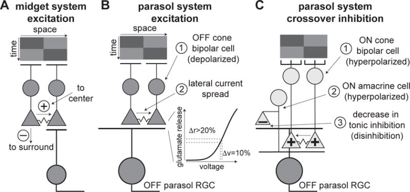
Figure 8.

Proposed circuitry for motion responses in midget and parasol ganglion cells. (A) Working model for midget ganglion cells. Dark bar (top) stimulates left then right bipolar cells in sequence. Output of left bipolar cell falls in midget cell receptive-field surround. Portion of depolarization in left bipolar cell spreads to right bipolar cell, potentiating right bipolar cell located in midget cell receptive-field center. (B) Working model for motion sensitivity in synaptic excitation to parasol cells. Dark bar (top) stimulates left then right bipolar cells. Portion of depolarization in left bipolar cell spreads to right bipolar cell. Due to the nonlinear relationship between voltage and synaptic release in the terminal, increasing the voltage of the terminal in the second bipolar cell by an extra 10% could increase glutamate release by >20% (right). The left bipolar cell, thus, primes the right bipolar cell to subsequent stimulation within a brief time interval. (C) Model for motion sensitivity in disinhibition via suppression of crossover inhibition. Dark bar sequences strongly hyperpolarize ON cone bipolar cells; motion sequences enhance suppression of ON bipolar cells through the same junctional mechanism described in (B), resulting in strong removal of glutamate release (or hyperpolarization via junctional coupling) to an ON-type amacrine cell. The hyperpolarized amacrine cell, in turn, decreases inhibitory transmitter release onto OFF parasol cell dendrites relative to gray background, resulting in disinhibition.
