Abstract
Neoadjuvant chemoradiation has become the standard of care for esophageal cancer, especially for middle third esophageal lesions and those with squamous histology. Although more and more thoracic surgeons and surgical oncologists have now shifted to video-assisted and robot-assisted thoracoscopic esophagectomy; there is still limited experience for the use of minimal-assisted approaches in patients undergoing surgery after neoadjuvant chemoradiation. Most surgeons have concerns of feasibility, safety, and oncological outcomes as well as issues related to difficult learning curve in adopting robotic esophagectomy in patients after chemoradiation. We present our initial experience of Robot-Assisted Mckeown Esophagectomy in 27 patients after neoadjuvant chemoradiation, from May 2013 to October 2014. All patients underwent neoadjuvant chemoradiation to a dose of 50.4 Gy/25Fr with concurrent weekly cisplatin, followed by reassessment with clinical examination and repeat FDG PET/CT 6 weeks after completion of chemoradiation. Patients with progressive disease underwent palliative chemotherapy while patients with either partial or significant response to chemoradiation underwent Robot-Assisted Mckeown Esophagectomy with esophageal replacement by gastric conduit and esophagogastric anastomosis in the left neck. Out of 27 patients, 92.5 % patients had stage cT3/T4 tumours and node-positive disease in 48.1 % on imaging. Most patients were middle thoracic esophageal cancers (23/27), with squamous histology in all except for one. All patients received neoadjuvant chemoradiation and subsequently underwent Robot Assisted Mckeown Esophagectomy. The average time for robot docking, thoracic mobilization and total surgical procedure was 13.2, 108.4 and 342.7 min, respectively. The procedure was well tolerated by all patients with only one case of peri-operative mortality. Average ICU stay was 6.35 days (range 3–9 days). R0 resection rate of 96.3 % and average lymph node yield of 18 could be achieved. Pathological node negativity rate (pN0) and complete response (pCR) were 66.6 and 44.4 %, respectively. In the initial cases, four patients had to be converted to open due technical reasons or intraoperative complications. The present study, with shorter operative times, similar ICU stay, overall low morbidity, and mortality and optimal oncological outcomes suggest that robot-assisted thoracic mobilization of esophagus in patients with prior chemoradiation is feasible and safe with acceptable oncological outcomes. It has a shorter learning curve and hence allows for a transthoracic minimally invasive transthoracic esophagectomy to more and more patients, otherwise unfit for conventional approach.
Keywords: RAME = robot assisted Mckeown esophagectomy, CTRT = concurrent chemoradiation, pCR = pathological complete response, MIE = Minimally invasive esophagectomy
Introduction
The incidence of esophageal cancer has rapidly increased in recent times. Radical esophagectomy with extended lymph node dissection plays a key role in multimodality management of esophageal cancer and is regarded as the best curative option. Although transthoracic esophagectomy through a right thoracotomy has been the standard surgery for esophageal carcinoma, it is often associated with higher rate of pulmonary complications, need for prolonged ventilator support and longer ICU and hospital stay [1]. Minimal invasive esophagectomy (MIE) has definite advantages as it avoids thoracotomy and decreases the incidence of pulmonary complications resulting from open surgery and leads to faster postoperative recovery [2]. Conventional open surgery also has a high mortality rate ranging from 4.8 to 16 % [3]. With the advent of minimal invasive esophagectomy, radical curative surgery can therefore be offered to a wide range of patients who would otherwise be unsuitable for surgery [4]. Although there are concerns about oncological adequacy and safety of minimal invasive esophagectomy, video-assisted thoracoscopic esophagectomy (VATS) remains the most popular approach for MIE. Several systematic reviews and meta-analysis have been published on the safety and efficacy of video-assisted thoracoscopic esophagectomy for carcinoma oesophagus, suggesting overall shorter hospital and ICU stay, rapid postoperative recovery and at least equivalent 3-year survivals, compared to conventional open surgery [5–8].
Currently, robotic approaches are becoming more and more popular in a wide range of oncological procedures in the field of surgical oncology. Although there are no comparative studies between robotic and conventional thoracoscopic esophagectomy, most retrospective studies have shown them to be equivalent in oncological outcomes and complication rates [9]. Robot-assisted esophagectomy provides a major technical advance over conventional video-assisted surgery in terms of improved 3D high-resolution vision, articulating instruments with better control and increased range of motion and dexterity and, moreover, a lesser dependence on assistant for control of instruments and camera vision. It also has a shorter learning curve compared to conventional laparoscopic or thoracoscopic surgery.
Inspite of these advantages, there is lot of hesitation among surgeons in performing robot-assisted or thoracoscopic esophagectomy in patients after neoadjuvant chemoradiation. There are concerns of adequate wide surgical resection, issues with lack of tactile sensation in minimal invasive approaches in post chemoradiation setting and the fear of increased post surgical morbidity. Overall, there is limited experience in robot-assisted thoracoscopic esophagectomy across the globe in patients receiving neoadjuvant chemoradiation.
We present our initial experience of 27 cases of robot-assisted Mckeown esophagectomy (RAME) in patients who have undergone preoperative concurrent chemoradiation (CTRT) for esophageal cancer.
Material and Methods
We retrospectively analysed data of our initial patients undergoing robot-assisted Mckeown esophagectomy after neoadjuvant CTRT between May 2013 and October 2014 with special emphasis on feasibility of robot-assisted esophagectomy in post neoadjuvant chemoradiation setting, immediate postoperative morbidity and surgical and oncologic outcome.
All patients underwent initial evaluation with upper GI endoscopy and biopsy for tissue diagnosis and whole body FDG18 PET/CT for staging. All patients were put on intensive nutrition supplementation, chest physiotherapy and incentive spirometry exercises. Patients underwent preoperative concurrent CTRT to a total dose of 50.4 Gy by 3D CRT/IMRT over 25 Fr with 4 cycles of concurrent weekly cisplatin. Response assessment was done at 6 weeks after completion of CTRT using whole body FDG18 PET/CT. PET response was categorized as significant response, partial response or progressive disease. All patients with partial or significant response underwent RAME after adequate pre-anaesthetic optimization, while patients with progressive disease were given palliative chemotherapy and best supportive case depending upon their performance status and symptoms.
Surgical Technique
Informed consent was taken prior to the procedure. Preoperative workup included Pulmonary Function Test (Spirometry and DLCo), 2D Echocardiography and routine blood investigations (CBC, RFT, LFT, TFT, blood coagulation profile and viral markers). Anaesthesia technique involved selective lung ventilation with double lumen endotracheal intubation (DLT) with routine intra-operative cardiac and pulmonary monitoring.
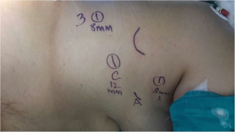
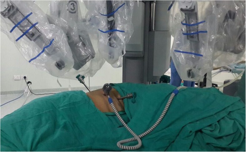
All surgeries were performed by a single surgical oncology team on the da Vinci® surgical robot from Intuitive Surgicals Inc. The patient was placed in left lateral semi-prone position with a 45° tilt towards the table. The right lung was allowed to collapse, and Verres’ needle inserted for CO2 insufflation up to a pressure of 6 mmHg. A 12-mm port was placed in 6th intercostal space 2 cm below and anterior to tip of scapula for placement of camera. Two 8-mm working robotic ports were placed in the 4th and 8th intercostal spaces at a hand breadth from the 1st port to achieve triangulation between camera and working ports (Fig. 1). An additional 12-mm assistant port was placed in the 6th intercostal space anteriorly for introduction of lung retractor, irrigation suction cannula and clip applicator. The patient cart was next brought in obliquely over the patient’s right shoulder at an angle of 15° and docking was done (Fig. 2). Permanent cautery spatula® and fenestrated bipolar® were used for dissection in the two working ports. The right lung was retracted anteriorly and the azygous vein was mobilized, clipped and divided. Entire thoracic oesophagus was mobilized with complete paraesophageal clearance to include a two field lymph node dissection. Haemostasis was secured and port sites were closed over a 28-F drain connected to a intercostal drain bag.
Fig. 1.
Port positions
Fig. 2.
Docking of robot
The patient was repositioned to supine for abdominal gastric tube mobilization, dissection of esophageal hiatus along with D2 nodal clearance through upper midline mini-laparotomy incision. The left neck was explored using transverse supraclavicular incision. Sternocleidomastoid clavicular head and strap muscles were divided and thyroid gland exposed after ligating and dividing the middle thyroid vein. Cervical oesophagus was dissected and mobilized and divided and delivered to the abdomen. A gastric tube was fashioned based on the right gastric and right gastroepiploic vessels and then pulled up into the left neck through esophageal hiatus. Side to side stapled or end to end hand sewn esophagogastric anastomosis was then performed in the left neck and neck wound was closed over No 12 Romovac® suction drain. Feeding jejunostomy was performed and the abdomen was closed with a right subhepatic drain in place. The left intercostal tube drain was placed prophylactically in all cases.
Results
Robot Assisted Mckeown Esophagectomy was attempted in 27 patients, 6 weeks after completion of neoadjuvant CTRT from May 2013 to October 2014. Twenty-three patients had middle third esophageal cancers with gastroesophageal junction tumour in one and lower third esophageal growth in the rest. Squamous cell carcinoma was the most common histology encountered. Clinicopathological stage was T3/T4 in 92.5 % cases (25/27) with metabolically active paraesophageal lymph nodes in 48.1 % cases (13/27) (Table 1). All patients tolerated the chemoradiation schedule well with grade 3/4 toxicity in only 7.4 % patients. Whole body PET/CT for response evaluation after 6 weeks of therapy revealed complete response in primary in 48 % cases and complete nodal response in 69 % cases.
Table 1.
Patient characteristics in patients undergoing RAME
| Patient characteristics | N = 27 | |
|---|---|---|
| Age | Mean (years) | 53.5 |
| Sex | Male/female | 17/10 |
| Histology | SCC/ADC | 26/1 |
| Tumour location | Mid/lower/GEJ | 23/3/1 |
| Clinical T stage | T1 | 0 |
| T2 | 2 | |
| T3 | 21 | |
| T4 | 4 | |
| Clinical N stage | N0 | 14 |
| N+ | 13 |
Preoperative cardiac and pulmonary evaluation and optimization was done for all the patients under supervision of a cardiologist and pulmonologist in view of prior radiation and chemotherapy. All patients underwent surgery by the same surgical team as described above. All patients tolerated single-lung ventilation. The time for docking of the robot in initial ten cases was 18–20 min, and it decreased to an average time of 9–12 min in subsequent cases. Four cases were converted to open thoracotomy due to difficult dissection for technical reasons in initial two cases, and due to intra-operative tracheal injury and bleeding in one case each. The tracheal injury was repaired after thoracotomy and the patient recovered well in the postoperative period. The average time for thoracic mobilization of the oesophagus was 108.4 min. Total operative time was 342.7 min for completion of entire procedure. Average estimated blood loss was 208.2 ml (Table 2).
Table 2.
Surgical outcomes of RAME
| Surgical outcomes | N = 27 |
|---|---|
| Duration | In minutes |
| Robotic setup time | 13.2(9–18) |
| Thoracic dissection time | 108.4 |
| Total operative time | 342.7 |
| Average blood loss | 208.2 ML |
| Stay | In days |
| ICU stay | 6.35 (range 3–9) |
| Hospital stay | 14.25 (range 8–21) |
| Postoperative events | Number (%) |
| Cardiac events | 1 (3.7 %) |
| Respiratory events | 2(7.4 %) |
| Chylothorax | 1(3.7 %) |
| Anastomotic leak | 3(11.1 %) |
| RLN injury | 2(7.4 %) |
| Anastomotic stricture | 1(3.7 %) |
| Perioperative mortality | 1(3.7 %) |
| Histopathology report | No. of pts |
| pCR | 12 |
| pT1 | 4 |
| pT2 | 2 |
| pT3 | 9 |
| R0/R1 resection | 26/1 |
| LN retrieved (mean) | 18 |
| pN0 | 18 |
| pN1 | 8 |
| pN2 | 1 |
| pN3 | 0 |
All patients were kept in intensive care unit and intermediate care ward with cardiac and respiratory monitoring for a mean of 6.35 days (range 3–9 days) as per our institutional protocol for esophageal surgery. Early ambulation, proactive chest physiotherapy, use of DVT stockings and early enteral feeding helped in the faster recovery of patients. Gastrograffin study was done on the 7th postoperative day and patients without any clinicoradiological evidence of anastomotic leak were started on oral feeding. All patients were discharged on oral diet and medication supplemented with jejunostomy feeds.
Postoperative complications included recurrent laryngeal nerve injury in two patients. Three patients had anastomotic leak, one recovered with conservative management while two patients required exploration and repair with buttressing with latissimus dorsi muscle flap. One patient developed thoracic duct leak with resultant chylothorax and was managed with re-exploration and thoracic duct ligation. There were two respiratory complications which improved with intensive care and regular endobronchial suctioning and lavage. One patient had prolonged hypotension and atrial fibrillation and ultimately succumbed inspite of aggressive management.
Final postoperative histopathology revealed complete response (pCR) in 12/27 patients (44.4 %) for patients undergoing RAME after neoadjuvant CTRT. Sixty percent of the remaining patients were pathologically pT3, and 66.6 % were node negative pathologically (pN0). Total R0 resection rate of 96.2 % could be achieved with an average lymph node yield of 18. Only one patient with mid-thoracic tumour, operated after induction therapy, had positive proximal margin.
Discussion
For esophageal cancers, three incision McKeown Esophagectomy is the accepted surgery for resection of middle and lower third esophageal tumours, as it allows for a wider proximal and radial margin and better nodal clearance. Transthoracic esophagectomy with a conventional posterolateral thoracotomy often results in higher complication rates and increased mortality. Thoracoscopic esophagectomy has gradually became popular as it avoids thoracotomy and its resultant respiratory and cardiac complications [10, 11]. Video-assisted thoracoscopic surgery for radical esophagectomy appeared as a boon for both thoracic surgeons and oncosurgeons due to lesser overall postoperative complications, better functional outcomes and reduced hospital and ICU stay, without compromising oncological outcomes as depicted by lymph node yield and locoregional control rates in several series [6, 9–13].
However, the intrinsic limitations of VATS (2D view, limited degree of freedom and hand-eye coordination leading to difficult mediastinal dissection) and higher operative time for adequate lymph node dissection have always been an area of concern [14]. Minimal invasive surgery in any field of oncology requires a certain learning curve and experience to get acquainted with the new technique. Several studies have suggested a learning curve of at least 30 to 66 cases to reach a plateau of improved results in terms of operative time, blood loss, recurrent laryngeal nerve preservation, morbidity and lymph node yield [12, 14–16]. With the beneficial effect of multimodality treatment, surgery after neoadjuvant chemoradiation is gradually becoming the standard of care for esophageal cancer with acceptable morbidity, mortality and survival benefit [17, 18].
Robot-assisted thoracoscopic surgery is becoming more and more acceptable for McKeown Esophagectomy around the globe, as it helps to overcome the above-mentioned lacunae of conventional thoracoscopic surgery. There is still a lot of hesitation in accepting the technology in post chemoradiation setting for radical esophagectomy. Several series have been published ascertaining the feasibility of conventional VATS Esophagectomy after CTRT [19, 20]. To our knowledge although there is enough evidence to support the feasibility and safety of robot-assisted transhiatal esophagectomy after CTRT [21], but there are only few small series with limited number of patients undergoing Robotic McKeown Esophagectomy after neoadjuvant CTRT. In the absence of big series, even our limited and early experience carries some significance. Most initial series have fewer cases of RAME after CTRT in their initial experience (14–36 %) [9, 22].
The semi prone lateral decubitus position used in our study allowed for easy retraction and access for ports along with robotic arms using single-lung ventilation. It also had the advantage of familiar anatomy to the surgeon and easy monitoring for anaesthetist and allowed for faster conversion to open (if required) with minimal change in position. Oblique position of the patient cart lets the completion of procedure without changing instrument or port position leading to reduction in operative time, as evident by mean duration for thoracic and total time of 108.4 and 342.7 min in our series; which is much less than other comparative initial series (thoracic surgical time 173–180 min and total surgical time 450–500 min) [9, 22–25]. With reduction in tumour size and nodal volume after chemoradiation, intrinsic benefits of robotics with shorter learning curve and the experience gained from the technical aspects mentioned in previous studies made the surgery feasible in shorter time in our series. Conversion rate of 15 % was comparable to the study by van Hillegersberg et al. [22], but a little higher than some other recent studies [23, 25].
The most common postoperative complication in earlier robotic esophagectomy series are hypotension or arrhythmias (4–36 %), respiratory events (4–21 %), chylothorax (7–14 %), anastomotic leak (14–33 %) and postoperative anastomotic stricture in 9.5 % patients [26, 27–32]. The overall major complication rate varied from 29 to 64 %, with low peri-operative mortality (4.5–20 %) in these studies. The ICU stay and total hospital stay varied from 1 to 9 days and 14 to 21 days respectively in most series. The peri-operative morbidity and mortality was no different in our study. Mean ICU stay of 6.35 days (range 3–9 days) and total hospital stay of 14.25 days (range 8–21 days) seems to be slightly higher in our series; this was due to exploratory thoracotomy for anastomotic leak in two cases and chylothorax in one case.
Most series on video assisted thoracoscopic or robotic esophagectomy have suggested equivalent oncological outcome in terms of lymph node yield and survival rates; total lymph node retrieved (range 12–38), R0 resection rate (76–100 %) [19, 20, 22–28]. In cases after preoperative CTRT, mean lymph node yield and R0 resection rates were 15 and 95 % respectively in VATS esophagectomy, and 15 and 91.3 % respectively in robotic transhiatal surgery. In our study, an acceptable mean lymph node retrieval number of 18 and R0 resection rate of 96.3 % favours the role of robot-assisted surgery after prior CTRT in terms of oncological safety. Pathological complete response of 44.4 % and pN0 rate of 66.6 % in our results need a special mention, as it may reflect into better loco regional control and improved survival with longer follow-up. The difference in surgical cost of Robot Assisted and Thoracoscopic Esophagectomy at our centre was not more than 10 %. Moreover Robot Assisted Esophagectomy had the advantage of shorter learning curve and obvious benefits of 3D Vision and easy instrument manoeuvrability compared to the straight stick instruments used in conventional laparoscopic and thoracoscopic surgery.
Robot-Assisted Mckeown Esophagectomy following neoadjuvant chemoradiation therefore seems to be a feasible, safe and oncologically acceptable procedure. Although our study does not have enough follow-up data to substantiate the benefit of robotic surgery in long-term survival, it has still paved the way for prospective future studies.
Compliance with Ethical Standards
Conflict of Interest
The authors declare that they have no competing interests.
Contributor Information
Ashish Goel, Email: dr_ashishgoel@yahoo.com.
Swati H. Shah, Email: drswati2002@gmail.com
Veda Padma Priya Selvakumar, Email: privedsri@gmail.com.
Shubha Garg, Email: shubhambbs99@gmail.com.
Kapil Kumar, Email: kdrkapil@yahoo.com.
References
- 1.Hulscher JBF, van Sandwick JW, de Boer AG, et al. Extended transthoracic resection compared with limited transhiatal resection for adenocarcinoma of the esophagus. N Engl J Med. 2002;347:1662–1669. doi: 10.1056/NEJMoa022343. [DOI] [PubMed] [Google Scholar]
- 2.Biere SS, van Berge Henegouwen MI, Kirsten WM, et al. Minimally invasive versus open oesophagectomy for patients with oesophageal cancer: a multicentre, open-label, randomised controlled trial. Lancet. 2012;379:1887–1892. doi: 10.1016/S0140-6736(12)60516-9. [DOI] [PubMed] [Google Scholar]
- 3.Dimick JB, Goodney PP, Orringer MB, et al. Specialty training and mortality after esophageal cancer resection. Ann Thorac Surg. 2005;80:282–286. doi: 10.1016/j.athoracsur.2005.01.044. [DOI] [PubMed] [Google Scholar]
- 4.Luketich JD, Alvelo-Rivera M, Buenaventura PO, et al. Minimally invasive esophagectomy: outcomes in 222 patients. Ann Surg. 2003;238:486–494. doi: 10.1097/01.sla.0000089858.40725.68. [DOI] [PMC free article] [PubMed] [Google Scholar]
- 5.Nagpal K, Ahmed K, Vats A, et al. Is minimally invasive surgery beneficial in the management of esophageal cancer? A meta-analysis. Surg Endosc. 2010;24:1621–1629. doi: 10.1007/s00464-009-0822-7. [DOI] [PubMed] [Google Scholar]
- 6.Sgourakis G, Gockel I, Radtke A, et al. Minimally invasive versus open esophagectomy: meta-analysis of outcomes. Dig Dis Sci. 2010;55:3031–3040. doi: 10.1007/s10620-010-1153-1. [DOI] [PubMed] [Google Scholar]
- 7.Biere SS, Cuesta MA, van der Peet DL, et al. Minimally invasive versus open esophagectomy for cancer: a systematic review and meta-analysis. Minerva Chir. 2009;64:121–133. [PubMed] [Google Scholar]
- 8.Verhage RJ, Hazebroek EJ, Boone J, et al. Minimally invasive surgery compared to open procedures in esophagectomy for cancer: a systematic review of the literature. Minerva Chir. 2009;64:135–146. [PubMed] [Google Scholar]
- 9.Weksler B, Sharma P, Moudgill N. Robot-assisted minimally invasive esophagectomy is equivalent to thoracoscopic minimally invasive esophagectomy. Dis Esophagus. 2012;25:403–409. doi: 10.1111/j.1442-2050.2011.01246.x. [DOI] [PubMed] [Google Scholar]
- 10.Gemmill EH, McCulloch P. Systematic review of minimally invasive resection for gastro-oesophageal cancer. Br J Surg. 2007;94:1461–1467. doi: 10.1002/bjs.6015. [DOI] [PubMed] [Google Scholar]
- 11.Ninomiya I, Okamoto K, Fujimura T, Fushida S, Osugi H, Ohta T. Oncologic outcomes of thoracoscopic esophagectomy with extended lymph node dissection: 10-year experience from a single center. World J Surg. 2014;38(1):120–130. doi: 10.1007/s00268-013-2238-8. [DOI] [PubMed] [Google Scholar]
- 12.Mao T, Fang WT, Gu ZT, Yao F, Guo XF, Chen WH. Comparative study of perioperative complications and lymphadenectomy between minimally invasive esophagectomy and open procedure. (Article in Chinese) Zhongua Wei Chang Wai KeZa Zhi. 2012;15(9):922–925. [PubMed] [Google Scholar]
- 13.Tsujimoto H, Takahata R, Nomura S, et al. Video-assisted thoracoscopic surgery for esophageal cancer attenuates postoperative systemic responses and pulmonary complications. Surgery. 2012;151(5):667–673. doi: 10.1016/j.surg.2011.12.006. [DOI] [PubMed] [Google Scholar]
- 14.Ninomiya I, Osugi H, Fujimura T, et al. Thoracoscopic esophagectomy with extended lymph node dissection in the left lateral position: technical feasibility and oncologic outcomes. Dis Esophagus. 2014;27(2):159–167. doi: 10.1111/dote.12071. [DOI] [PubMed] [Google Scholar]
- 15.Guo W, Zou YB, Ma Z, et al. One surgeon’s learning curve for video-assisted thoracoscopic esophagectomy for esophageal cancer with the patient in lateral position: how many cases are needed to reach competence? Surg Endosc. 2013;27(4):1346–1352. doi: 10.1007/s00464-012-2614-8. [DOI] [PubMed] [Google Scholar]
- 16.Xie X, Fu JH, Wanq JY, et al. Analysis of learning process of video-assisted minimally invasive esophagectomy for thoracic esophageal carcinoma. (Article in Chinese) Zhonghua Wei Chang Wai KeZa Zhi. 2012;15(9):918–921. [PubMed] [Google Scholar]
- 17.Sjoquist KM, Burmeister BH, Smithers BM, et al. Survival after neoadjuvant chemotherapy or chemoradiotherapy for resectable esophageal carcinoma: an updated meta-analysis. Lancet Oncol. 2011;12(7):681–692. doi: 10.1016/S1470-2045(11)70142-5. [DOI] [PubMed] [Google Scholar]
- 18.Van Hagen P, Hulshof MCCM, van Lanschot JJB, et al. Preoperative chemoradiotherapy for esophageal or junctional cancer. N Engl J Med. 2012;366:2074–2084. doi: 10.1056/NEJMoa1112088. [DOI] [PubMed] [Google Scholar]
- 19.Warner S, Chang YH, Paripati H, et al. Outcomes of minimally invasive esophagectomy in esophageal cancer after neoadjuvant chemoradiotherapy. Ann Thorac Surg. 2014;97(2):439–445. doi: 10.1016/j.athoracsur.2013.09.042. [DOI] [PubMed] [Google Scholar]
- 20.David BK, Rossidis G, Zoltecki RA, et al. Minimally invasive esophagectomy is safe and effective following neoadjuvant chemoradiation therapy. Ann Surg Oncol. 2011;18(12):3324–3329. doi: 10.1245/s10434-011-1702-7. [DOI] [PubMed] [Google Scholar]
- 21.Coker AM, Barajas Gamboa JS, Cheverie J, et al. Outcomes of robotic assisted transhiatal esophagectomy for esophageal cancer after neoadjuvant chemoradiation. J Laparoendosc Adv Surg Technol. 2014;24(2):89–94. doi: 10.1089/lap.2013.0444. [DOI] [PubMed] [Google Scholar]
- 22.van Hillegersberg R, Boone J, Draaisma WA, et al. First experience with robot-assisted thoracoscopic esophagolymphadenectomy for esophageal cancer. Surg Endosc. 2006;20:1435–1439. doi: 10.1007/s00464-005-0674-8. [DOI] [PubMed] [Google Scholar]
- 23.Kernstine KH, DeArmond DT, Shamoun DM, et al. The first series of completely robotic esophagectomies with three-field lymphadenectomy: initial experience. Surg Endosc. 2007;21:2285–2292. doi: 10.1007/s00464-007-9405-7. [DOI] [PubMed] [Google Scholar]
- 24.Anderson C, Hellan M, Kernstine K, et al. Robotic surgery for gastrointestinal malignancies. Int J Med Robot. 2007;3:297–300. doi: 10.1002/rcs.155. [DOI] [PubMed] [Google Scholar]
- 25.Kim DJ, Hyung WJ, Lee CY, et al. Thoracoscopic esophagectomy for esophageal cancer: feasibility and safety of robotic assistance in the prone position. J Thorac Cardiovasc Surg. 2010;139:53–59. doi: 10.1016/j.jtcvs.2009.05.030. [DOI] [PubMed] [Google Scholar]
- 26.Galvani CA, Gorodner MV, Moser F, et al. Robotically assisted laparoscopic transhiatal esophagectomy. Surg Endosc. 2008;22:188–195. doi: 10.1007/s00464-007-9441-3. [DOI] [PubMed] [Google Scholar]
- 27.Bodner J, Wykypiel H, Wetscher G, et al. First experiences with the da Vinci operating robot in thoracic surgery. Eur J Cardiothorac Surg. 2004;25:844–851. doi: 10.1016/j.ejcts.2004.02.001. [DOI] [PubMed] [Google Scholar]
- 28.Bodner JC, Zitt M, Ott H, et al. Robotic-assisted thoracoscopic surgery (RATS) for benign and malignant esophageal tumors. Ann Thorac Surg. 2005;80:1202–1206. doi: 10.1016/j.athoracsur.2005.03.061. [DOI] [PubMed] [Google Scholar]


