Abstract
Gossypiboma is the retained foreign body which is generally a cotton sponge/gauze after surgery. Incidence of gossypiboma is around one in 3000 to 5000 surgeries. This low incidence is mainly attributed to a low case reporting due to an associated medicolegal aspect. We are reporting a case of a 38 years old male, who presented with signs and symptoms of peritonitis. The patient had a history of open cholecystectomy 2 years back. A working diagnosis of perforation peritonitis was made, and the patient underwent exploratory laparotomy. Intraoperatively, a surgical sponge was present inside the ileal lumen causing intestinal obstruction with dense adhesion of bowel loops proximal to the site of obstruction with multiple ileal perforations. Even though the incidence of gossypiboma is very low, it should always be kept in mind as a cause of chronic abdominal pain and abdominal discomfort in a patient with previous abdominal surgery.
Keywords: Gossypiboma, Acute abdomen, Medicolegal, Peritonitis
Introduction
Gossypiboma—Gossypium (cotton) + -oma (tumor/growth). Foreign bodies (sponge, gauze, etc.) forgotten intra-abdominally is a cause of significant morbidity and mortality. They may remain asymptomatic for years or can present as early as 3–12 weeks and as late as 5–7 years following surgery [1–3]. These foreign bodies may cause adhesions, can initiate sepsis, or can be encapsulated. Further, these can erode the bowel and cause perforation, intra-abdominal sepsis, or a fistula formation. Often the foreign body after eroding the bowel loop can migrate intraluminally and with peristalsis reach the ileocecal junction and present as intestinal obstruction [4].
Case Presentation
A 38 years old male patient attended the surgical emergency with complaints of abdominal pain, vomiting, and abdominal distention for the last 3 days. There was a history of open cholecystectomy 2 years back.
On examination, he was dehydrated with pulse of 110 per minute and BP—104/70 mm of Hg.
Clinical examination of the abdomen revealed features of peritonitis with visible right subcostal scar. No lump was palpable. Other systemic examination was essentially normal.
X-ray of the abdomen showed multiple air-fluid levels, without any free gas under diaphragm. USG of the abdomen was suggestive of moderate inter-bowel fluid collection. His blood investigations showed deranged KFT while the rest was essentially normal.
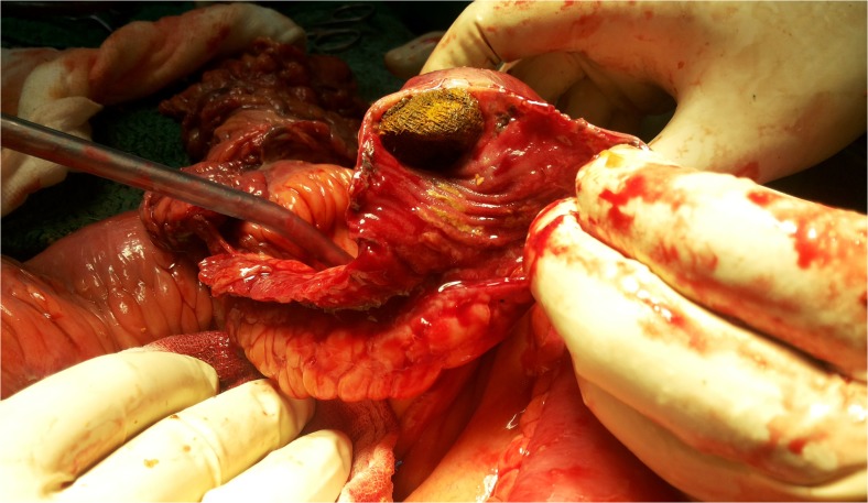
Patient underwent exploratory laparotomy for acute abdomen. Intraoperatively, around 500 ml of fecopurulent matter was present in the abdominal cavity. Loops of ileum were adhered to each other. On careful separation of bowel loops, a gangrenous small bowel loop was found with multiple perforations. A foreign body (mop) was found intraluminally in the gangrenous loop approximately 2 ft proximal to ileocecal junction (Fig. 1). Resection of the gangrenous small bowel with perforations was done, and a double-barrel ileostomy was made (Fig. 2). A thorough gut exploration could not suggest a probable site of entry. Postoperative recovery of the patient was uneventful.
Fig. 1.
Sponge seen inside the intestinal lumen
Fig. 2.
Resected segment of intestine with sponge
Discussion
Gossypiboma is a retained foreign body like a sponge/gauze after surgery. From the period of 1978–2011, 170 cases of gossypiboma have been reported [5]. As per the literature review from 2000 to 2010, 45 cases of transmural migration of gossypiboma were found. The most common site for the retained foreign body is the abdomen; other sites include pelvis, thorax, and vagina [6].
Gossypiboma can initiate two types of body response—exudative or aseptic fibrosis. Complications associated with gossypiboma include adhesions, fistula formation, abscess, or transmural migration [2]. The foreign body causes pressure necrosis of the bowel wall and migrates either partially or completely into the bowel lumen. Moreover, the perforation hence created closes spontaneously after the migration is complete [5]. The foreign body can then advance with bowel peristaltic activity up to the ileocecal junction and can present with symptoms of intestinal obstruction and other malabsorptive symptoms. More commonly, the retained foreign body can be either expelled out of the body through the wound or through a sinus tract.
The investigation of choice for diagnosis of gossypiboma and associated complications is CT scan. It shows a “spongiform pattern” having air bubbles [2]. Plain X-ray of the abdomen is of limited value until and unless the retained surgical sponge has a radiological marker [2].
Our patient presented with features suggestive of peritonitis. He had a history of open cholecystectomy 2 years back. X-ray of the abdomen and USG of the abdomen could not diagnose the retained foreign body, and it could only be diagnosed when patient underwent surgery.
Gossypiboma should be removed once it is diagnosed. Mainstay for removal remains the surgery while percutaneous extraction has also been reported as an alternative in some reports [7].
“Prevention is better than cure” is a notion which holds true for this condition, as it not only is associated with significant morbidity and mortality for patients; it also has a medicolegal implication. Gossypiboma, no doubt is distressful for patients but is equally embarrassing to a surgeon, can be a cause for significant mental suffering for both of them.
A baseline count of all the surgical stuff should be made preoperatively. This count should be counterchecked at the end of the operation before closing the abdomen and recorded in the case sheet.
It is also recommended to have a radiopaque marker on all surgical mops/sponges so that in case of any count discrepancy, an intraoperative X-ray may be performed.
Conclusion
Gossypiboma is a cause of significant morbidity and mortality for the patient. Even if the incidence of gossypiboma is very low, it should always be kept in mind as a cause of chronic abdominal pain and discomfort in a patient with history of previous abdominal surgery.
Compliance with Ethical Standards
Conflicts of Interest
The authors declare that they have no conflict of interest.
Consent
Written informed consent was obtained from the patient for publication of this case report and associated images.
Contributor Information
Himanshu Agrawal, Email: himagr1987@gmail.com.
Nikhil Gupta, Email: nikhil_ms26@yahoo.co.in.
Umesh Krishengowda, Email: umesh2906@gmail.com.
Arun Kumar Gupta, Email: drguptaarunkumar@gmail.com.
Dipankar Naskar, Email: drdipankarnask234@gmail.com.
C. K. Durga, Email: hodsurgeryrmlh@gmail.com
References
- 1.Rajagopal A, Martin J (2002) Gossypiboma—"a surgeon's legacy": report of a case and review of the literature. Dis Colon rectum 45:119–120 [DOI] [PubMed]
- 2.Manzella A, Filho PB, Albuquerque E, Farias F, Kaercher J. Imaging of gossypibomas: pictorial review. AJR Am J Roentgenol. 2009;193(6 Suppl):S94–101. doi: 10.2214/AJR.07.7132. [DOI] [PubMed] [Google Scholar]
- 3.Nizamuddin S. “Gossypiboma” an operative team’s dilemma. Pakistan J Surg. 2008;24:159–162. [Google Scholar]
- 4.Silva CS, Caetano MR, Silva EA, Falco L, Murta EF. Complete migration of retained surgical sponge into ileum without sign of open intestinal wall. Arch Gynecol Obstet. 2001;265:103–104. doi: 10.1007/s004040000141. [DOI] [PubMed] [Google Scholar]
- 5.Dux M, Ganten M, Lubienski A, Grenacher L. Retained surgical sponge with migration into the duodenum and persistent duodenal fistula. Eur Radiol. 2002;12:74–77. doi: 10.1007/s003300100980. [DOI] [PubMed] [Google Scholar]
- 6.Gawande AA, Studdert DM, Orav EJ, Brennan TA, Zinner MJ. Risk factors for retained instruments and sponges after surgery. N Engl J Med. 2003;348:229–235. doi: 10.1056/NEJMsa021721. [DOI] [PubMed] [Google Scholar]
- 7.Gencosmanoglu R, Inceoglu R. An unusual cause of small bowel obstruction: gossypiboma—case report. BMC Surg. 2003;3:6. doi: 10.1186/1471-2482-3-6. [DOI] [PMC free article] [PubMed] [Google Scholar]


