Introduction
Hemophagocytic lymphohistiocytosis (HLH) is a life-threatening disease associated with an overwhelming cytokine storm and severe inflammation. Although genetic mutations resulting in primary HLH are the most common cause in the pediatric population, adults often present with HLH secondary to infection, autoimmune disease, or malignancy. Among the secondary causes, lymphoma-associated hemophagocytic syndrome (LAHS), usually of the peripheral T-cell or Natural Killer (NK)-cell lineages is the most common and confers the poorest survival. Identification of this underlying secondary cause in adults often carries a significant diagnostic and therapeutic dilemma requiring the highest degree of clinical suspicion and awareness because it greatly affects patient outcome. Here we present 2 cases of LAHS that portend the atypical presentation and clinical dilemma associated with this disease, followed by a review and suggestions on a therapeutic approach.
Hemophagocytic lymphohistiocytosis, also called hemophagocytic syndrome is a rare, heterogeneous life-threatening disease characterized by uncontrolled activation of the normal immune system causing an overwhelming cytokine storm and severe inflammation.1 Although well-defined clinical criteria have been established, its rarity and extremely variable presentation often delay the diagnosis and timely management resulting in overall poor survival. It is most commonly associated with persistent fever, hepatosplenomegaly, hyperferritinemia, hypertriglyceridemia, or hypofibrinogenemia, hemocytopenia, and hemophagocytosis. Primary HLH is characterized by a known family history or genetic mutations, most commonly affecting the pediatric population, usually infants younger than 18 months of age.2,3 Several genetic mutations have been identified in primary HLH and it most commonly affects the perforin gene (PRF1). Incidence is estimated at 1.2 cases per 50,000 births.4 Although several genetic mutations have been identified in primary HLH, 5 distinct variants account for > 90% of the cases (unidentified gene defect, PRF1, UNC13D, Syntaxin 11 [STX11], Syntaxin Binding Protein-2 [STXBP-2]).5 Similar mutations have been described in adult-onset clinical HLH patients, however a secondary trigger such as infection, autoimmune disease, or malignancy can be identified in most of these patients, hence, most adult cases are grouped in a category of secondary HLH.6 Delay or failure to recognize the underlying etiology can have devastating consequences, because secondary causes can affect treatment decisions and survival.
Lymphoma-associated hemophagocytic syndrome is a relatively well-described subtype of secondary HLH that carries a relatively poor prognosis. Although commonly associated with T-cell lymphoma, B-cell lymphoma-associated cases have also been reported in the literature. Herein, we present 2 cases associated with lymphoma and highlight the fact that underlying lymphoma was difficult to initially diagnose in both cases, complicating therapeutic decisions.
Case Reports
Case 1
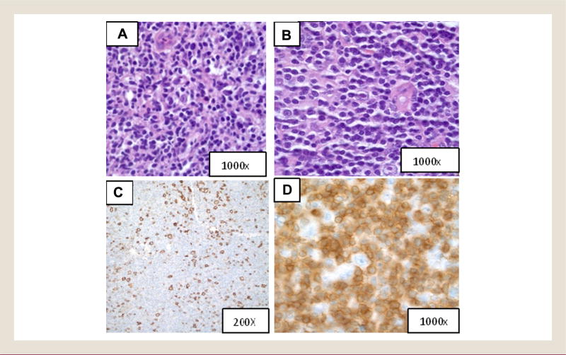
A 21-year-old previously healthy college student presented with a 2-week history of malaise, fatigue, and abdominal discomfort that progressed to jaundice and dark urine but no pruritus. On presentation, he had an abnormal liver profile with an alanine aminotransferase level of 2338 U/L, aspartate aminotransferase level of 2129 U/L, alkaline phosphatase level of 267 U/L, total bilirubin level of 8.4 mg/dL, direct bilirubin level of 6.5 mg/dL, albumin level of 3.7 g/dL, and an international normalized ratio (INR) of 1.4. He did admit to a modest 2-year social drinking history with approximately 12 beers on weekends. He denied taking any medications. Other laboratory workup revealed a hemoglobin level of 13.5 g/dL, platelet count of 148,000/µL, white blood cell count (WBC) of 3500/µL. A viral hepatitis panel, HIV, and alpha-feto protein tests (AFP) were normal. An Epstein-Barr virus (EBV) test was positive. Abdominal ultrasound showed increased and heterogenous hepatic echogenicity with splenomegaly. Symptoms resolved gradually without any treatment; however, he had persistent increased liver enzyme levels, resulting in evaluation by the outpatient hepatology team at our institution 8 months later. On physical examination, he had splenomegaly and spider angiomata and no evidence of lymphadenopathy, hepatomegaly, or ascites. Repeat workup at that time including viral studies, ferritin, iron studies, ceruloplasmin, autoimmune workup, alpha1-antitrypsin, and AFP, was normal. He had mild thrombocytopenia (platelet count of 118,000/µL) and abdominal duplex ultrasound showed increased hepatic echogenicity suggesting diffuse liver disease and splenomegaly of 15 cm. A liver biopsy showed that the hepatic parenchyma was effaced by marked chronic inflammation and proliferative ductules, favored to represent autoimmune hepatitis. Serologic workup for autoimmune hepatitis including antinuclear antibodies, anti-smooth muscle antibodies, and immunoglobulin (Ig) G were negative. With a diagnosis of autoimmune hepatitis based on the biopsy, treatment was started with azathioprine along with prednisone, obtaining excellent clinical and laboratory response with normalization of liver-related enzymes. After approximately 3 months of therapy, he presented with fevers and pancytopenia. Laboratory testing revealed a lactate dehydrogenase (LDH) level of 917 U/L, triglycerides of 229 mg/dL, ferritin of 5290 ng/mL, white count of 1400/µL (19% neutrophils), hemoglobin of 8.7 g/dL, platelet count of 58,000/µL, INR of 1.6, EBV quantity of 393 copies per microgram of DNA, decreased NK activity of 0% and increased level of soluble interleukin (IL)-2 receptors, fulfilling the clinical criteria of HLH. His physical exam revealed palpable cervical, axillary, and inguinal lymphadenopathy. Because of concern for HLH, he was admitted for further workup. A positron emission tomography (PET)-computed tomography (CT) scan was performed that demonstrated fluorine-18 fluorodeoxyglucose uptake throughout the bone marrow and lymph nodes. A bone marrow biopsy and cervical lymph node biopsy were performed, both revealing involvement by T cell-rich diffuse large B-cell lymphoma (Figure 1B–D). The previous liver biopsy was reviewed and showed similar immunohistochemical/morphologic features with infiltrating small CD3-positive (CD3+) T-cells, mixed with abundant CD68+ histiocytes and scattered CD20+ large atypical cells (compare Figure 1A with Figure 1B). He was treated with R-CHOP (rituximab, cyclophosphamide, doxorubicin, vincristine, and prednisone) for 6 cycles and achieved complete remission. Repeat bone marrow biopsy showed some hypocellularity with no evidence of lymphoma or myelodysplasia. However, he continued to show pancytopenia and mild liver dysfunction even several weeks after completion of therapy.
Figure 1.
(A) Liver Biopsy Showing Lymphocytic Infiltrate Containing Very Rare Large, Atypical Cells (H & E Stain, Magnification ×1000); (B) Lymph Node Biopsy Showing Effacement of Architecture by a Lymphocytic Infiltrate Identical to That Seen in (A) (H & E Stain, Magnification ×1000); (C) Lymph Node Biopsy Showing Scattered Large B Cells (CD20 Immunohistochemical Stain, Magnification ×200); (D) Lymph Node Biopsy Showing Abundant Small T Cells (CD3 Immunohistochemical Stain, Magnification ×1000)
Abbreviation: H & E = Hematoxylin and Eosin.
Case 2
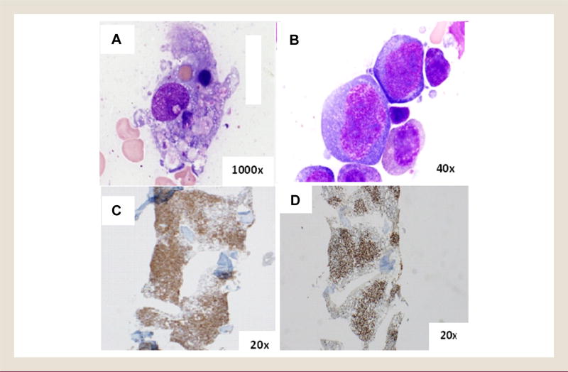
A 42-year-old Caucasian man with history of Crohn’s disease was admitted to the hospital with persistent fevers and hypotension, and developed hypoxemia during the hospitalization. He had recently started treatment with adalimumab for his Crohn’s disease and received a second dose just before hospitalization. Physical examination revealed no significant abnormalities like lymphadenopathy or enlarged liver or spleen. His workup showed pancytopenia, an increased ferritin level of 40,000 ng/mL, increased IL-2 receptor level, and a bone marrow biopsy showed scattered hemophagocytosis (Figure 2B). CT scan of the chest, abdomen, and pelvis also showed no evidence of lymphadenopathy or hepatosplenomegaly. Flow cytometry was unremarkable, and cytogenetics showed a normal male karyotype in 12 cells and the remaining 8 cells featured a complex karyotype with numerical and structural abnormalities including monosomy for chromosomes 4 and 6, gain of an X chromosome, loss of the Y chromosome, trisomy for chromosome 5, and an unbalanced t(1:11). Infectious workup was negative initially but repeat testing was positive for EBV DNA using polymerase chain reaction (PCR). The HLH-2004 protocol was started and he received etoposide, dexamethasone, cyclosporin, and intravenous immunoglobulin for 4 weeks. It was interrupted by development of thrombocytopenia, Crohn’s exacerbation, and pseudomonas sepsis. Repeat bone marrow biopsy performed 10 weeks after initiation of therapy because of pancytopenia and concerns for myelodysplasia showed a severe hypoplastic marrow and normal male karyotype in all 20 cells analyzed. He eventually recovered, became transfusion-independent, and returned to work. His ferritin and LDH levels normalized completely. His Crohn’s disease was inactive during this time and was managed only with monthly vitamin B12 injections and daily folic acid.
Figure 2.
(A) Original Bone Marrow Biopsy Showing Hemophagocytosis (Wright-Giemsa Stain of Marrow Aspirate, Magnification ×1000); (B) H & E Stain of the Bone Marrow Biopsy 1 Year Later Showing Diffuse Infiltrate of Large, Atypical Lymphocytes (Magnification ×40); (C) the Infiltrate Is Composed of T Cells (CD3 Immunohistochemical Stain, Magnification ×20); (D) T Cells are CD8-Positive (CD8 Immunohistochemical Stain, Magnification ×20)
Abbreviation: H & E = Hematoxylin and Eosin.
One year since the initial presentation, he developed monthly fevers for a few days at a time that responded to empiric antibiotics with no consistent change in ferritin and LDH levels. He was admitted to the hospital with a third episode of fever associated with worsening blood counts requiring a more extensive workup. Lumbar puncture, done because he complained of neck stiffness revealed herpes simplex virus meningitis and therefore valacyclovir treatment was initiated. Repeat EBV DNA using PCR was positive. A bone marrow biopsy as a part of a workup revealed involvement by T-cell lymphoproliferative disorder (CD8+ large lymphocytes showing CD30 and 3’UTR mRNA binding protein [TIA-1] reactivity). Cytogenetic studies again showed a complex karyotype similar to initial bone marrow biopsy but with some additional abnormalities suggesting clonal evolution. Clonal T-cell receptor (TCR) gene rearrangement was detected. Because of these findings, the diagnosis of a peripheral T-cell lymphoma (PTCL), not otherwise specified was favored (Figure 2B–D). Similar-appearing malignant T cells were found in the cerebrospinal fluid, and a PET scan showed extensive activity in the spleen, T1 vertebra, and bone marrow. Chemotherapy was initiated with Cyclophosphamide, Doxorubicin, Etoposide, Vincristine and Prednisone (CHOEP) and intrathecal methotrexate with the plan of alternating with systemic high-dose methotrexate. However, the patient developed multiple complications during this hospitalization including altered mental status, sepsis, and acute respiratory failure, requiring intubation. Despite attempts with aggressive therapy, he succumbed to his disease shortly thereafter.
Discussion
Etiology of HLH
Several genetic mutations causing immunologic defects have been described in primary HLH, including familial HLH (FHL)-1 (gene not identified but located on chromosome 9q21.3-22 region), FHL2 (perforin), FHL3 (mammalian uncoordinated [MUNC] 13-4 involved in packaging of cytolytic granzymes), Ras-related protein coding gene (RAB27A) (Griscelli syndrome), FHL4 (syntaxin 11), FHL5 (MUNC 18-2), less spontaneous activation of caspase-3, and inactivity of cytotoxic T lymphocyte-associated antigen-4 (LYST- Chediak-Higashi syndrome).7–12 Although rarely heterozygosity for HLH-associated gene mutations can be found in adults, secondary HLH is usually secondary to infection, autoimmune disease, or malignancy.4,13 In a single-institution case series study of 11 patients with HLH, all of them were noted to have genetic abnormalities that are pertinent to HLH, and included defects in FHL2 (6 patients), FHL3 (2 patients), FHL5 (1 patient), and X-linked lymphoproliferative disorder (XLP1) (2 patients).14 One large study including 1531 patients with a clinical diagnosis of HLH, of which 175 patients were ≥ 18 years of age, 14% of patients had missense and splice-site sequence variants in PRF1, MUNC13-4, and Syntaxin binding protein 2 (STXBP2). The A91V-PRF1 genotype was found in 12 of these patients (48%).15 Among infections, viral illness caused by EBV, Cytomegalovirus (CMV), herpes simplex virus, measles, varicella zoster, HHV 8, and HIV are most commonly associated. Of note, any of these can be detected in combination in HLH especially with HIV.13,16 Autoimmune diseases that have been associated with HLH include systemic lupus erythematosus, rheumatoid arthritis, Crohn’s disease, scleroderma, and Sjogren disease especially when these patients are given immunosuppressive therapy.17–20 In a retrospective study by Fries et al, 50 cases of macrophage activation syndrome (MAS), which is considered equivalent to secondary HLH, were found to be associated with inflammatory bowel disease, with most patients suffering from Crohn’s disease. All except 5 (10%) received immunosuppressive therapy and viral infections especially EBV and CMV were detected in 39 patients (78%). It is also important to note that 4 (8%) of these cases had a lymphoma present at the time of diagnosis. Thiopurines, specifically azathioprine or mercaptopurine, were most commonly associated with the development of HLH, but anti-tumor necrosis factor (TNF) therapy, specifically infliximab, has also been reported.20
Malignancies, especially T/NK cell lymphoma or leukemia, are another common cause of HLH. Patients with LAHS have a worse prognosis compared with other secondary HLH patients.21,22 In a study of 52 patients with 26 of them having LAHS, 88% died from multiorgan failure and disseminated intravascular coagulation with uncontrollable HLH despite aggressive treatment, whereas only 12% died of HLH not associated with lymphoma (although 1 patient had laryngeal cancer).22 In a study by Takahashi et al, no significant difference was found between LAHS and non-LAHS in the time from symptom onset to diagnosis and initial treatment. Hence, the greater mortality rate for LAHS patients cannot be attributed to the delay of lymphoma diagnosis and treatment alone. In a retrospective review of 159 cases of PTCL, 36 patients developed LAHS and had significantly worse outcomes compared with those who did not develop LAHS,23 the most common lymphoproliferative malignancy associated with HLH and lymphoma or leukemia of peripheral T-cell or NK-cell lineages.24–26 Cutaneous manifestations such as panniculitis in patients with HLH should raise the suspicion of an underlying T-cell lymphoma.27–29 Enteropathy-associated T-cell lymphoma has also been associated with secondary HLH and is believed to have a much worse prognosis if HLH develops in this context. In a single-institution case series of 15 patients with enteropathy-associated T-cell lymphoma, 6 patients developed HLH and all of them died within 3 months of diagnosis.30
When the diagnosis of HLH is revealed according to clinical and laboratory features, all possible underlying causes should be thoroughly investigated. Both of the cases described herein illustrate the difficulty in identifying an underlying etiology and emphasize the need for high clinical suspicion, especially of lymphoma. The first case was initially diagnosed as autoimmune hepatitis based on clinical symptoms and liver biopsy, which showed marked inflammation. Interestingly, the patient did not develop symptoms of HLH until being treated with azathioprine. Therefore, it is difficult to determine the sequence of events because lymphoma and use of immunosuppressant agents are associated with HLH. Furthermore, azathioprine has been associated with lymphoma development in retrospective analyses.31 It was not until liver biopsy samples were compared retrospectively with the bone marrow biopsy that lymphoma was confirmed as the likely underlying cause of HLH, although immunosuppression might have contributed as well. Fortunately, the patient described in Case 1 has done well with treatment of his lymphoma. It is also important to note that our first case described was of diffuse large B-cell lymphoma, which is relatively rarely associated with HLH. We can speculate that being T cell-rich might have driven the development of HLH.
The second case was even more challenging because of the known difficulty in the diagnosis of T-cell lymphomas. Crohn’s disease and immunosuppression was initially thought to be the reason for his HLH. Although to the best of our knowledge there has not been an obvious association reported between adalimumab in inflammatory bowel disease and secondary HLH, there is 1 reported association between adalimumab and MAS in a patient with rheumatoid arthritis. However, the latter patient also had infection with visceral leishmaniasis.32 Clonal abnormalities on the initial bone marrow biopsy was a feature inconsistent with HLH, however, lack of any morphologic and, flow-cytometric evidence of lymphoma and disappearance of the clone on the second bone marrow biopsy provided a false sense of security and response. As illustrated in this case, early diagnosis of lymphoma is difficult in the absence of usual features such as lymphadenopathy and splenomegaly; repeated bone marrow biopsies from different locations along with cytogenetic studies and imaging studies such as PET/CT scan might at times assist in diagnosis of an occult lymphoma.
Pathophysiology of HLH
Many of the clinical and laboratory features of HLH provide us insight into the pathogenesis of HLH. For an individual to develop HLH, a certain component of immunodeficiency (for example, decreased or absent NK cell function) coupled with uncontrolled immune activation (fever or organomegaly) with evidence of immunopathology at tissue levels (hemophagocytosis, cytopenia, hepatitis) is needed. In a normal immune system, inflammatory responses are controlled by several negative feedback mechanisms. For example chemicals like perforin and granzymes released by cytotoxic cells can drive apoptosis in antigen-presenting cells (APCs) and elimination of these APCs serve as a feedback for resolution of the inflammatory response to any specific stimulus. The proposed mechanism by which the mutation confers the disease, is the inability of the cytotoxic cells such as CD8+ lymphocytes and NK cells to release perforin and granzyme B, in response to a pathogen contained in APCs.33,34 Because elimination of APCs provides important feedback to further limit cytotoxic response, which is lacking in HLH because of ineffective cytotoxicity, a large amount of cytokines are released, further exacerbating the activation of T lymphocytes and histiocytes.35
Although not well understood, a similar dysregulation of the immune system can result in several manifestations of secondary HLH. For example, PTCL is one of the most common malignancies associated with HLH in immunocompetent patients.36 T cells cause uninhibited activation of macrophages that start phagocytosing normal blood cells, causing LAHS.37 T cells specifically are linked to the severe cytokine storm that might explain why patients with T-cell lymphomas fare worse than those with B-cell lymphomas.38 EBV infection is commonly seen in HLH caused by lymphoma. The (LMP1) Latent membrane protein 1 gene encoded by the EBV virus has been proposed to activate signaling lymphocyte activation molecule-associated protein leading to excessive T-cell activation and enhanced Th1 cytokine secretion which might lead to the development of HLH and X-linked lymphoproliferative disorder, therefore, possibly explaining the association of EBV and LAHS.39 EBV might also directly infect CD8+ T cells and can initiate clonal expansion by down-regulating CD5.40 We believe a more thorough search of an underlying lymphoproliferative disorder is warranted if a patient presents with HLH and detectable EBV infection because early diagnosis and management of the underlying malignancy will have a significant effect on the disease outcome.
Diagnostic Criteria for HLH
According to the criteria used in the HLH-2004 trial, an HLH-associated gene mutation or 5 of 8 are necessary for diagnosis of hemophagocytic syndrome.41 The 8 criteria include fever (> 38.5°C for 7 or more days), splenomegaly (> 3 cm below the costal margin), cytopenia in at least 2 cell lines (hemoglobin < 9, platelets < 100,000, and absolute neutrophil count (ANC) < 1000/µL), hypertriglyceridemia or hypofibrinogenemia (≥ 3 standard deviations), tissue demonstration of hemophagocytosis (in bone marrow, lymph node, or spleen), low or loss of NK cell activity, serum ferritin > 500 µg/L, and soluble CD25 > 2400 U/mL. As noted herein, because of its rarity and extremely variable clinical manifestations, diagnosis is often delayed and a modification to the HLH-2004 criteria proposes 3 of 4 clinical findings (fever, cytopenias, splenomegaly, hepatitis) in addition to 1 of 4 immunological findings (increased ferritin level, hemophagocytosis, hypofibrinogenemia, and decreased NK cell function).6,42
Management of Secondary HLH
Although the HLH-94 protocol or HLH-2004 protocol (etoposide and dexamethasone with cyclosporine with intent of allogeneic stem cell transplant after induction) has been established for primary HLH, the treatment for secondary HLH, especially LAHS, remains undefined and debatable. Because HLH is a rapidly fatal disease if not treated, Jordan et al recommend not to delay initial treatment while searching for underlying causes.41 Additionally, they propose treatment with immunochemotherapy aimed at controlling inflammation first, before changing to disease-specific treatment when inflammation markers normalize. However, these studies were mostly based in the pediatric population in which HLH is less likely to be due to secondary causes. We would argue that targeting the underlying cause, ie, the lymphoma itself, that caused the HLH, would be a more appropriate therapy upfront and might result in less relapse. The most appropriate upfront therapy is unclear, however, and should widely be based on the patient’s condition. Interestingly, some isolated cases and reports have identified a role for plasmapheresis in reducing the cytokine storm associated with HLH.43,44 This might be useful as soon as HLH is recognized while waiting for confirmation of underlying disease.
For the most part we agree with the approach of treating the cytokine storm initially, understanding that in some patients we might delay or miss the diagnosis of underlying lymphoma by readily treating HLH with immunochemotherapy, as was illustrated in case 2. Because he had severe inflammatory symptoms, we believe if we would not have initiated therapy the patient would not have survived his HLH at initial presentation. However, underlying lymphoma should be thoroughly ruled out using repeated testing and when recognized and the cytokine storm controlled, we would argue targeting the underlying cause. The appropriate upfront therapy should be based on the specific subtype of underlying lymphoma. Chemotherapy regimens such as the COP (cyclophosphamide, vincristine, and prednisone) regimen have been used in infection-associated secondary HLH with a 1-year overall survival rate of 67%.45 CHOP (cyclophosphamide, doxorubicin, vincristine, and prednisone) when used in 17 adults (7 patients with underlying lymphoma) without stem cell transplant led to a 2-year overall survival of 44%.46 In a study by Xie et al, of 159 PTCL patients, patients with LAHS who received intensive chemotherapy appeared to have an improved overall survival, however, it was not statistically significant compared with CHOP therapy alone.23 Allogeneic stem cell transplantation has shown promising results in children with a 3-year survival rate as high as 70% with primary HLH.47,48 However, data for stem cell transplant in adults is extremely limited and should largely be based on underlying etiology.49,50 Aggressive chemotherapy using an HLH protocol while searching for underlying etiology followed by allogeneic stem cell transplant is believed to be the best approach in high-risk pediatric patients especially with cytogenetic abnormalities, but further studies are warranted to validate this approach in adults.51,52
Often patients with B-cell lymphoma have a favorable outcome compared with patients with T-cell lymphoma.38,53 IgH or TCR clonality made no difference in outcome in 1 study, but low EBV viral load demonstrated a greater clinical response and longer overall survival compared with higher viral load.54
Conclusion
Lymphoma-associated hemophagocytic syndrome is a rare but devastating presentation of lymphoma and portends a very poor prognosis. Early recognition is imperative to treat the underlying cause because the disease is invariably rapidly fatal. Occult malignancy might be present at the time of diagnosis and therefore a vigorous search using thorough and repeated testing for secondary causes is important in adults. If HLH is associated with EBV, a vigorous search should be carried out because the combination is most often associated with underlying malignancy. Further studies are needed to better understand the most appropriate front-line treatment for LAHS. Aggressive chemotherapy followed by allogeneic stem cell transplantation appears to result in better survival in the high-risk pediatric population, but its role in adults remains to be established.
Clinical Practice Points.
Lymphoma-associated hemophagocytic syndrome is a disease with an extremely poor prognosis and can be easily missed at diagnosis.
Identifying and treating the underlying lymphoma early is of utmost importance because that directly affects outcome.
Thorough and repeated testing for the underlying cause is imperative.
Further studies are necessary to better understand the most appropriate front-line therapy.
Allogeneic bone marrow transplant has shown promising results in children and needs to be studied further in adults.
Footnotes
Disclosure
The authors have stated that they have no conflicts of interest.
References
- 1.Larroche C, Mouthon L. Pathogenesis of hemophagocytic syndrome (HPS) Autoimmun Rev. 2004;3:69–75. doi: 10.1016/S1568-9972(03)00091-0. [DOI] [PubMed] [Google Scholar]
- 2.Reiner AP, Spivak JL. Hematophagic histiocytosis. A report of 23 new patients and a review of the literature. Medicine (Baltimore) 1988;67:369–88. [PubMed] [Google Scholar]
- 3.Aricò M, Janka G, Fischer A, et al. Hemophagocytic lymphohistiocytosis. Report of 122 children from the International Registry. FHL Study Group of the Histiocyte Society. Leukemia. 1996;10:197–203. [PubMed] [Google Scholar]
- 4.Sung L, King SM, Carcao M, Trebo M, Weitzman SS. Adverse outcomes in primary hemophagocytic lymphohistiocytosis. J Pediatr Hematol Oncol. 2002;24:550–4. doi: 10.1097/00043426-200210000-00011. [DOI] [PubMed] [Google Scholar]
- 5.Gholam C, Grigoriadou S, Gilmour KC, Gaspar HB. Familial haemophagocytic lymphohistiocytosis: advances in the genetic basis, diagnosis and management. Clin Exp Immunol. 2011;163:271–83. doi: 10.1111/j.1365-2249.2010.04302.x. [DOI] [PMC free article] [PubMed] [Google Scholar]
- 6.Henter JI, Horne A, Aricó M, et al. HLH-2004: diagnostic and therapeutic guidelines for hemophagocytic lymphohistiocytosis. Pediatr Blood Cancer. 2007;48:124–31. doi: 10.1002/pbc.21039. [DOI] [PubMed] [Google Scholar]
- 7.Göransdotter Ericson K, Fadeel B, Nilsson-Ardnor S, et al. Spectrum of perforin gene mutations in familial hemophagocytic lymphohistiocytosis. Am J Hum Genet. 2001;68:590–7. doi: 10.1086/318796. [DOI] [PMC free article] [PubMed] [Google Scholar]
- 8.Meeths M, Chiang SC, Wood SM, et al. Familial hemophagocytic lymphohistiocytosis type 3 (FHL3) caused by deep intronic mutation and inversion in UNC13D. Blood. 2011;118:5783–93. doi: 10.1182/blood-2011-07-369090. [DOI] [PubMed] [Google Scholar]
- 9.zur Stadt U, Schmidt S, Kasper B, et al. Linkage of familial hemophagocytic lymphohistiocytosis (FHL) type-4 to chromosome 6q24 and identification of mutations in syntaxin 11. Hum Mol Genet. 2005;14:827–34. doi: 10.1093/hmg/ddi076. [DOI] [PubMed] [Google Scholar]
- 10.Côte M, Ménager MM, Burgess A, et al. Munc18-2 deficiency causes familial hemophagocytic lymphohistiocytosis type 5 and impairs cytotoxic granule exocytosis in patient NK cells. J Clin Invest. 2009;119:3765–73. doi: 10.1172/JCI40732. [DOI] [PMC free article] [PubMed] [Google Scholar]
- 11.Ménasché G, Pastural E, Feldmann J, et al. Mutations in RAB27A cause Griscelli syndrome associated with haemophagocytic syndrome. Nat Genet. 2000;25:173–6. doi: 10.1038/76024. [DOI] [PubMed] [Google Scholar]
- 12.Rubin CM, Burke BA, McKenna RW, et al. The accelerated phase of Chediak-Higashi syndrome. An expression of the virus-associated hemophagocytic syndrome? Cancer. 1985;56:524–30. doi: 10.1002/1097-0142(19850801)56:3<524::aid-cncr2820560320>3.0.co;2-z. [DOI] [PubMed] [Google Scholar]
- 13.Grossman WJ, Radhi M, Schauer D, Gerday E, Grose C, Goldman FD. Development of hemophagocytic lymphohistiocytosis in triplets infected with HHV-8. Blood. 2005;106:1203–6. doi: 10.1182/blood-2005-03-0950. [DOI] [PMC free article] [PubMed] [Google Scholar]
- 14.Sieni E, Cetica V, Piccin A, et al. Familial hemophagocytic lymphohistiocytosis may present during adulthood: clinical and genetic features of a small series. PLoS One. 2012;7:e44649. doi: 10.1371/journal.pone.0044649. [DOI] [PMC free article] [PubMed] [Google Scholar]
- 15.Zhang K, Jordan MB, Marsh RA, et al. Hypomorphic mutations in PRF1, MUNC13-4, and STXBP2 are associated with adult-onset familial HLH. Blood. 2011;118:5794–8. doi: 10.1182/blood-2011-07-370148. [DOI] [PMC free article] [PubMed] [Google Scholar]
- 16.Chen TL, Wong WW, Chiou TJ. Hemophagocytic syndrome: an unusual manifestation of acute human immunodeficiency virus infection. Int J Hematol. 2003;78:450–2. doi: 10.1007/BF02983819. [DOI] [PubMed] [Google Scholar]
- 17.Wong KF, Hui PK, Chan JK, Chan YW, Ha SY. The acute lupus hemophagocytic syndrome [published erratum appears in: Ann Intern Med 1991; 114:993] Ann Intern Med. 1991;114:387–90. doi: 10.7326/0003-4819-114-5-387. [DOI] [PubMed] [Google Scholar]
- 18.Dhote R, Simon J, Papo T, et al. Reactive hemophagocytic syndrome in adult systemic disease: report of twenty-six cases and literature review. Arthritis Rheum. 2003;49:633–9. doi: 10.1002/art.11368. [DOI] [PubMed] [Google Scholar]
- 19.Morris JA, Adamson AR, Holt PJ, Davson J. Still’s disease and the virus-associated haemophagocytic syndrome. Ann Rheum Dis. 1985;44:349–53. doi: 10.1136/ard.44.5.349. [DOI] [PMC free article] [PubMed] [Google Scholar]
- 20.Fries W, Cottone M, Cascio A. Systematic review: macrophage activation syndrome in inflammatory bowel disease. Aliment Pharmacol Ther. 2013;37:1033–45. doi: 10.1111/apt.12305. [DOI] [PubMed] [Google Scholar]
- 21.Takahashi N, Miura I, Chubachi A, Miura AB, Nakamura S. A clinicopathological study of 20 patients with T/natural killer (NK)-cell lymphoma-associated hemophagocytic syndrome with special reference to nasal and nasal-type NK/T-cell lymphoma. Int J Hematol. 2001;74:303–8. doi: 10.1007/BF02982065. [DOI] [PubMed] [Google Scholar]
- 22.Takahashi N, Chubachi A, Kume M, et al. A clinical analysis of 52 adult patients with hemophagocytic syndrome: the prognostic significance of the underlying diseases. Int J Hematol. 2001;74:209–13. doi: 10.1007/BF02982007. [DOI] [PubMed] [Google Scholar]
- 23.Xie W, Hu K, Xu F, et al. Clinical analysis and prognostic significance of lymphoma-associated hemophagocytosis in peripheral T cell lymphoma. Ann Hematol. 2013;92:481–6. doi: 10.1007/s00277-012-1644-6. [DOI] [PMC free article] [PubMed] [Google Scholar]
- 24.Kleynberg RL, Schiller GJ. Secondary hemophagocytic lymphohistiocytosis in adults: an update on diagnosis and therapy. Clin Adv Hematol Oncol. 2012;10:726–32. [PubMed] [Google Scholar]
- 25.Gallipoli P, Drummond M, Leach M. Hemophagocytosis and relapsed peripheral T-cell lymphoma. Eur J Haematol. 2009;82:246. doi: 10.1111/j.1600-0609.2008.01167.x. [DOI] [PubMed] [Google Scholar]
- 26.Warnnissorn N, Kanitsap N, Kulkantrakorn K, Assanasen T. Natural killer cell malignancy associated with Epstein-Barr virus and hemophagocytic syndrome. J Med Assoc Thai. 2007;90:982–7. [PubMed] [Google Scholar]
- 27.Glaudemans AW, Slart RH, Pruim J. Panniculitis-like T-cell lymphoma detected by positron emission tomography/computed tomography scanning in a patient with haemophagocytic syndrome. Eur J Haematol. 2011;87:379. doi: 10.1111/j.1600-0609.2011.01655.x. [DOI] [PubMed] [Google Scholar]
- 28.Blom A, Beylot-Barry M, D’Incan M, Laroche L. Lymphoma-associated hemophagocytic syndrome (LAHS) in advanced-stage mycosis fungoides/Sézary syndrome cutaneous T-cell lymphoma. J Am Acad Dermatol. 2011;65:404–10. doi: 10.1016/j.jaad.2010.05.029. [DOI] [PubMed] [Google Scholar]
- 29.Miura T, Kawakami Y, Sato M, Ohtsuka M, Yamamoto T. Hemophagocytic syndrome occurred in a patient with subcutaneous panniculitis-like T-cell lymphoma without overt skin lesion: successful treatment with steroid pulse therapy. J Dermatol. 2011;38:1113–5. doi: 10.1111/j.1346-8138.2010.01178.x. [DOI] [PubMed] [Google Scholar]
- 30.Amiot A, Allez M, Treton X, et al. High frequency of fatal haemophagocytic lymphohistiocytosis syndrome in enteropathy-associated T cell lymphoma. Dig Liver Dis. 2012;44:343–9. doi: 10.1016/j.dld.2011.10.008. [DOI] [PubMed] [Google Scholar]
- 31.Pasternak B, Svanström H, Schmiegelow K, Jess T, Hviid A. Use of azathioprine and the risk of cancer in inflammatory bowel disease. Am J Epidemiol. 2013;177:1296–305. doi: 10.1093/aje/kws375. [DOI] [PubMed] [Google Scholar]
- 32.Moltó A, Mateo L, Lloveras N, Olivé A, Minguez S. Visceral leishmaniasis and macrophagic activation syndrome in a patient with rheumatoid arthritis under treatment with adalimumab. Joint Bone Spine. 2010;77:271–3. doi: 10.1016/j.jbspin.2010.01.011. [DOI] [PubMed] [Google Scholar]
- 33.Risma K, Jordan MB. Hemophagocytic lymphohistiocytosis: updates and evolving concepts. Curr Opin Pediatr. 2012;24:9–15. doi: 10.1097/MOP.0b013e32834ec9c1. [DOI] [PubMed] [Google Scholar]
- 34.Mazodier K, Marin V, Novick D, et al. Severe imbalance of IL-18/IL-18BP in patients with secondary hemophagocytic syndrome. Blood. 2005;106:3483–9. doi: 10.1182/blood-2005-05-1980. [DOI] [PMC free article] [PubMed] [Google Scholar]
- 35.Janka GE, Lehmberg K. Hemophagocytic lymphohistiocytosis: pathogenesis and treatment. Hematology Am Soc Hematol Educ Program. 2013:605–11. doi: 10.1182/asheducation-2013.1.605. [DOI] [PubMed] [Google Scholar]
- 36.Jaffe ES, Costa J, Fauci AS, Cossman J, Tsokos M. Malignant lymphoma and erythrophagocytosis simulating malignant histiocytosis. Am J Med. 1983;75:741–9. doi: 10.1016/0002-9343(83)90402-3. [DOI] [PubMed] [Google Scholar]
- 37.Ascani S, Zinzani PL, Gherlinzoni F, et al. Peripheral T-cell lymphomas. Clinicopathologic study of 168 cases diagnosed according to the R.E.A.L. Classification. Ann Oncol. 1997;8:583–92. doi: 10.1023/a:1008200307625. [DOI] [PubMed] [Google Scholar]
- 38.Yu JT, Wang CY, Yang Y, et al. Lymphoma-associated hemophagocytic lymphohistiocytosis: experience in adults from a single institution. Ann Hematol. 2013;92:1529–36. doi: 10.1007/s00277-013-1784-3. [DOI] [PubMed] [Google Scholar]
- 39.Toga A, Wada T, Sakakibara Y, et al. Clinical significance of cloned expansion and CD5 down-regulation in Epstein-Barr Virus (EBV)-infected CD8+ T lymphocytes in EBV-associated hemophagocytic lymphohistiocytosis. J Infect Dis. 2010;201:1923–32. doi: 10.1086/652752. [DOI] [PubMed] [Google Scholar]
- 40.Takagi S, Masuoka K, Uchida N, et al. High incidence of haemophagocytic syndrome following umbilical cord blood transplantation for adults. Br J Haematol. 2009;147:543–53. doi: 10.1111/j.1365-2141.2009.07863.x. [DOI] [PubMed] [Google Scholar]
- 41.Jordan MB, Allen CE, Weitzman S, Filipovich AH, McClain KL. How I treat hemophagocytic lymphohistiocytosis. Blood. 2011;118:4041–52. doi: 10.1182/blood-2011-03-278127. [DOI] [PMC free article] [PubMed] [Google Scholar]
- 42.Filipovich AH. Hemophagocytic lymphohistiocytosis (HLH) and related disorders. Hematology Am Soc Hematol Educ Program. 2009:127–31. doi: 10.1182/asheducation-2009.1.127. [DOI] [PubMed] [Google Scholar]
- 43.Yanagiya N, Takahashi N, Nakae H, Kume M, Chubachi A, Miura I. Plasma exchange and continuous hemodiafiltration as an initial treatment for diffuse large B-cell lymphoma-associated hemophagocytic syndrome [in Japanese] Rinsho Ketsueki. 2002;43:35–40. [PubMed] [Google Scholar]
- 44.Coman T, Dalloz MA, Coolen N, et al. Plasmapheresis for the treatment of acute pancreatitis induced by hemophagocytic syndrome related to hypertriglyceridemia. J Clin Apher. 2003;18:129–31. doi: 10.1002/jca.10056. [DOI] [PubMed] [Google Scholar]
- 45.Hu Y, Xu J, Wang L, Li J, Qiu H, Zhang S. Treatment of hemophagocytic lymphohistiocytosis with cyclophosphamide, vincristine, and prednisone. Swiss Med Wkly. 2012;142:w13512. doi: 10.4414/smw.2012.13512. [DOI] [PubMed] [Google Scholar]
- 46.Shin HJ, Chung JS, Lee JJ, et al. Treatment outcomes with CHOP chemotherapy in adult patients with hemophagocytic lymphohistiocytosis. J Korean Med Sci. 2008;23:439–44. doi: 10.3346/jkms.2008.23.3.439. [DOI] [PMC free article] [PubMed] [Google Scholar]
- 47.Horne A, Janka G, Maarten Egeler R, et al. Haematopoietic stem cell transplantation in haemophagocytic lymphohistiocytosis. Br J Haematol. 2005;129:622–30. doi: 10.1111/j.1365-2141.2005.05501.x. [DOI] [PubMed] [Google Scholar]
- 48.Ouachée-Chardin M, Elie C, de Saint Basile G, et al. Hematopoietic stem cell transplantation in hemophagocytic lymphohistiocytosis: a single-center report of 48 patients. Pediatrics. 2006;117:e743–50. doi: 10.1542/peds.2005-1789. [DOI] [PubMed] [Google Scholar]
- 49.Machaczka M, Nahi H, Karbach H, Klimkowska M, Hägglund H. Successful treatment of recurrent malignancy-associated hemophagocytic lymphohistiocytosis with a modified HLH-94 immunochemotherapy and allogeneic stem cell transplantation. Med Oncol. 2012;29:1231–6. doi: 10.1007/s12032-011-9963-3. [DOI] [PubMed] [Google Scholar]
- 50.Kunitomi A1, Kimura H, Ito Y, et al. Unrelated bone marrow transplantation induced long-term remission in a patient with life-threatening Epstein-Barr virus-associated hemophagocytic lymphohistiocytosis. J Clin Exp Hematop. 2011;51:57–61. doi: 10.3960/jslrt.51.57. [DOI] [PubMed] [Google Scholar]
- 51.Imashuku S, Hibi S, Tabata Y, et al. Outcome of clonal hemophagocytic lymphohistiocytosis: analysis of 32 cases. Leuk Lymphoma. 2000;37:577–84. doi: 10.3109/10428190009058510. [DOI] [PubMed] [Google Scholar]
- 52.Han AR, Lee HR, Park BB, et al. Lymphoma-associated hemophagocytic syndrome: clinical features and treatment outcome. Ann Hematol. 2007;86:493–8. doi: 10.1007/s00277-007-0278-6. [DOI] [PubMed] [Google Scholar]
- 53.Balwierz W, Czogała M, Czepko E. Anaplastic large cell lymphoma associated with hemophagocytic lymphohistiocytosis: a case report and review of the literature [in Polish] Przegl Lek. 2010;67:436–8. [PubMed] [Google Scholar]
- 54.Ahn JS, Rew SY, Shin MG, et al. Clinical significance of clonality and Epstein-Barr virus infection in adult patients with hemophagocytic lymphohistiocytosis. Am J Hematol. 2010;85:719–22. doi: 10.1002/ajh.21795. [DOI] [PubMed] [Google Scholar]


