Abstract
Aloe vera (L.) Burm.f. is a highly important and extensively cultivated medicinal plant and that is also extensively used in the cosmetic industry. It has been frequently reported to suffer from Alternaria leaf spot disease in various parts of the world. Various fungicides used to combat this disease, have deleterious effects on the environment and on pharmacologically important constituents of Aloe vera. To avoid the harmful effects of fungicides an ecofriendly approach has been adopted here. A weekly survey was conducted during 2013–2015 in and around North 24 Parganas (West Bengal) to obtain the percentage of disease index (PDI). For biological control of the disease, screening of the antagonistic efficacy of biocontrol agents was carried out through the in vitro dual-culture-plate method and scanning electron microscopy (SEM) was used to study the mechanism. The in vitro effects of fungicides on the radial growth of the pathogen were evaluated through the poison food method and were compared with potent antagonistic fungi. Field application of potent antagonistic fungi was conducted through the dip-and-spray method. The results showed that, the PDI peaked during the hot and humid conditions of May to September (76.57%–98.57%) but decreased during the winter, December-January (35.71–46.66%). Trichoderma asperellum exerted the greatest inhibition of the radial growth of A. brassicae acting through non volatile (70.39%) and volatile metabolites (72.17%). A SEM study confirmed the hyperparasitic nature of T. asperellum through hyphal coiling-T. asperellum was similar to 2% blitox-50 (73.92%) and better than 2% bavistin (59.77%) (in vitro). In agricultural field trials (2013–15), Trichoderma application restricted the disease to the smallest area (PDI 24.00–29.33%) in comparison to untreated plots (73.33%). In conclusion, saplings treated with the dip method (108 spores / mL) and sprayed 4 times with a spore suspension of biocontrol agents such as T. asperellum, T. viride and T. harzianum, standardized at a rate of 2.5 L / plot (36 sq ft) (108 spores/ mL) are suggested for the ecofriendly management of this epidemic leaf spot disease of Aloe vera in agricultural fields.
Introduction
Aloe vera (L.) Burm.f. (Aloe barbadensis) belongs to the family Aloeaceae and is an extensively cultivated medicinal plant worldwide, ranging from tropical to temperate regions. Many herbal drugs and drinks have been formulated from A. vera for the maintenance of good health. In the cosmetic industry. Aloe spp. are used in the production of soap, shampoo, hair wash, tooth paste and body creams [1]. Additionally, A. vera gel has been reported to be very effective for the treatment of sores and wounds, skin cancer, skin disease, colds and coughs, constipation, piles, asthma, ulcer, diabetes and various fungal infections [1,2,3,4]. The plant Aloe vera has been frequently reported to have Alternaria leaf spot disease, both in India and in various other parts of the world. [5,6,7,8,9]. Many fungal pathogens are responsible for the production of mycotoxins that alter the potentiality of this highly important medicinal plant [10]. Chemical control of these diseases is not an ideal solution to this situation, as chemicals themselves can exert adverse effects on the pharmacologically important and other economically important products of medicinally essential plants. Therefore, biological control is best suited to the scenario. Generally Thiram, Arasan[11], Dithane M-45, Bavistin, Dithane Z-78, Difoltan, Blitox-50 and Bordeaux mixture [12,13] are used to combat Alternaria disease. However, the prolonged use of these chemicals is environmentally hazardous and toxic to humans. Non-chemical or eco-friendly methods are now popular in developed countries (such as the USA and -U.K.). Seed priming of tomato with plant growth promoting rhizobacteria (PGPR) such as Pseudomonas aeruginosa, P. putida, Bacillus subtilis, B.cereus, Azotobacter chroococcum are reported to increase seed germination, seedling vigor, fruit size, chlorophyll content, IAA production, and rhizospheric phosphate solubilization, which in turn improve plant health and growth. These PGPR strains induce plants’ defences through upregulation of defense-related enzymes such asperoxidise, polyphenol oxidase, glucanase and chitinase along with the production of HCN, siderophore which act as the inducers against Alternaria-derived early blight disease in tomato and / or other plant disease resistance involving Systemic Acquired Resistance (SAR) / Induced Systemic Resistance (ISR) mechanisms[14]. Seed treatment with the combination of 3 mg/mL of crude oligosacchharides from the cell wall of Alternaria solani and a specific strain of Bacillus subtilis, TN_Vel-35, showed significant increases in seed germination and,- seedling vigor, along with enhanced accumulation of defense-related enzymes such aspolyphenol oxidase and peroxidase in comparison to control tomato plants and thus improves plant defense against early blight disease of tomato [15]. A variety of alternative approaches have been adopted among which the use of fungal biocontrol agents such as Trichoderma and Beauveria has been widely implemented for the management of fungal diseases on crop plants. The application of Trichoderma has proven fruitful against many soil borne and foliar pathogens [16,17,18]. Recently formulations from T. viride, T. harzianum, and T. virens have been frequently used as soil applications that, alone or in combination, provided maximum efficacy against diseases of cereals, vegetables, pulses, spices and fruits [19,20,18].
Therefore, the main objectives of this work are the in vitro control of the causal organism of Aloe leaf spot disease by the antagonistic efficacy of various biocontrol agents such as Trichoderma viride, T. harzianum, T. asperellum, T. longibrachiatum and Beauveria bassiana and application of potential biocontrol agents to fields, focusing on a lab-to-land approach to combat the devastating leaf spot disease.
Results and discussion
Symptoms of the disease
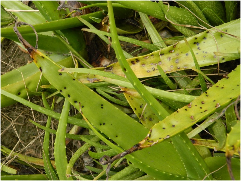
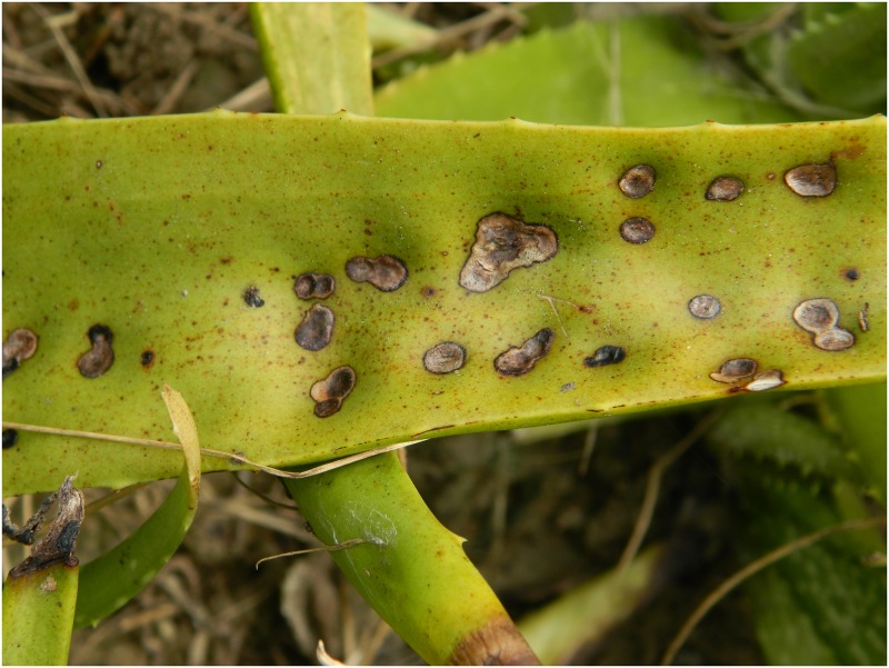
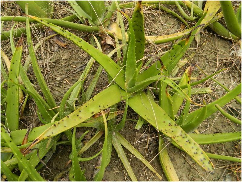
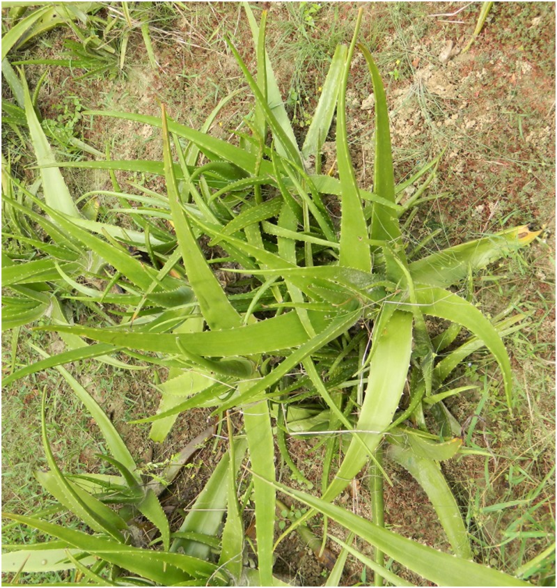
The symptoms appear on the leaves in the form of small dark brown necrotic spots on both sides that gradually become larger, eventually covering an area of 2–8 cm in diameter. The spots gradually coalesce and the infected areas transform from dark brown to black. The leaf surface becomes covered with numerous such lesions, which in turn become rotten and dried within 4-7days, and each lesion finally develops into a central cup shaped depression with a depth of 5–8 mm. (Figs 1 and 2), indicating the presence of leaf spot [5].
Fig 1. An Aloe vera plant showing symptoms of leaf spot.
Fig 2. A close-up view of leaf spot symptoms on an Aloe vera leaf.
On the basis of the microscopic characteristics of pathogen cultures, PCR amplification of ITS1-5.8S-ITS2 from the rDNA of the isolated pathogen, and gene sequencing through the BLAST analysis of a 510-bp sequence, 100% homology with the Alternaria brassicae strain HYMS01 (GenBank Accession No. JX857165.1) from NCBI was obtained. The gene sequence was submitted to the NCBI gene bank, was incorporated into GenBank under accession no. KJ022772.1 and was published in NCBI Genbank on March, 23rd, 2014 [5].
Through asurvey conducted in and around several regions of North 24 Parganas, specifically, Barasat, Naihati, Basirhat, Mohishbathan, Barrackpore, Nilganj, Haroa, Basanti, Duttapukur, Bongaon, Habra, Kalyani, Halishahar, Taki, Hingalganj, Nahata and Gopalnagar from 2013 to 2015, leaf spot disease of Aloe vera was recorded in all the locations under survey at all times of year.
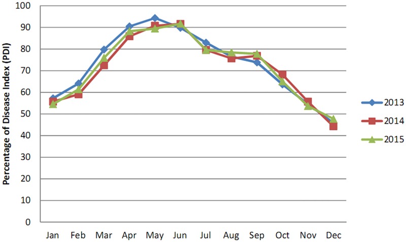
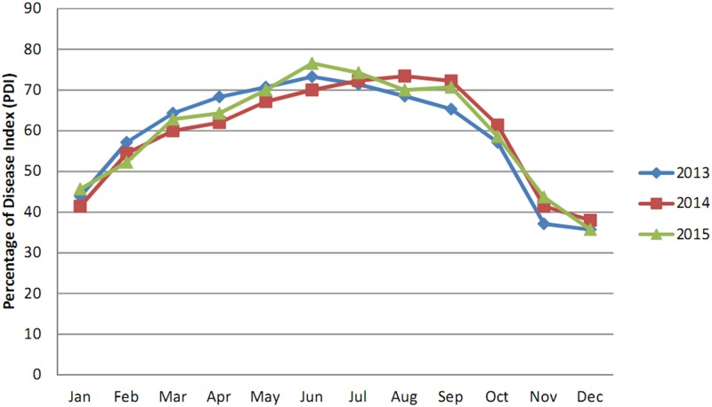
According to the data in Table 1, the percentage of disease index (PDI) for Aloe leaf spot disease was greatest during May,- 2013 (94.31%) in Nilganj where as during December,-2014, it decreased to 44.31%. At the Agri Horticultural Society of India, Alipore, the effect of the disease was greatest during June,- 2015 (76.57%) and mildest during December, 2013 and December,-2015 (35.71%) (Figs 3 and 4).
Table 1. Percentage of disease index (PDI) for leaf spot disease of Aloe vera in Nilganjandat the Agri Horticultural Society (AHS) of India, Alipore.
| Month | Nilganj | Pool data **SD+/- |
AHS, Alipore | Pool data **SD+/- |
||||
|---|---|---|---|---|---|---|---|---|
| 2013 | 2014 | 2015 | 2013 | 2014 | 2015 | |||
| Jan | 57.27(49.14) | 55.68(48.22) | 54.54(47.58) | 55.83(48.33) 0.640* |
43.87(41.44) | 41.42(39.82) | 45.71(42.53) | 43.66(41.32) 1.114* |
| Feb | 64.09(53.13) | 59.09(50.18) | 61.36(51.53) | 61.51(51.65) 1.206* |
57.14(49.08) | 54.42(47.52) | 52.25(46.26) | 54.60(47.64) 1.156* |
| Mar | 79.77(63.22) | 72.50(58.37) | 75.90(60.60) | 76.05(60.67) 1.983* |
64.28(53.25) | 60.00(50.77) | 62.85(52.42) | 62.37(52.12) 1.031* |
| Apr | 90.45(71.95) | 85.90(67.94) | 88.18(69.82) | 88.17(69.82) 1.640* |
68.28(55.67) | 62.00(51.94) | 64.28(53.25) | 64.85(53.61) 1.545* |
| May | 94.31(76.19) | 90.75(70.24) | 89.45(71.00) | 91.50(73.05) 2.705* |
70.71(57.23) | 67.14(55.00) | 70.00(56.79) | 69.28(56.29) 0.965* |
| Jun | 89.77(71.23) | 91.59(73.05) | 91.69(73.15) | 91.01(72.54) 0.884* |
73.28(58.82) | 70.00(56.79) | 76.57(61.00) | 73.28(58.82) 1.719* |
| Jul | 82.95(65.57) | 79.54(63.08) | 79.54(63.08) | 80.67(63.87) 1.174* |
71.42(57.67) | 72.30(58.24) | 74.28(59.47) | 72.66(58.44) 0.751* |
| Aug | 76.5(61.00) | 75.65(60.40) | 78.40(62.31) | 76.85(61.21) 0.797* |
68.48(55.80) | 73.42(58.95) | 70.00(56.79) | 70.63(57.17) 1.315* |
| Sep | 73.86(59.21) | 76.81(61.21) | 77.80(61.89) | 76.15(60.73) 1.138* |
65.28(53.85) | 72.28(58.18) | 70.71(57.23) | 69.42(56.42) 1.858* |
| Oct | 63.63(52.71) | 68.18(55.61) | 65.00(53.73) | 65.60(54.09) 1.203* |
57.14(49.08) | 61.42(51.59) | 58.57(49.89) | 59.04(50.18) 1.045* |
| Nov | 54.54(47.58) | 55.68(48.22) | 53.63(47.06) | 54.61(47.64) 0.474* |
37.14(37.52) | 41.42(40.05) | 43.71(41.38) | 40.75(39.64) 1.601* |
| Dec | 45.77(42.53) | 44.31(41.73) | 47.72(43.68) | 45.93(42.65) 0.800* |
35.71(36.69) | 38.00(38.06) | 35.71(36.69) | 36.47(37.11) 0.646* |
| CD (p 0.05) SE ± |
12.60 6.08 |
CD (p 0.05) SE ± |
12.64 6.10 |
|||||
*Values within parentheses denote the angular transformation value.
** Indicates SD± value.
Fig 3. Monthly disease progression curve of Aloe leaf spot disease from January, 2013 to December, -2015 in Nilganj.
Fig 4. Monthly disease progression curve of Aloe leaf spot disease from January, -2013 to December,-2015 at the Agri Horticultural Society, Alipore.
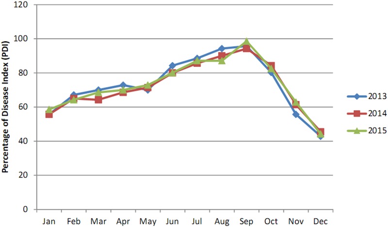
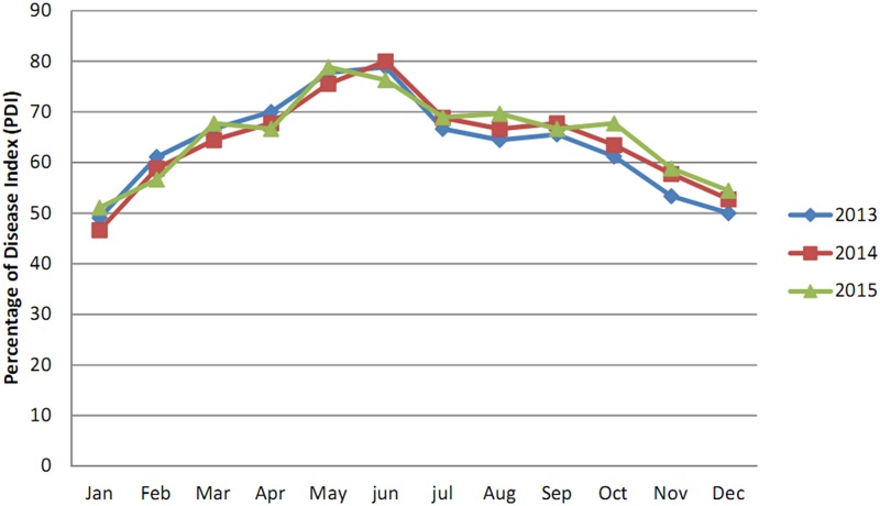
The data presented in Table 2 reveal that the PDI for Aloe leaf spot disease reached its peak during, May2015 (98.57%) in the State Pharmacopoeial Laboratory and Pharmacy for Medicine, Nadia where as during, December2013 it decreased to 42.85%. In Narendrapur the effect of the disease was highest during June;-2014 (80.00%) and lowest during January; 2014 and December;-2015 (46.66%) (Figs 5 and 6).
Table 2. Percentage of disease index (PDI) for leaf spot disease of Aloe vera at the State Pharmacopoeial Laboratory and Pharmacy for Medicine, Nadia (State Pharma), and Narendrapur.
| Months | State Pharma, Nadia | Narendrapur | ||||||
|---|---|---|---|---|---|---|---|---|
| 2013 | 2014 | 2015 | Pool data **SD+/- |
2013 | 2014 | 2015 | Pool data **SD+/- |
|
| Jan | 55.71(48.27) | 55.71(48.27) | 58.57(49.89) | 56.66(48.79) 0.763* |
49.11(44.46) | 46.66(43.05) | 51.11(45.63) | 48.96(44.37) 1.054* |
| Feb | 67.14(55.00) | 65.00(53.73) | 64.28(53.25) | 65.47(53.97) 0.738* |
61.11(51.41) | 58.88(50.07) | 56.66(48.79) | 58.88(50.07) 1.069* |
| Mar | 70.00(56.79) | 64.28(53.25) | 68.57(55.86) | 67.61(55.30) 1.498* |
66.66(54.70) | 64.44(53.37) | 67.77(55.37) | 66.29(54.45) 0.831* |
| Apr | 72.85(58.56) | 68.57(55.86) | 70.00(56.79) | 70.47(57.04) 1.120* |
70.00(55.79) | 67.77(55.37) | 66.66(54.70) | 68.14(55.61) 0.553* |
| May | 70.00(56.79) | 71.42(57.67) | 72.85(58.56) | 71.42(57.67) 0.722* |
77.77(61.82) | 75.55(60.33) | 78.88(62.58) | 77.40(61.62) 0.935* |
| Jun | 84.28(66.58) | 80.00(63.44) | 80.14(63.51) | 81.47(64.45) 1.465* |
78.88(62.58) | 80.00(63.44) | 76.33(60.87) | 78.40(62.31) 1.068* |
| Jul | 88.57(70.18) | 85.71(67.78) | 87.14(68.95) | 87.14(68.95) 0.980* |
66.66(54.70) | 68.88(56.04) | 68.98(56.11) | 68.17(55.61) 0.648* |
| Aug | 94.28(76.06) | 90.00(71.56) | 87.14(68.95) | 90.47(71.95) 2.946* |
64.44(53.37) | 66.66(54.70) | 69.66(56.54) | 66.92(54.88) 1.299* |
| Sep | 95.71(78.03) | 94.28(76.06) | 98.57(82.96) | 96.18(78.61) 2.930* |
65.55(54.03) | 67.77(55.37) | 66.66(54.70) | 66.66(54.70) 0.547* |
| Oct | 80.28(63.58) | 84.28(66.58) | 82.57(65.27) | 82.37(65.12) 1.228* |
61.20(51.47) | 63.44(52.77) | 67.77(55.37) | 64.13(53.19) 1.621* |
| Nov | 55.71(48.27) | 61.42(51.59) | 62.85(52.42) | 59.99(50.71) 1.793* |
53.33(46.89) | 57.77(49.43) | 58.88(50.07) | 56.66(48.79) 1.373* |
| Dec | 42.85(40.86) | 45.50(42.42) | 44.28(41.67) | 44.21(41.67) 0.637* |
50.00(45.00) | 52.77(46.55) | 54.44(47.52) | 52.40(46.38) 1.038* |
| CD (p 0.05) SE± |
15.56 7.52 |
CD (p 0.05) SE± |
9.72 4.69 |
|||||
*Values within parentheses denote the angular transformation value.
** Indicates SD± value.
Fig 5. Monthly disease progression curve of Aloe leaf spot disease from January,-2013 to December, 2015 at the State Pharmacopical Laboratory and Pharmacy for Medicine, Nadia.
Fig 6. Monthly disease progression curve of Aloe leaf spot disease from January, -2013 to December, -2015 in Narendrapur.
This study indicated that the maximum disease intensity was occurred between April and September in all four studied areas. This result was probably due to high temperature and moisture as this pathogen is mostly active in wet seasons and in areas with relatively high rainfall [21].
Similarly in a survey conducted on fungal diseases of medicinal plants in Maharashtra State, scientists observed leaf spot disease of Aloe vera in the Osmanabad district of Maharashtra [7]. They reported that three pathogens were associated with the disease, namely, Alternaria alternata, A. tenuissima and Fusarium sp. According to the authors, the disease was found during the rainy season and winter but absent in summer. However, in this survey, occurrence of the disease was observed throughout the year. This may be due to differences in geographical and climatic factors among the survey areas.
In vitro control of the pathogen
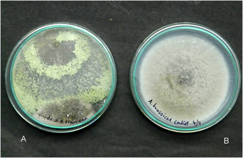
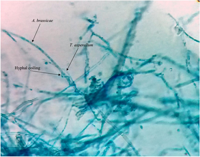
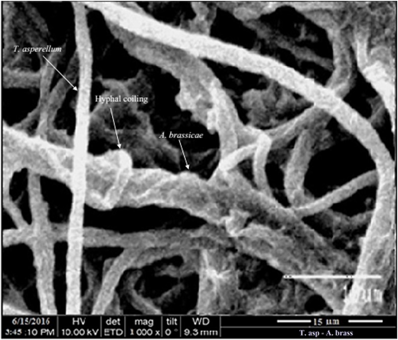
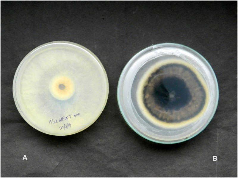
The pathogen Alternaria brassicae, isolated from Aloe vera was treated with fungal antagonists such as Trichoderma viride, T. harzianum, T. asperellum, T. longibrachiatum and Beauveria bassiana. The results presented in Table 3 reveal that the antagonist T. asperellum was recorded with a maximum percentage of inhibition of radial growth (PIRG) of 70.39% through non volatile metabolites (Fig 7) followed by T. viride (67.22%), T. harzianum (65.92%), T. longibrachiatum (64.80%) and Beauveria bassiana (64.61%) in a dual-culture-method. Our compound microscopy (Fig 8) and scanning electron microscopy (SEM) studies (Fig 9) showed features such as hyphal coiling, cellular distortion, and vacuolation that confirmed the hyperparasitism of Trichoderma over A. brassicae.
Table 3. Non volatile effects of the fungal biocontrol agents on the radial growth of Alternaria brassicae.
| BCA | Average radial growth (cm) of pathogen | Percentage of inhibition of radial growth (PIRG) of antagonistic fungi over A. brassicae (after 7 days) |
|---|---|---|
| T. asperellum | 2.65♦ | 70.39 a** (56.98)# |
| T. harzianum | 3.05 | 65.92 b (54.27) |
| T. viride | 2.92 | 67.22 ab (55.06) |
| T. longibrachiatum | 3.15 | 64.80 b (53.61) |
| Beauveria bassiana | 3.16 | 64.61 b (53.49) |
| Control (untreated) | 8.95 | |
| CD (p = 0.05) 1.876 SE ± 0.676 | ||
♦For each set of experiments 3 replicates were performed.
**Note: In the same column the same letter indicates that values statistically the same while different letters indicate that they are significantly different. Mean values followed by a different letter indicate significant differences (P = 0.05) according to Duncan’s multiple range test.
#values within parentheses denote the angular transformation value.
Fig 7. Colony of A. brassicae completely overgrown by the mycelial growth of T. asperellum (A); and, control plate of A. brassicae (B).
Fig 8. Hyphae of T. asperellum coils over the hyphae of A. brassicae (450X).
Fig 9. Scanning electron microscopic view of the hyphal coiling of the hyperparasitic biocontrol agent T. asperellum over the pathogen A. brassicae.
The data presented in Table 4 revealed that the antagonist T. asperellum was recorded with maximum PIRG of 72.17% through volatile metabolites (Fig 10) followed by T. longibrachiatum (65.32%), T. harzianum (63.31%), T. viride (59.47%) and B. bassiana (33.46%).
Table 4. Volatile effect of the fungal biocontrol agents on radial growth of Alternaria brassicae.
| BCA | Average radial growth (cm) | Percentage of Inhibition of Radial growth (PIRG) of antagonistic fungi over A. brassicae (after 7 days) |
|---|---|---|
| T. asperellum | 2.30♦ | 72.17 (58.12)# a** |
| T. harzianum | 3.03 | 63.31 (52.71)b |
| T. longibrachiatum | 2.86 | 65.32 (53.91) b |
| T. viride | 3.35 | 59.47 (50.42) c |
| Beauveria bassiana | 5.50 | 33.46 (35.30)d |
| Control (untreated) | 8.26 | |
| CD (p = 0.05) 1.717 SE ± 0.618 | ||
♦For each set of experiment 3 replicates were performed.
**Note: In the same column, the same letter indicates that the values are statistically equivalent, while different letters indicate that they are significantly different. Mean values followed by different letters indicate significant differences (P = 0.05) according to Duncan’s multiple range test.
#Values within parentheses denote the angular transformation value.
Fig 10. (A) A. brassicae treated with T. asperellum (B) Control.
Detection of volatile substances
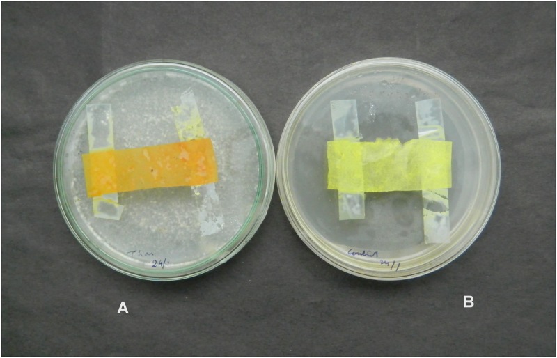
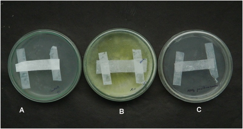
The data presented in Table 5 show that among the five fungal antagonists, only T. harzianum (Fig 11) produced HCN, where as T. viride, T. asperellum, T. longibrachiatum and B. bassiana were unable to produce HCN. The role of HCN (hydrogen cyanide) production by biocontrol agents in the biological control of pathogens has been reported by many authors [22] and a positive correlation between HCN production in vitro and plant protection has been reported [23]. The direct inhibition of fungi by HCN was thought to be one of the main mechanisms of action. In addition to the biocontrol efficacy of HCN production in rhizosphere numerous reports are also available on the stimulation of root length and root hair germination in tobacco plants[24]. The bacterium Pseudomonas fragi CS11RH1 (MTCC8984), produces hydrogen cyanide (HCN), and wheat seeds bacterized with this isolate showed significant increases in the percent germination, rate of germination, plant biomass and nutrient uptake as seedlings. On the other hand, among the five fungal antagonists, four were recorded to produce ammonia (NH3) although the production of ammonia was more evident for T. harzianum (Fig 12), T. viride, T. longibrachiatum and T. asperellum than for Beauveria bassiana. The production of ammonia gas as an important biocontrol tool was also characterized in the interaction between the nitrogen fixing bacterium Enterobacter cloacae and pathogens such as Pythium ultimum and Rhizoctonia solani [25]. The liberated volatile substances secreted by biocontrol agents accumulate in the soil where they limit the growth and germination of a wide range of pathogens in agricultural fields, a process that is also favored by mulching techniques [26]. The production of diffusible and volatile metabolites by Trichoderma spp. was also reported previously[27; 28; 29]. Meena et. al.[30] reported that volatile metabolites of T. harzianum and T. viride induced 40% and 35% growth inhibition respectively of Alternaria alternata,. They further analyzed volatile compounds produced by- Trichoderma spp. Through the GC-MS technique and abundance of glacial acetic acid (45.32%) and propyl-benzene (41.75%) were reported as distinguishable antifungal volatile metabolites.
Table 5. Production of volatile substances by biocontrol agents.
| Fungal antagonist | HCN Production | NH3 Production |
|---|---|---|
| T. harzianum | + | + |
| T. viride | - | + |
| T. asperellum | - | + |
| T. longibrachiatum | - | + |
| Beauveria bassiana | - | +/- |
(+) indicates the production of volatile substances, and (–) indicates no production of volatile substances. For each set of experiments 3 replicateswereperformed.
Fig 11.
(A) Plate inoculated with T. harzianum. (B) Control.
Fig 12.
(A) Negative control. (B) Plate inoculated with T. asperellum. (C) Positive control.
Parasitism of fungi particularly mycoparasitism is a special mode of existence for many species. Since the discovery that Trichoderma has great potential for biocontrol [31], many researchers working with Trichoderma noticed that hyphae of the antagonists parasitized hyphae of other fungi ‘in vitro’ and brought about several morphological changes such as coiling, haustorium formation, disorganization of host cell contents and penetration of the host [32]. Classical evidence of the mycoparasitic activity of Trichoderma through phase contrast and electron microscopywas provided previously through advanced microscopic studies [33,34]. Our compound microscopy (Fig 8) and SEM studies (Fig 9) showed hyphal coiling, cellular distortion, and vacuolation, among other features and these findings are supported by previous studies [31,32,33]. T. viride and T. harzianum [35,36,37,38,39,40,41] showed the potential of in vitro antagonistic activity against the important plant pathogens such as Sclerotinia sclerotiorum, Pyricularia oryzae, Bipolaris oryzae, Alternaria solani. A. alternata. Phytophthora colocasiae, P. parasitica var. piperina, Pythium aphanidermatum and Colletotrichum gloeosporioides. Antibiosis, mycoparasitism and competition are the main mechanisms of biological control [42,43]. The production of diffusible and volatile metabolites by Trichoderma spp. was reported previously [27,28,29]. Trichoderma strains exert biocontrol against fungal phytopathogens either indirectly, by competing for nutrients and space, modifying the environmental conditions, or promoting plant growth and defense mechanisms and antibiosis, or directly, by mechanisms such as hyperparasitism. The activation of each mechanism implies the production of specific compounds and metabolites such as plant growth factors, hydrolytic enzymes, siderophores, antibiotics and carbon-nitrogen permeases [44]. Pathogen inhibition in co-cultures begins soon after contact with the antagonist [45]. Trichoderma spp. adhere exactly on other fungal pathogenic hyphae, where they coil around them and degrade the cell walls. This action of parasitism restricts the development and activity of pathogenic fungi. Additionally, or together with mycoparasitism, some Trichoderma species release antibiotics [46]. While analyzing the biocontrol—plant pathogen interaction, Trichoderma virens spores or cell-free culture filtrate was reported to regulate the growth and the defense responses of tomato against Fusarium wilt disease of tomato (C.O.- Fusarium oxysporum f. sp. lycopersici) through induction of higher levels of salicylic acid and jasmonic acid in the host; those phytohormones in turn served as inducers of various pathogenesis related proteins, resulting in significantly lower disease incidence [18]. Plant immunization with rhizospheric fungi such as Trichoderma harzianum resulted in resistance of tomato against bacterial wilt disease through increased production of defense-related enzymes such as peroxidase, phenylalanine ammonia lyase, β-1,3-glucanase along with early emergence and increased vigor of the fruit [47]. Nagaraju et. al.[48] evaluated the efficacy of seven different rhizospheric fungi from various regions of Southern India for plant growth promotion and disease resistance against sunflower downy mildew disease(C.O.- Plasmopara halstedii). According to those authors, priming seed with a conidial suspension of PGPF at 1 × 10⁸CFU mL-1 significantly increased seed germination and seedling vigor, as well as reducing downy mildew disease severity by 61%, compared to the untreated control. T. asperellum was recorded to show prolific activity in controlling Fusarium wilt in chickpea (75.25% and 67.15% of crop protection in two consecutive years) and to act as an excellent growth promoting factor in a mini pot trial. The field application of T. hamatum and T. koningii can increase crop productivity up to 300% [49]. Similar results were also reported by some other researchers in green houses for seed treatment with Trichoderma spores [50]. The activity of biocontrol agents could also reduce the soil concentrations of substances that inhibit to plant growth [51]. Thus, the plant growth promotion may be due to the production of plant hormones or the increased uptake of nutrients by the plant [52], control of other pathogens at subinfectious levels and/or strengthening plants own defense mechanisms[53].
Effect of fungicides on the radial growth of the pathogen
The data presented in Table 6 demonstrate that the most potent biocontrol agent T. asperellum showed the highest PIRG against A. brassicae in vitro (70.39%), which was highly comparable to 2% blitox-50 (73.92%) and much better than 2% bavistin (59.77%). Additionally, T. harzianum (65.91%) and T. viride (67.22%) also exerted significant in vitro control over the leaf spot pathogen A. brassicae. More importantly bavistin and blitox-50 are toxic to the environment and are carcinogenic. Therefore, in terms of environmental hazards, application of T. asperellum not only provided a more or less comparable result with 2% blitox-50 but also provided a much better result than 2% bavistin in in vitro conditions.
Table 6. In vitro comparison between applications of fungicides and of the most potent biocontrol agents.
| Treatment | Average radial growth (cm) of A. brassicae | Average PIRG of A. brassicae |
|---|---|---|
| Bavistin (2% conc.) | 3.60 | 59.77 (50.59) c |
| Blitox-50 (2% conc.) | 2.33 | 73.92 (59.28) a |
| T. asperellum | 2.65 | 70.39 (56.98) a |
| T. harzianum | 3.05 | 65.91 (54.27) b |
| T. viride | 2.92 | 67.22 (55.06) b |
| Control (untreated) | 8.95 | |
| CD (p = 0.05) 2.068 SE ± 0.745 | ||
For each set of experiments, 3 replicates were performed. Note: In the same column the same letter indicates that values are statistically equivalent, while different letters indicate that they are significantly different. Mean values followed by different letters indicate significant differences (P = 0.05) according to Duncan’s multiple range test. Data presented in the parentheses represent the angular transformation value
Field trial for the in vivo study
Pool data presented in Table 7 revealed that based on analysis of three years (2013, 2014 and 2015) of data, the percentage of disease index (PDI) values in control plots were much higher (73.33%) (Fig 13) than those in their treated counterparts (Fig 14). Plots treated with T. asperellum only showed in 24.00% PDI (Fig 14). Statistically similar results were obtained in plots treated with T. viride (25.33%) and T. harzianum (29.33%). Therefore, there was a significant decrease in terms of pool data of PDI as a result of the application of fungal biocontrol agents T. asperellum, T. viride or T. harzianum in spore suspension at a rate of 108 spores / mL. This is the first attempt to apply T. asperellum, T. viride or T. harzianum (108 spores/ mL) as biocontrol agents at the standardized rate of 2.5 L / plot (36 sq ft) against this epidemic leaf spot disease of Aloe vera in a comprehensive approach to the ecofriendly management of this disease.
Table 7. Field trial of biocontrol agents on Aloe vera through mini plot design.
| Treatments | 2013 | 2014 | 2015 | Pool data |
|---|---|---|---|---|
| T. asperellum | 28.00 * (31.95)♦ | 20.00 (26.56) | 24.00 (29.33) | 24.00 (29.33) b° |
| T.harzianum | 28.00 (31.95) | 32.00 (34.45) | 28.00 (31.95) | 29.33 (32.77) b |
| T. viride | 24.00 (29.33) | 28.00 (31.95) | 24.00 (29.33) | 25.33 (30.20) b |
| Untreated | 72.00 (58.05) | 68.00 (55.55) | 80.00 (63.44) | 73.33 (58.89) a |
| CD (p = 0.05) SE ± |
5.86 2.40 |
|||
♦Data within parentheses denotes the angular transformation value.
°Note: In the same column, the same letter indicates that values are statistically equivalent, while different letters indicate that they are significantly different. Mean values followed by different letters indicate significant differences (P = 0.05) according to Duncan’s multiple range test.
* For each set of experiments, 3 replicates were performed.
Fig 13. Untreated saplings of Aloe vera showing many leaf spots.
Fig 14. Aloe vera treated with a spore suspension of T. asperellum (108 spores/mL) showing a negligible number of leaf spots.
In conclusion, among the biocontrol agents tested by using the dual-culture-plate method, T. asperellum showed the highest PIRG, 70.39%. The hyperparasitic nature of the biocontrol agents was confirmed by hyphal coiling, as well as cellular changes such as granulation, distortion and vacuolation induced by T. asperellum, T. harzianum and T. viride over A. brassicae, phenomena that were confirmed through compound microscopy as well as SEM. When analyzing the effect of volatile metabolites on the radial growth of pathogens, T. asperellum again showed a maximum PIRG of 72.17%. T. asperellum showed the highest PIRG against A. brassicae in vitro (70.39%), a value that was quite comparable to that of2% blitox-50 (73.92%) and even better than 2% bavistin (59.77%) treatment for the same pathogen. More importantly bavistin and blitox-50 are toxic to the environment and are carcinogenic. Treating saplings via the dip method (108 spores / mL) and spraying 4 times with a spore suspension standardized at a rate of 2.5 L / plot (36 square feet) is suggested for managing this epidemic leaf spot disease of Aloe vera in widespread agricultural fields.
Material and methods
Study of disease occurrence and intensity
Four agricultural farmers fields such as i) Nilganj, North 24-Parganas, ii) the State Pharmacopoeial Laboratory and Pharmacy for Medicine, Govt. of W.B., Nadia, iii) the Agri Horticultural Society of India, Alipore and iv) Narendrapur were selected for this purpose. Although specific permissions are not needed, but we have taken the permission as follows-
Nilgunj, North 24-Parganas- private farmers field permission has been taken
State Pharmacopical Laboratory and Pharmacy for Medicine, Govt. of W.B., Nadia permission from the director has been taken
The Agri Horticultural Society of India, Alipore- permission from joint secretary has been taken
Narendrapur. private farmers field and permission has taken
Moreover, we declare that the field studies did not involved endangered or protected species.
An extensive survey was also carried out in a few selected areas of North 24 Parganas to record the disease occurrence of Aloe vera from January 2013 to December 2015. An intensive survey during the same time period was conducted in order to record the percentage of disease index (PDI) by monitoring the cultivation of Aloe vera in the 4 selected areas of West Bengal. The fields were visited once per week for the entire study duration to evaluate the PDI. For the study of PDI, three plots from each of the four areas were chosen and 50 plants per plot were randomly selected and tagged. Tagged plants were visited, once per week from January 2013 to December 2015. The disease was rated on the following scale: 0 = 1%, 1 = 10%; 2 = 25%; 3 = 40% 4 = 60%, 5 = 70%, 6 = 90% and 7 = 95%. The PDI was calculated by using the following formula.
where A = Total number of infected plants; and B = Total number of selected plants.
Characterization and identification of the pathogen
The isolation, purification and identification of the pathogen were previously conducted phenotypically in our laboratory following published keys; as well as by molecular identification. Genomic DNA of fungi was extracted by the modified CTAB method [54]. Internal transcribed spacer regions (ITS1-5.8S-ITS2 of rDNA) from genomic DNA were amplified by PCR using a DNA amplification reagent kit manual (GeNei) along with the fungus- specific forward primer ITS-1 F (CTTGGTCATTTAGAGGAA-GTAA) [55] and the reverse primer ITS-4 (TCCTCCGCTTATTGATA-TGC)[56]. The sequencing of PCR product was carried out [57] in the Sci Genome Labs Pvt Ltd, Kerala, India; the sequence obtained was submitted to NCBI GenBank and the accession number was obtained. The sequence analysis was carried out using the BLAST bioinformatic tool of NCBI in earlier work from this laboratory [5].
Antagonistic fungi used
Previously, in our laboratory, four species of Trichoderma and one species of Beauveria were phenotypically isolated, characterized and identified following published keys [58,59,60] and through DNA extraction [54], PCR amplification of ITS regions using ITS forward [55] and reverse primer [56], and sequencing of the PCR product (ITS1-5.8s-ITS2 of rDNA) [57]. They were published in NCBI GenBank as Trichoderma asperellum (GenBank accession no KY966018.1), Trichoderma viride (GenBank accession no KY966032.1), T. harzianum (GenBank accession no KY966015.1), and B. bassiana (GenBank accession no KM604668.1). Trichoderma longibrachiatum was phenotypically characterized, identified and deposited into IMTECH, Chandigarh, India under accession no. MTCC11582).
Antagonistic potentiality test of isolated antagonistic fungi(dual culture plate method)
Pathogens and antagonists were grown on separate sterilized Petri dishes containing PDA medium. After 5 days, 5-mm discs of young vigorously growing cultures of both pathogens and antagonists were uniformly cut out by using a cork borer and placed on opposite sides of a 9-cm diameter sterilized Petri dish containing 15 mL of PDA and incubated in a BOD incubator at 28± 1°C for 7 days. A plate containing the pathogen served as the control. After 7 days, the radial growth of the pathogen was recorded for each test plate. Antagonistic activity was measured by percentage of inhibition of radial growth (PIRG). The PIRG was calculated with the following formula[61].
A = Mean diameter of growth in the control and B = Mean diameter of growth in a given test.
Effect of the volatile metabolites of antagonistic fungi on the radial growth of pathogens
To investigate the inhibitory effect of volatile components released by the antagonistic fungi or bacteria, the “inverted plate technique” (Dennis and Webster, 1971) was adopted. First, 15 mL of PDA medium was poured into the lower half of a Petridish. Fifteen mL of PDA medium was poured into the upper half of a Petri dish and both halves were allowed to cool and solidify. Next, at the centre of the upper half, a 5-day-old pathogen cultures of 5 mm in diameter was aseptically inoculated, and at the center of the lower half, a 5-mm-diameter 5-day-old culture of an antagonistic fungus was aseptically inoculated. Then, the upper and lower halves of the Petri dish were fixed to each other by paraffin film. In the control set, only the upper half was inoculated. After 7 days the radial growth of the pathogen was measured. The PIRG was calculated with the following formula [61].
A = Mean diameter of growth in the control and B = Mean diameter of growth in a given treatment.
Detection of volatile substances
Hydrogen cyanide (HCN) production
Production of HCN was qualitatively tested according to the method of Wei et.al., [62]. The fungal antagonist was inoculated in TSA medium supplemented with the amino acid glycine (4.4g /L of medium). A strip of sterilized filter paper saturated with a solution containing 0.5% picric acid and sodium carbonate (2%) was placed in the upper half of a Petridish. Each Petridish was then sealed with paraffin and incubated at 30°C for 7 days. A change in color of the filter paper from yellow to brown indicates HCN production. A control set was also incubated with the same experimental condition for each set to determine the color change.
Ammonia (NH3) production
The production of NH3 was qualitatively tested qualitatively according to the method of Wei et.al.[62]. The antagonist was inoculated in 15 mL of PDA medium in sterilized Petridishes. A strip of sterilized paper saturated with a solution of Hg2(Cl2)2 was placed in the upper half of the Petridish. The Petridishes were then sealed with paraffin and incubated at 28°C for 7 days. A change in colour of the filter paper from white to blackish indicates ammonia production. Two control sets were also incubated with the same experimental condition to determine the color change. One contained sterile PDA medium but was left uninoculated (regarded as the negative control) and in the second control set, 15 mL of sterile NH4OH was poured instead of the PDA medium to match the color change (regarded as positive control) in the treated sets as NH4OH liberates ammonia gas, which produces about the color change of the filter paper from white to blackish.
Effect of fungicides on the radial growth of the pathogen
Isolated pathogens were subjected to in vitro efficacy on the radial growth of the pathogen with the application of bavistin (2%) and blitox-50 (2%) following the principle of the poison food method. In this assay, 5mm discs of young vigorously growing cultures of pathogens were placed on a 9-cm-diameter sterilized Petridish containing 15 mL of PDA medium mixed with a particular fungicide at the indicated concentration and incubated in a BOD incubator at 28 ± 1°C for 7 days. Control sets were inoculated with 5-mm discs of young vigorously growing cultures of pathogens placed at the center of a plate and incubated under the same conditions but without the fungicide. After 7 days radial growth of the pathogens was recorded for both fungicide treated and control plates. The fungicide activity of each treatment was measured by PIRG. The PIRG was calculated with the following formula [61].
A = Mean diameter of growth in the control and B = Mean diameter of growth in a given treatment.
Field trial for in vivo study
Preparation of spore suspension of fungus for field study
The isolated most potent biocontrol agents were grown in sterilized 1-L conical flasks, each containing 50 g of sterilized substrate (rice husk) and 40 mL of sterile water for 30 days for mass growth. Then, autoclaved distilled water was poured into the conical flasks, and they were shaken with the help of a vortex to prepare the stock spore suspension. The intended spore concentration (108 spores/ mL) was prepared from the stock spore solution with the help of hemocytometer counting.
Mini plot design and cultivation techniques
Saplings of Aloe vera were planted within 6 ft × 6 ft mini plots. Each mini plot was separated by a 1 ft border. In each plot 25 saplings were planted and tagged, with 5 plants in each row and spacing of 1 ft, between each plant and between each row. A total of 12 plots were arranged in a randomized block design (RBD) and maintained for three consecutive years from 2013 to 2015. For treated plots the saplings were dipped in a spore suspension of T. asperellum, T. viride or T. harzianum (108 spores / mL) for 10 hours before planting. Three plots were kept as untreated controls. Spore suspensions of T. asperellum, T. viride and T. harzianum (each at 108 spores / mL) were sprayed using a knapsack sprayer for the biocontrol of the pathogen in the field at the rate of 2.5 L / plot (three plots for each treatment) 4 times at a 7 day interval for the season. All plants were well maintained with regular irrigation and weeding. In control (untreated) plots, sterile water was sprayed 4 times at a 7 day interval for the season. Continuous monitoring was performed every day during the season in order to identify any symptoms on untreated as well as treated sets. For the study of the PDI per plot, tagged plants were visited regularly, once per week throughout the season for three consecutive years. The PDI was calculated by using the following formula in untreated and treated sets.
A = Total number of infected plants; B = total number of selected plants.
Supporting information
(DOCX)
(DOCX)
(DOCX)
(DOCX)
(DOCX)
Acknowledgments
The authors are grateful to Prof. Ananda Mohan Chakraborty, Distinguished University Prof. Deptt. of Microbiology and Immunology, University of Illinois, USA. for essential advice during this research work. We are also thankful to the principal of RKMVC College for laboratory facilities.
Data Availability
All relevant data are within the paper and its Supporting Information files.
Funding Statement
This work was supported by AYUSH, National Medicinal Plant Board of India, Ministry of Health & Family Welfare, Government of India, Project Sanction No: Z.18017/187/CSS/R & D/WB-01/2012-13-NMPB, to GSKG. The funder had no role in study design, data collection and analysis, decision to publish, or preparation of the manuscript.
References
- 1.Daodu T. Aloe vera, the miracle healing plant. Health Field Corporation, Lagos: 2000. [Google Scholar]
- 2.Djeraba A, Quere P. In vivo macrophage activation in Chicken with acemannan, a complex carbohydrate extracted from Aloe vera. Int. J. Immunopharmacol. 2000; 22(5):365–372. [DOI] [PubMed] [Google Scholar]
- 3.Olusegun A. One hundred medicinal uses of Aloe vera. Good health Inc; Lagos: 2000. [Google Scholar]
- 4.Davis RH, Moro NP. Aloe vera and gibbrellin, anti-inflammatory activity in diabetes. J. Am. Paediatr. Med. Assoc. 1989; 79(1): 24–26. [DOI] [PubMed] [Google Scholar]
- 5.Ghosh SK, Banerjee S. First report of Alternaria brassicae leaf spot disease of Aloe vera and its disease inensity in West Bengal. European Journal of Biotechnology and Bioscience. 2014; 2(1):37–43. [Google Scholar]
- 6.Silva WLDE, Singh R. First Report of Alternaria Causing Leaf Spot on Aloe vera in Lousinia. Plant Disease. 2012; 96(9):1379.1–1379.1. [DOI] [PubMed] [Google Scholar]
- 7.Chavan SP, Korekar SL. A Survey of some Medicinal Plants for Fungal Diseases from Osmanabad District of Maharashtra State. Recent Research in Science and Technology.2011;3(5): 15–16. [Google Scholar]
- 8.Bajwa R, Mukhtar I, Mushtaq S. New report of Alternaria alternata causing leaf spot of Aloe vera in Pakistan. Can J Plant Pathol. 2010;32(4): 490–492. [Google Scholar]
- 9.Kamalakannan A, Gopalakrishnan C, Rebnuka R, Kalpana K. First report of Alternaria alternata causing leaf spot on Aloe barbadensis in India. Australasian Plant Dis. Notes. 2008; 3: 110–11. [Google Scholar]
- 10.World Health Organization (WHO). Mycotoxins, Environment, Health Criteria No. 11, Geneva, 1979. [Google Scholar]
- 11.Sahni ML, Singh RP. Bioassay of tetramethylthiuramdisulphide fungicide. Indian Phytopathol. 1967; 20:71–73. [Google Scholar]
- 12.Sinha PP, Prasad RK. Chemical management of Alternaria blight of cauliflower seed crop. Ind J Mycol Plant Pathol. 1989;19:204–205. [Google Scholar]
- 13.Hussaini SH, Singh H. Efficacy of different fungicides against Alternaria blight of radish seed crop. Plant Disease Res. 1989; 4: 105–107. [Google Scholar]
- 14.Anupama NB, Jogaiah S, Ito S-i., Amruthesh KN, Tran L.S.P. Improvement of growth, fruit weight and early blight disease protection of tomato plants by rhizosphere bacteria is correlated with their beneficial traits and induced biosynthesis of antioxidant peroxidase and polyphenol oxidase, Plant Science. 2015; 231: 62–73. doi: 10.1016/j.plantsci.2014.11.006 [DOI] [PubMed] [Google Scholar]
- 15.Anupama N, Murali M, Jogaiah S, Amruthesh KN. Crude oligosaccharides from Alternaria solani with Bacillus subtilis enhance defensive activity against early blight disease of tomato. Asian Journal of Science and Technology. 2014; 5(7): 412–416. [Google Scholar]
- 16.Sudisha J, Niranjana SR, Umesha S, Prakash HS, Shekar S H. Transmission of seed-borne infection of muskmelon by Didymella bryoniae and effect of seed treatments on disease incidence and fruit yield. Biol Cont. 2006; 37: 196–205. [Google Scholar]
- 17.Mostafa A, Abdel-Motaal F, El-Sayed M, Jogaiah S, Shigyo M, Ito S-i et al. Dissection of Trichoderma longibrachiatum-induced defense in onion (Allium cepa L.) against Fusarium oxysporum f. sp. cepa by target metabolite profiling. Plant Science. 2016; 246: 128–138. doi: 10.1016/j.plantsci.2016.02.008 [DOI] [PubMed] [Google Scholar]
- 18.Jogaiah S, Abdelrahman M, Ito S-I, Tran LSP. Different mechanisms of Trichoderma virens-mediated resistance in tomato against Fusarium wilt involve the jasmonic and salicylic acid pathways. Mol. Plant Path. 2017; doi: 10.1111/mpp.12571 [DOI] [PMC free article] [PubMed] [Google Scholar]
- 19.Sharma P, Sharma M, Raja M, Shammugam V. Status of Trichoderma Research in India: A review. Indian Phytopathology. 2014; 67 (1):1–19. [Google Scholar]
- 20.Pandian RTP, Raja M, Kumar A, Sharma P. Morphological and Molecular characterization of Trichoderma asperellum strain Ta 13. Indian Phytopathology. 2016; 69(3):297–303. [Google Scholar]
- 21.Humpherson-Jones FM, Phelps Kathleen. Climatic factors influencing spore production in Alternaria brassicae and A. brassicicola. Annals Appl. Bio. 1989;114: 449–458. [Google Scholar]
- 22.Ramette A, Frapolli M, Défago G, Moënne-Loccoz Y. Phylogeny of HCN synthase encoding hcnBC genes in biocontrol Fluorescent Pseudomonads and its relationship with host plant species and HCN synthesis ability. Molecular Plant-Microbe Interactions. 2003;16 (Suppl 6): 525–535. [DOI] [PubMed] [Google Scholar]
- 23.Laville J, Blumer C, Schroetter CV, Gaia V, Defago G, Keel C et al. Characterization of the hcnABC gene cluster encoding hydrogen cyanide synthase and anaerobic regulation by ANR in the strictly aerobic biocontrol agent Pseudomonas fluorescens CHA10. Journal of Bacteriology. 1998; 180:3187–3196. [DOI] [PMC free article] [PubMed] [Google Scholar]
- 24.Voisard C, Keel C, Haas D, Defago G. Cyanide production by Pseudomonas fluorescence helps suppress black root rot of tobacco under gnotobiotic conditions. EMBO J. 1989; 8:351–358. [DOI] [PMC free article] [PubMed] [Google Scholar]
- 25.Howell CR, Beier RC, Stipanovic RD. Production of ammonia by Enterobacter cloacae and its possible role in the biological control of Pythium pre- emergence damping-off by the bacterium. Phytopathology. 1988;78:1075–1078. [Google Scholar]
- 26.Bliss DE. The destruction of Armillaria mellea in Citrus soil. Phytopathology. 1951; 41: 665–683. [Google Scholar]
- 27.Ghildyal A, Pandey A. Isolation of cold tolerant antifungal strains of Trichoderma sp. from glacial sites of Indian Himalayan Region. Research Journal of Microbiology. 2008; 3 (8): 559–64. [Google Scholar]
- 28.Dennis C, Webster J. Antagonistic properties of species groups of Trichoderma II. Production of non-volatile antibiotics. Trans. Br. Mycol. Soc. 1971; 57: 41–48. [Google Scholar]
- 29.Ubalua A, Oti E, 2007. Antagonistic properties of Trichoderma viride on post harvest cassava root rot pathogens. African Journal of Biotechnology. 6(21):2447–450. [Google Scholar]
- 30.Meena M, Swapnil P, Zehra A, Dubey MK, Upadhyay RS. Antagonistic assessment of Trichoderma spp. by producing volatile and non-volatile compounds against different fungal pathogens. Archives in Phytopathology and Plant Protection. 2017; (http://www.tandfonline.com/doi/full/10.1080/03235408.2017.1357360).
- 31.Weindling R. Studies on lethal principle effective in the parasitic action of Trichoderma ligorum on Rhizoctonia solani and other fungi, Phytopathology. 1934; 24: 1153–1179. [Google Scholar]
- 32.Papavizas GC. Trichoderma and Gliocladium: biology, ecology and potential for biocontrol. Ann. Rev. Phytopathol. 1985; 23: 23–54. [Google Scholar]
- 33.Durell LW. Hyphal invatin by Trichoderma viride. Mycopathological and Mycological Applications. 1986; 35: 138–144. [Google Scholar]
- 34.Chet I, Harman GE, Baker R. Trichoderma hamatum: its hyphal interactions with Rhizoctonia solani and Pythium spp. Microb Ecol. 1981; 7: 29–38. doi: 10.1007/BF02010476 [DOI] [PubMed] [Google Scholar]
- 35.Pan. S. and Ghosh S.K. Antagonistic Potential of some soil fungi on Phytophthora colocasiae Racib. J. Mycopath. Res. 1997; 35 (2): 153–157. [Google Scholar]
- 36.Ghosh SK. Fungal antagonists of Phytophthora parasitica var piperina. Journal of Mycopathological Research. 2000; 38(2): 113–115. [Google Scholar]
- 37.Ghosh SK. Biocontrol of damping off disease of chili caused by Pythium aphanedermatum. Journal of Mycopathological Research. 2002; 40(2): 117–119. [Google Scholar]
- 38.Ghosh SK, Chakraborty N. In vitro biological control of Colletotrichum gloeosporioides, causal organism of anthracnose of Sarpagandha (Roulvolfia serpentine). The Agriculture and Biolology Journal of North America. 2012; 3 (8): 306–310. [Google Scholar]
- 39.Gveroska B, Ziberoski J. Trichoderma harzianum as a biocontrol agent against Alternaria alternata on tobacco. Applied Technologies and Innovations. 2012; 7(2):67–76. [Google Scholar]
- 40.Prabhakaran N, Prameeladevi T, Sathiyabama M, kamil D. Screening of Different Trichoderma Species against agriculturally important plant pathogens. Journal of Environmental Biology. 2014; 36: 191–198. [PubMed] [Google Scholar]
- 41.Tagaram N, Rani SA, Hindumathi A and Reddy NB. In vitro evaluation of Trichoderma viride and Trichoderma harzianum for its efficasy against Alternaria alternata, the leaf spot pathogen on Senna plant. IOSR Journal of Pharmacy and Biological Sciences. 2015;10(6): 145–147. [Google Scholar]
- 42.Umamaheswari B, Thakore B, More T. Post-harvest management of ber (Ziziphus mauritiana Lamk) fruit rot (Alternaria alternata (Fr.) Keissler. using Trichoderma species, fungicides and their combinations, Crop Protection. 2009; 28(6):525–32. [Google Scholar]
- 43.Benitez T, Rincon AM, Limon MC, Codon AC. Biocontrol mechanisms of Trichoderma strains. International Microbiology. 2004; 7: 249–260. [PubMed] [Google Scholar]
- 44.Almeida F, Cerqueira F, Silva R, Uhoa C, Lima A. Mycoparatism studies of Trichoderma harzianum strains against Rhizoctonia solani: Evaluation of coiling and hydrolytic enzyme production. Biotechnol Lett. 2007; 29(8):1189–193. doi: 10.1007/s10529-007-9372-z [DOI] [PubMed] [Google Scholar]
- 45.Harman GE, Trichoderma for biocontrol of plant pathogens: From basic research to comercialized products. Conference on Biological Control, Cornell community. April 11–13. 1996.
- 46.Ghosh SK, Pal S. Growth Promotion and Fusarium Wilt Disease Management Ecofriendly in Chickpea by Trichoderma asperellum. International Journal of Curent Research andcademic Review. 2017;5(1):14–26. [Google Scholar]
- 47.Jogaiah S, Mostafa A, Tran LSP, Ito S-I. Characterization of rhizosphere fungi that mediate resistance in tomato against bacterial wilt disease. J. of Experimental Botany. 2013; 64: 3829–3842. [DOI] [PubMed] [Google Scholar]
- 48.Nagaraju A, Jogaiah S, Mahadeva murthy S, Ito S-I. Seed priming with Trichoderma harzianum isolates enhances plant growth and induces resistance against Plasmopara halstedii, an incitant of sunflower downy mildew disease. Aust. J. Plant Path. 2012; 41: 609–620. [Google Scholar]
- 49.Chet I, Inbar J, Hadar Y. Fungal Antagonists and Mycoparasites In: The Mycota, Environmental and Microbial Relationships, Wicklow D.T. and Soderstrom B. (Eds.).Vol. 4, Springer-Verlag, Berlin, Germany; 1997. pp 165–184. [Google Scholar]
- 50.Windham M, Elad T, Baker R. A mechanical for increased plant growth induced by Trichoderma sp. Phytopathology. 1986; 76: 518–521 [Google Scholar]
- 51.Chet I, Brotman Y, Viterbo A. Trichoderma an environment-friendly biocontrol agent of plant disease. Proceedings of the 5th International conference on Biopesticides: Stakeholders perspectives. New Delhi. 2009.
- 52.Baker R. Biological control: An overview. Can J Plant Pathol. 1986; 8: 218–221. [Google Scholar]
- 53.Zimand G, Elad Y, Chet I. Effect of Trichoderma harzianum on Botrytis cinerea pathogenicity. Phytopathology. 1996; 86: 1255–1260. [Google Scholar]
- 54.Chutima R, Dell B, Vessabutr S, Busaban B, Lumyong S. Endophytic fungi from Pectielis susannae (L.) Rafin. (Orchidaceae), a threatened terrestrial orchid in Thailand. Mycorrhiza. 2010; 21 (3): 221–229. doi: 10.1007/s00572-010-0327-1 [DOI] [PubMed] [Google Scholar]
- 55.Grades M, Bruns TD. ITS primers with enhanced specificity of basidiomycetes: application to the identification of mycorrhizae and rusts. Mol Ecol. 1993;2:113–118. [DOI] [PubMed] [Google Scholar]
- 56.White T J, Bruns T, Lee S, Taylor J. Amplification and direct sequencing of fungal ribosomal RNA genes for phylogenetics In PCR Protocols: A guide to Methods and Applications(ed. Innis M. A., Gelfand D. H., Sninsky J. J. & White T. J.), pp. 315–322. Academic Press: San Diego, U.S.A, 1990. [Google Scholar]
- 57.Kullnig-Gradinger CM, Szakacs G, Kubicek CP. Phylogeny and evolution of the fungal genus Trichoderma: a multigene approach. Mycol Res. 2002; 106:757–767. [Google Scholar]
- 58.Domsch KH, Gams W, Anderson TH. Compendium of soil Fungi. Vol.–I and II, Academic Press, London: 1980. [Google Scholar]
- 59.Parkinson D, Grey TKG, Williams ST. Methods for studying the ecology of soil microorganism. Blackwell Sci. Pub; Oxford and Edinburg: 1971. [Google Scholar]
- 60.Nagamani A, Kunwar IK, Manoharachary C. Hand book of Soil Fungi. I.K. International, New Delhi: 2006. [Google Scholar]
- 61.Topps JH, Wain RL. Investigation on fungicides. 111. The fungi toxicity of 3- and 5-alkyl salicylanilide and P-chloronilines. Ann Appl Biol. 1957; 45 (3): 506–511. [Google Scholar]
- 62.Wei G, Kloepper JW, Tuzun S. Induction of systemic resistance of cucumber to Colletotrichum orbiculare by select strains of plant growth-promoting rhizobacteria. Phytopathology. 1991; 81:1508–1512. [Google Scholar]
Associated Data
This section collects any data citations, data availability statements, or supplementary materials included in this article.
Supplementary Materials
(DOCX)
(DOCX)
(DOCX)
(DOCX)
(DOCX)
Data Availability Statement
All relevant data are within the paper and its Supporting Information files.


