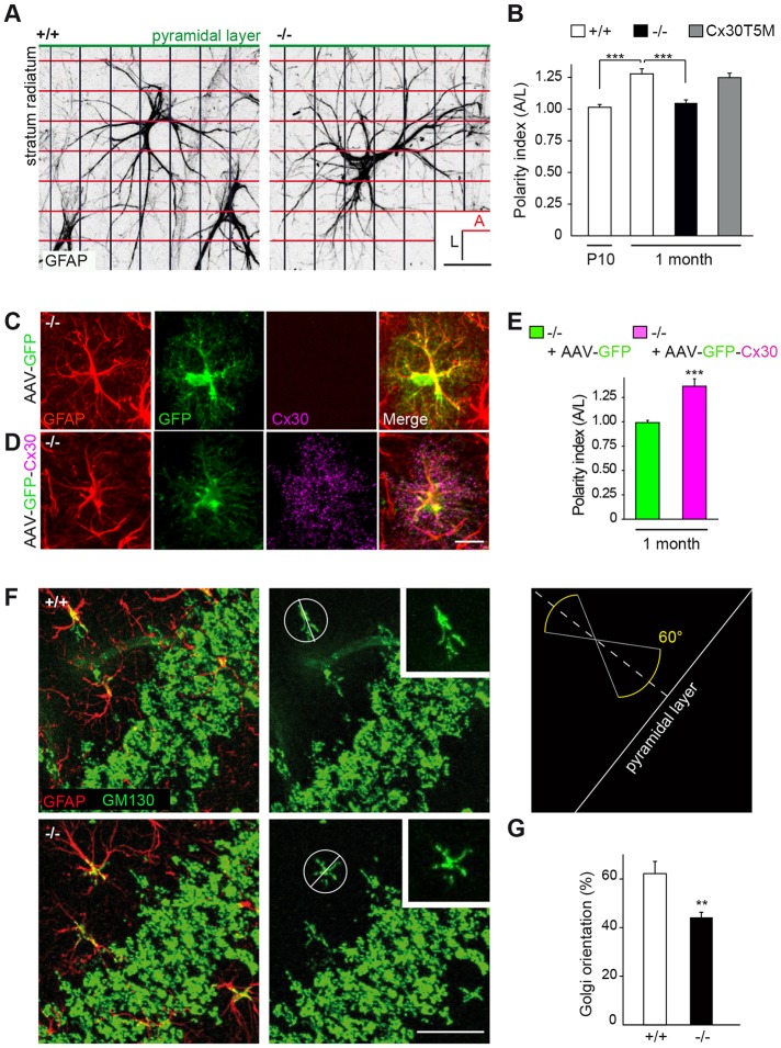
Fig. 6.
Cx30 sets the polarity of hippocampal astrocytes in vivo. (A) Schematic representation of grid-baseline analysis for orientation quantification of GFAP-labeled CA1 s.r. astrocytes. Scale bar: 20 µm. (B) Quantitative analysis of astrocytes polarity with respect to the pyramidal cell layer. Cx30 was found to be required for the proper perpendicular orientation of developing astrocytes (+/+ P10, n=43 cells; +/+ 1 month, n=44 cells; −/− 1 month, n=49 cells; Cx30T5M 1 month, n=47 cells). (C,D) Cx30 expression in astrocytes from the hippocampal CA1 s.r. region of Cx30−/− mice (1-month-old) infected with AAV-GFP (C) or AAV-GFP-Cx30 (D), as shown by GFAP, GFP and Cx30 labeling in hippocampal sections. Scale bar: 15 µm. (E) Quantitative analysis of astrocytes polarity in −/− mice showing that wild-type polarity is rescued in s.r. astrocytes by restoring Cx30 expression (n=32 cells), but not by expressing GFP alone (n=34 cells). (F) Immunohistochemical labeling of GM130 (green) and GFAP (red) in the CA1 area of the hippocampus. The diagram illustrates the quantification used to estimate the angular orientation of astrocyte Golgi with regards to the pyramidal layer. Scale bar: 30 µm. (G) Percentage of cells with their Golgi apparatus oriented perpendicularly (±30°) to the pyramidal layer (+/+, n=216 cells; −/−, n=262 cells). Cx30 deficiency decreased the proportion of CA1 astrocytes with their Golgi oriented perpendicularly to the pyramidal layer. Asterisks indicate statistical significance (***P<0.001; **P<0.01).

