Abstract
Background
Differentiating between dysthyroid optic neuropathy (DON), which requires urgent therapy to prevent blindness, and moderate-to-severe Graves orbitopathy (GO) remains challenging. There is no pathognomonic feature of DON in either ophthalmological or radiological examinations.
Objectives
Our aim was to investigate the prevalence of radiological signs of DON in magnetic resonance imaging (MRI) in patients with moderate-to-severe and very severe GO.
Methods
Two researchers reassessed MRI scans of 23 consecutive patients (46 eyes) with active, moderate-to-severe GO and 14 patients (23 eyes) with very severe GO. Typical signs of DON in MRI include apical crowding and optic nerve stretching. These were evaluated in the eyes of both groups of patients. Lack of cerebrospinal fluid in the optic nerve sheath as well as muscle index values were also studied. These clinical evaluations and laboratory results were then compared between groups.
Results
At least one of the typical radiological features of DON was found in 22 (96%) and 16 (35%) eyes with very severe and moderate-to-severe GO, respectively. Each occurred statistically more often in patients with very severe GO. There were no ophthalmological signs of very severe GO observed in the group of patients with moderate-to-severe GO during the study or its subsequent follow-up (234 weeks).
Conclusions
MRI is a useful tool in evaluating very severe GO. However, features typical for DON are also found in up to 35% of eyes in patients with active, moderate-to-severe GO. Therefore, ophthalmological evaluation seems to be most important in the recognition of very severe GO.
Keywords: Dysthyroid optic neuropathy, Graves orbitopathy, Very severe Graves orbitopathy, Graves disease, Magnetic resonance imaging
Introduction
Dysthyroid optic neuropathy (DON) is the impairment of optic nerve function caused by Graves orbitopathy (GO) in the presence of pertinent radiological alterations such as optic nerve compression at the orbital apex by enlarged extraocular muscles (apical crowding) or optic nerve stretching [1]. The European Group on Graves' Orbitopathy (EUGOGO) classifies DON as very severe GO [2]. DON occurs in 3–5% of GO patients [3]. Presently, there is no pathognomonic feature of very severe GO in either ophthalmological or radiological examination and a set diagnostic criteria have not yet been established [1]. Recognition, therefore, is based on the presence of a compilation of signs typical for DON. Additionally, ophthalmological diseases such as glaucoma, cataract, high myopia, or corneal exposition can affect visual function, further complicating diagnosis. Distinguishing DON from the less severe forms of GO is challenging. Ultimately this may lead to delays in therapy which should be urgent in order to avoid complications such as permanent loss of vision. In several studies, the proclivity of various computed tomography (CT) features for DON diagnosis have been tested [4, 5, 6, 7, 8, 9]. Magnetic resonance imaging (MRI) seems to provide better visualization of the optic nerve and soft tissue details than CT, and nowadays is also more widely available [10]. Therefore, there is a growing need to investigate the usefulness of MRI as a tool in diagnosing DON. The aim of our paper was to analyse and compare the prevalence of typical radiological signs of DON in patients with moderate-to-severe and very severe GO.
Materials and Methods
We conducted a retrospective study evaluating the MRI scans of 14 consecutive patients (23 eyes) diagnosed with very severe GO and 23 patients (46 eyes) diagnosed with active, moderate-to-severe GO who were treated in the Department of Endocrinology from 2009 to 2015. We excluded from the study patients with prior treatment with orbital decompression or orbital radiotherapy, and patients with other diseases affecting visual function. Informed consent was provided by all patients. Clinical characteristics of the studied groups are shown in Table 1. The study protocol was approved by the institute's committee on human research.
Table 1.
Clinical characteristics of patients with very severe GO (n = 14) and moderate-to-severe GO (n = 23)
| Patients with very severe GO | Patients with moderate-to-severe GO | p value* | |
|---|---|---|---|
| Graves disease | 11 | 20 | 0.83** |
| Hashimoto disease | 3 | 2 | 0.55** |
| Euthyroid Graves disease | 0 | 1 | |
| Female | 10 (71) | 15 (65) | 0.10** |
| Age, years | 57 (31–81) | 52 (24–76) | 0.10 |
| Active smokers | 8 (57) | 9 (39) | 0.64** |
| Duration of thyroid disease, weeks | 118 (9–805) | 75 (10–802) | 0.50 |
| Duration of GO, weeks | 31 (13–805) | 43 (11–478) | 0.80 |
| Duration of DON, weeks | 9 (1–25) | – | – |
| TSH (ref. range 0.270–4.200 µIU/mL) | 1.24 (0.005–14.1) | 1.27 (0.005–5.8) | 0.77 |
| fT3 (ref. range 3.1–6.8 pmol/L) | 4.46 (3.2–12.9) | 5.07 (3.7–8) | 0.50 |
| fT4 (ref. range 12–22 pmol/L) | 16.97 (7.1–23.2) | 16.94 (9.9–24.2) | 0.38 |
| TBII (ref. range <1.75 IU/L) | 14 (1.0–25) | 6.9 (0.6–40) | 0.05 |
| TPOAb (ref. range <34 IU/mL) | 51 (7–600) | 121 (7–6,000) | 0.27 |
| TgAb (ref. range <115 IU/mL) | 23 (5–4,000) | 38 (10–4,000) | 0.50 |
| CAS | 4 (2–6) | 4 (3–7) | 0.45 |
| Proptosisa, mm | 21 (16–23.5) | 22 (18–27) | 0.29 |
Data are presented as n (%) or median (range), as appropriate. GO, Graves orbitopathy; DON, dysthyroid optic neuropathy; fT3, free triiodothyronine; fT4, free thyroxine; TBII, TSH-binding inhibitory immunoglobulins; TPOAb, thyroid peroxidase antibodies; TgAb, thyroglobulin antibodies; CAS, clinical activity score.
Mann- Whitney U test or otherwise, as indicated
χ2 test.
Proptosis was measured with a Hertel exophthalmometer.
Patients
The diagnosis of DON was based on at least 2 signs such as (i) deterioration of visual acuity (VA) (< 1.0), (ii) loss of colour vision (more than 2 errors in Ishihara plates), (iii) optic disc swelling and/or visual field defect, and (iv) relative afferent pupillary defect. Five patients with DON had unilateral optic nerve involvement. Patients were treated with high doses of intravenous methylprednisolone pulses (ivMP) as first-line therapy (1 g/day for 3 consecutive days) and with orbital decompression as second-line therapy in cases which lacked improvement [11, 12]. In some of the patients additional treatment was necessary (ivMP, other orbital decompression) [11].
The diagnosis of moderate-to-severe GO was based on EUGOGO recommendations [2]. Evaluation of GO activity was based on the clinical activity score (CAS); GO was assessed as active if CAS was ≥3/7 [2, 13, 14]. Patients were treated according to EUGOGO recommendations with ivMP pulses (6 × 0.5 g followed by 6 × 0.25 g on a weekly schedule) [2]. Therapy with ivMP was accompanied by concomitant prednisone treatment (starting from 30 mg per day with tapering lasting 3 months), and in some cases with orbital radiotherapy.
MRI
MRI was performed using a 1.5-T unit (Siemens Magnetom Avanto, Erlangen, Germany). Routine orbital protocol consisted of the following unenhanced thin-slice sequences: axial T2 turbo spin echo (TSE) and T2 TSE fat saturated, coronal T2 TSE and T2 TSE fat saturated, axial T1 SE, coronal T1 SE, and sagittal T1 SE for right and left orbit. The sequences were characterized by the following parameters: slice thickness of 2 mm, matrix 256 × 256, FoV = 140 and 160 mm. MRI scans were reassessed by 2 researchers – a trained neuroradiologist and a clinician with 3 years of experience in diagnosing DON. Our researchers were not aware of the diagnoses (moderate-to-severe or very severe GO); however, they reached mutual, independent consensus. The following parameters were analysed:
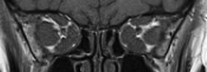
Apical crowding – evaluated on coronal T1W images as described by Nugent et al. [7] (Fig. 1).
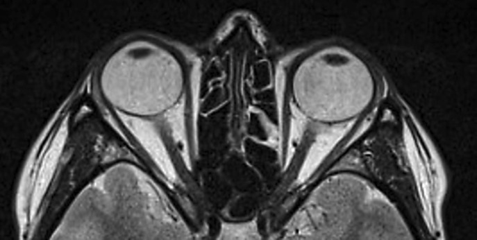
Optic nerve stretching – using a line on the axial T2W section to best depict the course of the optic nerve between the posterior globe and the orbital apex (Fig. 2). Optic nerve stretching was recognized when the optic nerve was lying in a straight line with no natural curves.
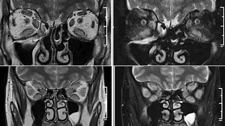
Assessment of fluid presence in the optic nerve sheath on T2W images, on both axial and coronal cross-sections (Fig. 3).
Muscle index (MI) – using the method described in CT by Barrett et al. [4] to calculate MI on coronal T1W images halfway between the posterior globe and the orbital apex. The horizontal MI was expressed as the percentage of orbital width occupied by the lateral and medial rectus muscles. Vertical MI was expressed as the percentage of the height of the orbit occupied by the inferior rectus and the superior muscle group. The larger of these two values was considered as the final MI and used in data analysis.
Fig. 1.
Coronal T1W image: apical crowding of the muscles.
Fig. 2.
Axial T2W section shows stretching of the optic nerve.
Fig. 3.
T2W images show the presence (upper axial and coronal cross-sections) and lack (lower cross-sections) of fluid in the optic nerve sheath.
Laboratory Evaluations
Thyroid stimulating hormone (TSH), free triiodothyronine, and free thyroxine concentrations, as well as TSH-binding inhibitory immunoglobulins (TBII), thyroid peroxidase antibodies (TPOAb), and thyroglobulin antibodies (TgAb), were measured using an electro-chemiluminescence immunoassay performed on a Cobas 6000 Analyzer from Roche Diagnostics (Mannheim, Germany).
Outcome Analysis
We compared the groups with moderate-to-severe and very severe GO considering age, sex, cigarette smoking, duration of thyroid disease and GO, CAS, proptosis, TSH, TBII, TPOAb, and TgAb concentrations, and the presence of typical signs of DON in MRI. The sensitivity and specificity of MI values, including 50, 55, and 60%, and the correlation between MI and the presence of apical crowding was calculated.
Statistical Analysis
Median values were used to present continuous variables, while categorical variables were expressed as numbers or percentage values. Comparisons between continuous data were performed with the Mann-Whitney U test. A χ2 test was used to analyse the differences between categorical data. Statistical significance was established for results with a p value < 0.05. To assess the correlation between data we used the Pearson product-moment correlation. All analyses were made with the statistical software R version 3.0.3.
Results
All patients with moderate-to-severe GO had bilateral GO in the active phase. There were no ophthalmological signs of DON observed before therapy, or during treatment and follow-up, which lasted 234 weeks (from 103 to 293 weeks). The clinical characteristics of the group with very severe GO are presented in Table 2.
Table 2.
Ophthalmological characteristics of the eyes affected with very severe GO (n = 23)
| Eyes, n (%) | |
|---|---|
| Reduction of VAa | 23 (100) |
| VA | |
| 0.9 | 1 (4) |
| 0.8 | 5 (22) |
| 0.7 | 2 (9) |
| 0.6 | 3 (13) |
| 0.5 | 7 (31) |
| 0.3 | 1 (4) |
| 0.2 | 4 (17) |
| Impaired colour sensitivity | 12 (52) |
| Optic disc swelling or defect in visual field | 10 (43) |
| RAPD | 2 (9) |
GO, Graves orbitopathy; VA, visual acuity; RAPD, relative afferent pupillary defect.
Mean reduction of VA 0.5.
Comparing clinical and laboratory data between patients with moderate-to-severe and very severe GO before therapy, we found a higher concentration of TBII in patients with very severe GO (p = 0.05). There were no statistically significant differences in age, sex, duration of thyroid disease, CAS, proptosis, TPOAb, or TgAb between groups (Table 1).
The radiological signs in MRI typical for DON were found both in patients with very severe GO and some patients with moderate-to-severe GO. One of the radiological features of DON (optic nerve stretching or apical crowding) was found in 22 (96%) eyes with very severe GO and in 16 (35%) eyes with moderate-to-severe GO. Each of them occurred in patients with very severe GO statistically more often than in patients with moderate-to-severe GO (Table 3).
Table 3.
Comparison of radiological features of the eyes with very severe GO (n = 23) and moderate-to-severe GO (n = 46)
| Eyes with very severe GO, n (%) | Eyes with moderate-to-severe GO, n (%) | p value | Sensitivity, % | Specificity, % | PPV, % | NPV, % | |
|---|---|---|---|---|---|---|---|
| Apical crowding | 16 (70) | 15 (33) | 0.01* | 70 | 67 | 52 | 82 |
| Optic nerve stretching Lack of cerebrospinal fluid in | 17 (74) | 9 (20) | 0.00004* | 74 | 80 | 65 | 86 |
| optic nerve sheath | 13 (57) | 11 (24) | 0.01* | 57 | 76 | 54 | 78 |
| MI | 18 (78) | 21 (46) | 0.10** | 78 | 54 | 46 | 83 |
| ≥50% | |||||||
| ≥55% | 15 (65) | 12 (26) | 0.97** | 65 | 74 | 56 | 81 |
| ≥60% | 10 (43) | 7 (15) | 0.32** | 43 | 85 | 59 | 75 |
GO, Graves Orbitopathy; MI, muscular index; PPV, positive predictive value; NPV, negative predictive value.
χ2 test
Mann-Whitney U test.
Moreover, we observed a lack of cerebrospinal fluid in the optic nerve sheath in 13 (57%) eyes with very severe GO and in 11 (24%) eyes with moderate-to-severe GO. The difference was statistically significant.
Statistically significant correlations were also found between the presence of very severe GO and a high MI value (p = 0.009). Table 3 shows the prevalence of orbits with MI values of 50, 55, and 60% in both groups, with sensitivity and specificity noted for each value. The best sensitivity to specificity ratio was found for an MI value of 55%. Additionally, we found moderate positive correlations between a high MI value and the presence of apical crowding (r = 0.62).
Discussion
Diagnosis of DON is a demanding task and there is still a lack of unequivocal recognition criteria. Both ophthalmological and radiological evaluations play an important role in this process. The difficulties in distinguishing moderate-to-severe from very severe GO may lead to sight-threatening delays in treatment. In the EUGOGO survey, McKeag et al. [15] suggest that impaired colour vision and optic disc swelling, together with radiological evidence of apical optic nerve compression, are clinical features frequently used to make the diagnosis of DON across Europe. In previous papers authors diagnosed DON based on a combination of ophthalmological and radiological features, usually with at least one obligatory sign. In the study by Currò et al. [16], there was at least 1 feature characteristic of DON in CT and at least 2 features in ophthalmological examination; Mourits et al. [17] observed obligatory apical crowding in CT and 1 or more clinical signs of DON, and Wakelkamp et al. [18] demonstrated VA < 0.63 plus either apical crowding in CT and/or clinical signs of DON. We diagnosed DON in the present study if a patient fulfilled at least 2 criteria: deterioration of VA and/or visual field defect and/or loss of colour vision and/or optic disc swelling and relative afferent pupillary defect, in the absence of any other explanation. A diagnosis dependent on several factors typical of DON, without any single obligatory feature, seems to be rational and effective.
The main objective of our analysis was to investigate the prevalence of radiological signs of DON in patients with very severe and active, moderate-to-severe GO. All but one patient with very severe GO had at least one of the typical features in MRI: apical crowding or stretching of the optic nerve. The prevalence of these features was 70 and 74% of eyes, respectively. In the group with active, moderate-to-severe GO, we found at least one of the radiological features typical for DON in up to 35% of eyes. Their prevalence was 33 and 20% of eyes, respectively. There were no ophthalmological signs of DON observed within this group either before treatment, during treatment, or during the subsequent follow-up (234 weeks). Therefore, we did not recognize very severe GO in any of these patients. Each of the typical signs of DON was found statistically significant more often in patients with very severe GO than in patients with moderate-to-severe GO.
Very few studies have directly used MRI to assess signs of DON in moderate-to-severe and very severe GO, which is why there is no data with which to compare our results. MRI is more informative than CT studies, allowing optimal assessment of the optic nerve, its sheath, and surrounding soft tissue [19]. However, both CT and MRI are essential to detect the apical crowding, muscular diameter, bony orbital angles, and intracranial fat prolapse, which are highly correlated with DON [1]. Dodds et al. [20] compared the diameter of the optic nerve sheath at different positions from the posterior globe to the pre-chiasmal region in the MRI scans of three groups of patients (with DON, GO without DON, and control group). The optic nerve diameter was significantly smaller in the group with DON; however, other features typical of DON were not assessed.
The prevalence of radiological signs of DON in CT examination of patients with moderate-to-severe and very severe GO was investigated in several studies. Nugent et al. [7] found severe apical orbital crowding in 66% of orbits with DON but in only 12% of orbits without DON. An EUGOGO survey found the prevalence of apical crowding analysed mostly in CT to be 95 and 43% in patients with definite DON and without DON, respectively [15]. The number of radiologically evaluated eyes in the group without DON was 14 among 22 eyes. Neigel et al. [21] revised CT in patients with GO with and without neuropathy and found a prevalence of severe apical crowding in 67 and 13%, respectively.
We also found a lack of cerebrospinal fluid in the optic nerve sheath in eyes with very severe and moderate-to-severe GO in 57 and 24% of eyes, respectively. This difference was considered statistically significant. The occurrence of this feature was not studied previously, and is worth considering including in future DON diagnostic criteria.
Besides apical crowding, many authors find MI noteworthy in the diagnosis of DON. In a retrospective study first described by Barrett et al. [4], the authors concluded that an MI of 67% could be a diagnostic threshold for DON. Subsequent studies found MI values ranging from 50 to 67% [6, 22, 23] to be a good predictor of DON. However, obtaining perfectly oriented coronal cuts and defining the precise position of the coronal cut along the length of the orbit is complicated. In our study, we found that MI of 55% is the ratio of sensitivity to specificity. We also observed a moderate positive correlation between a high MI value and apical crowding, which is easier to evaluate and more practical, from a radiological perspective.
MRI and CT are valuable tools used in patients with GO, but they are not always necessary. In a typical clinical case, it is rather unlikely that a therapeutic decision would be substantially influenced by additional information obtained from imaging [24]. The indications for orbital imaging are (i) atypical presentation, (ii) non-thyroidal aetiology, and (iii) clinical suspicion of optic nerve compression. We found that our patients with moderate-to-severe GO with radiological features of DON did not go on to develop very severe GO over 4 years of observation. In the ATOR study, older age of onset of GO, restricted oculomotility, strabismus, reduced palpebral aperture, and active GO were predictors for later development of DON in GO [25]. A definite systemic risk factor for DON is pre-existing vasculopathy in diabetes, hypertension, or smoking [1]. Additionally, serum thyroid stimulating immunoglobulin levels may be a useful diagnostic tool in identifying patients with early-onset DON [26]. Careful clinical evaluation and follow-up of patients with GO is crucial. Dayan and Dayan [27] drew similar conclusions in their study previously presented in a simple diagnostic algorithm for DON. Special attention must be paid, especially in those with these pre-existing conditions, given their higher risk of developing DON later in life. Orbital imaging, however, seems to be superfluous to regular ophthalmological examination and control.
Limitations
Our study can be criticized for its relatively small number of patients within both groups. In the group with very severe GO this is mainly due to its rarity of prevalence, which is a constraint faced by many other studies as well.
Conclusions
Ophthalmological evaluation seems to be the most important aspect in the recognition of very severe GO. Lack of ophthalmological signs of DON in patients with GO can exclude a diagnosis of DON regardless of MRI evaluation. Radiological manifestations of DON can be heterogeneous; the most common features are optic nerve stretching and apical crowding. Radiological features typical for DON may be found in up to 35% of eyes in patients with active, moderate-to-severe GO.
Disclosure Statement
The authors declare no conflict of interest which could be perceived as a prejudice against the impartiality of this study.
Funding Sources
This research did not receive any specific grant from funding agencies in the public, commercial, or non-profit sectors.
Acknowledgements
We would like to show our gratitude to Zuzanna Żurecka for her support in the study.
References
- 1.Boschi A, Currò N. Management of very severe Graves' orbitopathy: dysthyroid optic neuropathy and corneal breakdown. In: Wiersinga WM, Kahaly GJ, editors. Graves' Orbitopathy: A Multidisciplinary Approach – Questions and Answers. ed 3, revised and expanded edition. Basel: Karger; 2017. pp. 193–201. [Google Scholar]
- 2.Bartalena L, Baldeschi L, Boboridis K, Eckstein A, Kahaly GJ, Marcocci C, Perros P, Salvi M, Wiersinga WM. The 2016 European Thyroid Association/European Group on Graves' Orbitopathy Guidelines for the Management of Graves' Orbitopathy. Eur Thyroid J. 2016;5:9–26. doi: 10.1159/000443828. [DOI] [PMC free article] [PubMed] [Google Scholar]
- 3.Wiersinga W, Bartalena L. Epidemiology and prevention of Graves' ophthalmopathy. Thyroid. 2002;12:855–860. doi: 10.1089/105072502761016476. [DOI] [PubMed] [Google Scholar]
- 4.Barrett L, Glatt HJ, Burde RM, Gado MH. Optic nerve dysfunction in thyroid eye disease: CT. Radiology. 1988;167:503–507. doi: 10.1148/radiology.167.2.3357962. [DOI] [PubMed] [Google Scholar]
- 5.Gonçalves AC, Silva LN, Gebrim EM, Monteiro ML. Quantification of orbital apex crowding for screening of dysthyroid optic neuropathy using multidetector CT. AJNR Am J Neuroradiol. 2012;33:1602–1607. doi: 10.3174/ajnr.A3029. [DOI] [PMC free article] [PubMed] [Google Scholar]
- 6.Monteiro M, Gonçalves AC, Silva C, Moura J, Ribeiro C, Gebrim E. Diagnostic ability of Barrett's index to detect dysthyroid optic neuropathy using multidetector computed tomography. Clinics (Sao Paulo) 2008;63:301–306. doi: 10.1590/S1807-59322008000300003. [DOI] [PMC free article] [PubMed] [Google Scholar]
- 7.Nugent RA, Belkin RI, Neigel JM, Rootman J, Robertson WD, Spinelli J, Graeb DA. Graves orbitopathy: correlation of CT and clinical findings. Radiology. 1990;177:675–682. doi: 10.1148/radiology.177.3.2243967. [DOI] [PubMed] [Google Scholar]
- 8.Gonçalves AC, Silva LN, Gebrim EM, Matayoshi S, Monteiro ML. Predicting dysthyroid optic neuropathy using computed tomography volumetric analyses of orbital structures. Clinics (Sao Paulo) 2012;67:891–896. doi: 10.6061/clinics/2012(08)06. [DOI] [PMC free article] [PubMed] [Google Scholar]
- 9.Chan LL, Tan HE, Fook-Chong S, Teo TH, Lim LH, Seah LL. Graves ophthalmopathy: the bony orbit in optic neuropathy, its apical angular capacity, and impact on prediction of risk. AJNR Am J Neuroradiol. 2009;30:597–602. doi: 10.3174/ajnr.A1413. [DOI] [PMC free article] [PubMed] [Google Scholar]
- 10.Hiromatsu Y, Eguchi H, Tani J. Management of Graves' ophthalmopathy by using orbital magnetic resonance imaging. Nihon Rinsho. 2012;70:1932–1937. [PubMed] [Google Scholar]
- 11.Miśkiewicz P, Rutkowska B, Jablonska A, Krzeski A, Trautsolt-Jeziorska K, Kecik D, Milczarek-Banach J, Pirko-Kotela K, Samsel A, Bednarczuk T. Complete recovery of visual acuity as the main goal of treatment in patients with dysthyroid optic neuropathy. Endokrynol Pol. 2016;67:166–173. doi: 10.5603/EP.a2016.0018. [DOI] [PubMed] [Google Scholar]
- 12.Ruchała M, Sawicka-Gutaj N. Advances in the pharmacological treatment of Graves' Orbitopathy. Expert Rev Clin Pharmacol. 2016;9:981–989. doi: 10.1586/17512433.2016.1165606. [DOI] [PubMed] [Google Scholar]
- 13.Mourits MP, Prummel MF, Wiersinga WM, Koornneef L. Clinical activity score as a guide in the management of patients with Graves' ophthalmopathy. Clin Endocrinol (Oxf) 1997;47:9–14. doi: 10.1046/j.1365-2265.1997.2331047.x. [DOI] [PubMed] [Google Scholar]
- 14.Mourits MP, Koornneef L, Wiersinga WM, Prummel MF, Berghout A, van der Gaag R. Clinical criteria for the assessment of disease activity in Graves' ophthalmopathy: a novel approach. Br J Ophthalmol. 1989;73:639–644. doi: 10.1136/bjo.73.8.639. [DOI] [PMC free article] [PubMed] [Google Scholar]
- 15.McKeag D, Lane C, Lazarus JH, Baldeschi L, Boboridis K, Dickinson AJ, Hullo AI, Kahaly G, Krassas G, Marcocci C, Marinò M, Mourits MP, Nardi M, Neoh C, Orgiazzi J, Perros P, Pinchera A, Pitz S, Prummel MF, Sartini MS, Wiersinga WM, European Group on Graves' Orbitopathy (EUGOGO) Clinical features of dysthyroid optic neuropathy: a European Group on Graves' Orbitopathy (EUGOGO) survey. Br J Ophthalmol. 2007;91:455–458. doi: 10.1136/bjo.2006.094607. [DOI] [PMC free article] [PubMed] [Google Scholar]
- 16.Currò N, Covelli D, Vannucchi G, Campi I, Pirola G, Simonetta S, Dazzi D, Guastella C, Pignataro L, Beck-Peccoz P, Ratiglia R, Salvi M. Therapeutic outcomes of high-dose intravenous steroids in the treatment of dysthyroid optic neuropathy. Thyroid. 2014;24:897–905. doi: 10.1089/thy.2013.0445. [DOI] [PubMed] [Google Scholar]
- 17.Mourits MP, Kalmann R, Sasim IV. Methylprednisolone pulse therapy for patients with dysthyroid optic neuropathy. Orbit. 2001;20:275–280. doi: 10.1076/orbi.20.4.275.2612. [DOI] [PubMed] [Google Scholar]
- 18.Wakelkamp IM, Baldeschi L, Saeed P, Mourits MP, Prummel MF, Wiersinga WM. Surgical or medical decompression as a first-line treatment of optic neuropathy in Graves' ophthalmopathy? A randomized controlled trial. Clin Endocrinol (Oxf) 2005;63:323–328. doi: 10.1111/j.1365-2265.2005.02345.x. [DOI] [PubMed] [Google Scholar]
- 19.Müller-Forell W, Kahaly GJ. Neuroimaging of Graves' orbitopathy. Best Pract Res Clin Endocrinol Metab. 2012;26:259–271. doi: 10.1016/j.beem.2011.11.009. [DOI] [PubMed] [Google Scholar]
- 20.Dodds NI, Atacha AW, Birchall D, Jackson A. Use of high-resolution MRI of the optic nerve in Graves' ophthalmopathy. Br J Radiol. 2009;82:541–544. doi: 10.1259/bjr/56958444. [DOI] [PubMed] [Google Scholar]
- 21.Neigel JM, Rootman J, Belkin RI, Nugent RA, Drance SM, Beattie CW, Spinelli JA. Dysthyroid optic neuropathy. The crowded orbital apex syndrome. Ophthalmology. 1988;95:1515–1521. doi: 10.1016/s0161-6420(88)32978-7. [DOI] [PubMed] [Google Scholar]
- 22.Giaconi JA, Kazim M, Rho T, Pfaff C. CT scan evidence of dysthyroid optic neuropathy. Ophthal Plast Reconstr Surg. 2002;18:177–182. doi: 10.1097/00002341-200205000-00005. [DOI] [PubMed] [Google Scholar]
- 23.Birchall D, Goodall KL, Noble JL, Jackson A. Graves ophthalmopathy: intracranial fat prolapse on CT images as an indicator of optic nerve compression. Radiology. 1996;200:123–127. doi: 10.1148/radiology.200.1.8657899. [DOI] [PubMed] [Google Scholar]
- 24.Pitz S, Müller-Forell W. Orbital imaging. In: Wiersinga WM, Kahaly GJ, editors. Graves' Orbitopathy: A Multidisciplinary Approach – Questions and Answers. ed 3, revised and expanded edition. Basel: Karger; 2017. pp. 61–73. [Google Scholar]
- 25.Khong JJ, Finch S, De Silva C, Rylander S, Craig JE, Selva D, Ebeling PR. Risk factors for Graves' orbitopathy; the Australian Thyroid-Associated Orbitopathy Research (ATOR) Study. J Clin Endocrinol Metab. 2016;101:2711–2720. doi: 10.1210/jc.2015-4294. [DOI] [PubMed] [Google Scholar]
- 26.Ponto KA, Diana T, Binder H, Matheis N, Pitz S, Pfeiffer N, Kahaly GJ. Thyroid-stimulating immunoglobulins indicate the onset of dysthyroid optic neuropathy. J Endocrinol Invest. 2015;38:769–777. doi: 10.1007/s40618-015-0254-2. [DOI] [PubMed] [Google Scholar]
- 27.Dayan CM, Dayan MR. Dysthyroid optic neuropathy: a clinical diagnosis or a definable entity? Br J Ophthalmol. 2007;91:409–410. doi: 10.1136/bjo.2006.110932. [DOI] [PMC free article] [PubMed] [Google Scholar]


