Abstract
Metabotropic GABAB receptors mediate slow inhibitory effects presynaptically and postsynaptically through the modulation of different effector signalling pathways. Here, we analysed the distribution of GABAB receptors using highly sensitive SDS-digested freeze-fracture replica labelling in mouse cerebellar Purkinje cells. Immunoreactivity for GABAB1 was observed on presynaptic and, more abundantly, on postsynaptic compartments, showing both scattered and clustered distribution patterns. Quantitative analysis of immunoparticles revealed a somato-dendritic gradient, with the density of immunoparticles increasing 26-fold from somata to dendritic spines. To understand the spatial relationship of GABAB receptors with two key effector ion channels, the G protein-gated inwardly rectifying K+ (GIRK/Kir3) channel and the voltage-dependent Ca2+ channel, biochemical and immunohistochemical approaches were performed. Co-immunoprecipitation analysis demonstrated that GABAB receptors co-assembled with GIRK and CaV2.1 channels in the cerebellum. Using double-labelling immunoelectron microscopic techniques, co-clustering between GABAB1 and GIRK2 was detected in dendritic spines, whereas they were mainly segregated in the dendritic shafts. In contrast, co-clustering of GABAB1 and CaV2.1 was detected in dendritic shafts but not spines. Presynaptically, although no significant co-clustering of GABAB1 and GIRK2 or CaV2.1 channels was detected, inter-cluster distance for GABAB1 and GIRK2 was significantly smaller in the active zone than in the dendritic shafts, and that for GABAB1 and CaV2.1 was significantly smaller in the active zone than in the dendritic shafts and spines. Thus, GABAB receptors are associated with GIRK and CaV2.1 channels in different subcellular compartments. These data provide a better framework for understanding the different roles played by GABAB receptors and their effector ion channels in the cerebellar network.
Keywords: Electron microscopy, Cerebellum, GABAB receptors, Potassium channels, Calcium channels, Purkinje cells, Freeze-fracture replica immunolabelling, Synapses, Quantification, Parallel fibre, Active zone
Introduction
GABAB receptors are the G protein-coupled receptors for GABA, the main inhibitory neurotransmitter in the brain, and through coupling to different intracellular signal transduction mechanisms they mediate slow inhibitory postsynaptic potentials (IPSPs) (Bettler et al. 2004; Gassmann and Bettler 2012). Functional GABAB receptors are obligate heterodimers composed of GABAB1 and GABAB2 subunits, and they are implicated in a number of disorders, including cognitive impairments, nociception, anxiety, depression and epilepsy (Bettler et al. 2004; Luján and Ciruela 2012; Luján et al. 2014). Depending on their subcellular localisation, GABAB receptors exert distinct regulatory effects on synaptic transmission (Gassmann and Bettler 2012; Luján and Ciruela 2012). Stimulation of postsynaptic GABAB receptors generally triggers inhibition of adenylate cyclase and activation of G protein-gated inwardly rectifying K+ (GIRK/Kir3) channels, leading to cell hyperpolarisation (Kaupmann et al. 1998). Presynaptic GABAB receptors, however, suppress neurotransmitter release by depressing Ca2+ influx via P/Q-type and N-type voltage-gated Ca2+ (CaV) channels (Huston et al. 1995; Takahashi et al. 1998; but see Zhang et al. 2016). There is now substantial evidence showing that GABAB receptors, their cognate G proteins and downstream effectors are organised as macromolecular complexes (Clancy et al. 2005; David et al. 2006; Jaén and Doupnik 2006; Fowler et al. 2007; Fernández-Alacid et al. 2009; Ciruela et al. 2010; Laviv et al. 2011; Fajardo-Serrano et al. 2013; Schwenk et al. 2016). This data favours the idea that the spatial proximity of the interacting proteins seems to be a general mechanism to ensure that signalling is specific and fast.
In situ hybridization and immunohistochemical studies have shown that Purkinje cells (PCs), the output neurons of the cerebellar cortex, are the neuron type with the highest levels of GABAB receptors (Bowery et al. 1987; Chu et al. 1990; Turgeon and Albin 1993; Kaupmann et al. 1997; Bischoff et al. 1999; Fritschy et al. 2004; Luján and Shigemoto 2006). Although electrophysiological and pharmacological studies have characterised pre- and postsynaptic inhibitory functions of GABAB receptors in PCs (Batchelor and Garthwaite 1992; Dittman and Regehr 1996; Vigot and Batini 1997), the spatial relationship of GABAB and their effector ion channels in various subcellular compartments of central neurons remains mostly unknown. Consistent with the functional coupling of GABAB receptors with GIRK and CaV channels, immunohistochemical studies have shown that PCs have high density of GIRK channels (Aguado et al. 2008; Fernández-Alacid et al. 2009) and CaV2.1 (P/Q-type) channels (Kulik et al. 2004; Indriati et al. 2013). These ion channels have been detected at postsynaptic sites along dendrites and spines of PCs, as well as presynaptically at parallel fibre terminals (Kulik et al. 2004; Aguado et al. 2008; Fernández-Alacid et al. 2009; Indriati et al. 2013).
To visualise the two-dimensional distribution of GABAB receptors along the surface of PCs, as well as their spatial relationship with GIRK2 and CaV2.1 channels, we used the freeze-fracture replica immunogold labelling (SDS-FRL) method, a highly sensitive and quantitative immunoelectron microscopic technique (Masugi-Tokita and Shigemoto 2007). This approach allowed us to examine the numbers, densities, and co-localization of these functionally coupled signalling proteins at post- and pre-synaptic membranes, allowing us to evaluate in a quantitative fashion the compartment-dependent association and segregation of GABAB receptors and effector channels.
Materials and methods
Animals
Three adult C57BL/6J mice obtained from the Animal House Facility of the National Institute for Physiological Sciences (NIPS, Okazaki, Japan) were used in this study for immunoelectron microscopic analyses. For Co-IP, three adult C57BL/6J mice obtained from the Animal House Facility of the Universitat de Barcelona, as well as four wild type and four GABAB1 knockout mice (Schuler et al. 2001) from the Institute of Physiology, University of Basel, and three wild type, three GIRK2 knockout (Signorini et al. 1997) and three GIRK3 knockout (Torrecilla et al. 2002) mice from the University of Minnesota. Care and handling of animals prior to and during experimental procedures were in accordance with Japanese and European Union regulations (86/609/EC), and the protocols were approved by the local Animal Care and Use Committee.
Antibodies and chemicals
The primary antibodies used were: rabbit anti-GABAB1 (B17, aa. 525–539 of mouse GABAB1; Kulik et al. 2002), guinea pig anti-CaV2.1 (GP-Af810; aa. 361–400 of mouse CaV2.1; Frontier Institute Co., Japan; Indriati et al. 2013), guinea pig anti-GIRK2 (GP-Af830; aa. 390–421 of mouse GIRK2; Frontier Institute Co., Japan; Aguado et al. 2008), rabbit anti-GIRK2 (Rb-Af280; aa. 390–421 of mouse GIRK2; Frontier Institute Co., Japan; Aguado et al. 2008), and rabbit anti-GIRK3 polyclonal (Rb-Af750; aa. 358–389 of mouse GIRK3; Frontier Institute Co., Japan; Aguado et al. 2008) polyclonal antibodies. ChromPure Rabbit IgG (011-000-003, Jackson ImmunoResearch Laboratories, Inc., West Grove, PA, USA) was used control IgG for coimmunoprecipitation experiments. The characteristics and specificity of the anti-GABAB1 antibody have been described elsewhere (Luján and Shigemoto 2006; Vigot et al. 2006). The characteristics and specificity of the anti-GIRK2 and anti-GIRK3 antibodies have been described elsewhere (Aguado et al. 2008; Fernández-Alacid et al. 2009). We have provided here further information on their specificity in the cerebellum using SDS-FRL. Indeed, to validate the specificity of the immunoreactions, GIRK2 knockout (KO) and GIRK3 KO mice were used. The pattern of immunoreactivity for GIRK2 and GIRK3 observed in the cerebellar cortex of wild-type mice was completely missing in that of the corresponding KO mice (see below). Secondary antibodies conjugated to 5 or 10 nm gold particles were purchased from British Biocell International (BBI, Cardiff, UK).
Co-immunoprecipitation
A membrane suspension from the cerebella was obtained as described (Burgueño et al. 2003). In brief, membrane extracts were solubilised with radio-immunoprecipitation assay (RIPA) buffer (50 mM Tris–HCl (pH 7.4), 100 mM NaCl, 1% Triton-X 100, 0.5% sodium deoxycholate, 0.2% SDS and 1 mM EDTA) for 30 min on ice. The solubilised extract was then centrifuged at 13,000×g for 30 min and the supernatant (1 mg/mL) was processed for immunoprecipitation, each step of which was conducted with constant rotation at 0–4 °C. The supernatant was incubated overnight with the indicated antibody. Then 50 µL of TrueBlot™ anti-rabbit Ig IP Beads (eBioscience, San Diego, CA, USA) were added and the mixture was incubated overnight. Subsequently, the beads were washed with ice-cold RIPA buffer and aspirated to dryness with a 28-gauge needle. Then, 100 µL of sodium dodecyl sulphate-polyacrylamide gel electrophoresis (SDS-PAGE) sample buffer (0.125 M Tris–HCl pH 6.8, 4% SDS, 20% glycerol, and 0.004% bromophenol blue) was added to each sample. Immune complexes were dissociated by adding fresh dithiothreitol (DTT) (50 mM final concentration) and heating to 90 °C for 10 min. Proteins were resolved by SDS-PAGE on 7% polyacrylamide gels and then transferred to PVDF membranes using a semi-dry transfer system. The membranes were probed with the indicated primary antibody and a horseradish-peroxidase (HRP)-conjugated anti-guinea pig IgG or anti-rabbit IgG (Thermo Fisher Scientific, IL, USA). Immunoreactive bands were visualised using the chemiluminescence SuperSignal West Pico Chemiluminescent Substrate (Thermo Fisher Scientific Inc., Waltham, MA, USA) and detected in an Amersham Imager 600 (GE Healthcare Europe GmbH, Barcelona, Spain).
SDS-digested freeze-fracture replica labelling (SDS-FRL) technique
SDS-FRL was performed with some modifications to the original method described previously (Fujimoto 1995). Animals were anesthetised with sodium pentobarbital (50 mg/kg, i.p.) and perfused transcardially with 25 mM PBS for 1 min, followed by perfusion with 2% paraformaldehyde in 0.1 M phosphate buffer (PB) for 12 min. The cerebella were dissected and cut into sagittal slices (130 µm) using a Microslicer (Dosaka, Kyoto, Japan) in 0.1 M PB. Next, we trimmed cerebellar slice middle lobules containing the molecular, PC and granule cell layers, and immersed them in graded glycerol of 10–30% in 0.1 M PB at 4 °C overnight. Slices were frozen using a high-pressure freezing machine (HPM010, BAL-TEC, Balzers). Slices were then fractured into two parts at − 120 °C and replicated by carbon deposition (5 nm thick), platinum (60° unidirectional from horizontal level, 2 nm), and carbon (15–20 nm) in a freeze-fracture replica machine (JFD II, JEOL). Replicas were transferred to 2.5% SDS and 20% sucrose in 15 mM Tris buffer (pH 8.3) for 18 h at 80 °C with shaking to dissolve tissue debris. The replicas were washed three times in 50 mM Tris-buffered saline (TBS, pH 7.4), containing 0.05% bovine serum albumin (BSA), and then blocked with 5% BSA in the washing buffer for 1 h at room temperature. Next, the replicas were washed and reacted with a polyclonal rabbit antibody for GABAB1 (5 μg/mL), a polyclonal guinea pig antibody for GIRK2 (8 μg/mL) and a rabbit antibody for GIRK3 (8 μg/mL), at 15 °C overnight. Following three washes in 0.05% BSA in TBS and blocking in 5% BSA/TBS, replicas were incubated in secondary antibodies conjugated with 10-nm gold particles overnight at room temperature. When the primary antibody was omitted, no immunoreactivity was observed. After immunogold labelling, the replicas were immediately rinsed three times with 0.05% BSA in BS, washed twice with distilled water, and picked up onto grids coated with pioloform (Agar Scientific, Stansted, Essex, UK). Co-localization of GABAB1 with effector ion channels was examined by double labelling with guinea pig antibodies against GIRK2 (Fernández-Alacid et al. 2009) and CaV2.1 (Indriati et al. 2013). For double labelling of GABAB1 with GIRK2 or CaV2.1, replicas were first reacted with the GABAB1 antibody (5 μg/mL) and then anti-rabbit secondary antibody, followed by incubation with the GIRK2 (8 μg/mL) or CaV2.1 (8 μg/mL), antibodies and appropriate anti-guinea pig secondary antibody. After immunogold labelling, replicas were rinsed three times with 0.05% BSA/TBS, washed with TBS and distilled water, and picked up onto grids coated with pioloform (Agar Scientific).
Development of automatic in-house software
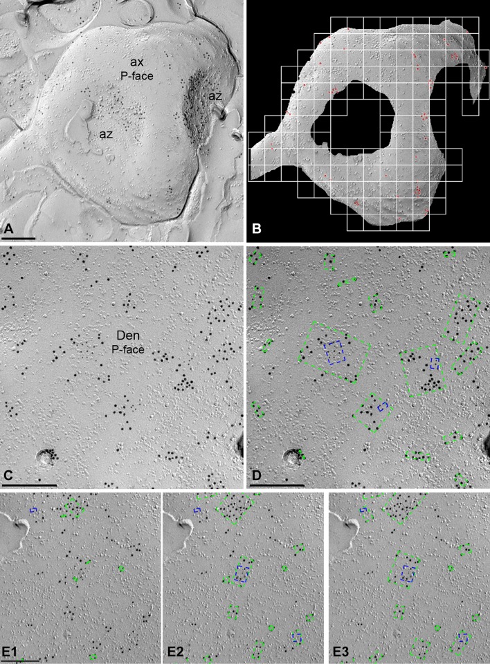
We have developed GPDQ (Gold Particle Detection and Quantification), a software tool that performs automated and semi-automated detection of gold particles present in a given compartment of the cerebellum. The tool is interactive, allowing the user to supervise the process of segmentation and counting, modifying the appropriate parameters and validating the results as needed. It is also modular, which permits new functions to be implemented if required. We have also focused on usability, implementing a user-friendly interface to minimise the learning curve for the tool, and on portability, to make it accessible to a wide range of users (Fig. 1).
Fig. 1.
Development and operation of the GPDQ software used for quantitative analyses of immunoparticle distribution. a Image of an axon terminal (ax) with two active zones (az) in the molecular layer of the cerebellum immunolabelled for GABAB1 (10 nm) on the P-face. b To determine the density of immunoparticles, we first selected manually the contour of the compartment under study, and then the software calculates the area of the profile and the number of immunoparticles (red dots) per profile. c Image of a dendritic shaft (Den) of Purkinje cell double-labelled for GABAB1 (10 nm) and CaV2.1 (5 nm) on the P-face in the molecular layer of the cerebellum. d The software determines the clustering according to the distance among gold particles, both for 10 nm (green dotted rectangles) and for 5 nm (blue dotted rectangles), establishing the number of immunoparticles and the distance among them. The dotted rectangles define the bounding box of the points on each cluster. e1–e3 Clusters of immunoparticles were detected based on distance determined by standard deviation (SD) of NND. Mean (e1), mean + 1SD (e2) and mean + 2SD (e3) were tried and finally the mean + 2SD was chosen for data analysis. Scale bars: a–e 0.2 μm
GPDQ has been implemented with MATLAB and Image Processing Toolbox 9.3 (The MathWorks, Inc., Natick, MA, USA). It is currently divided into four main modules: particle detection, analysis and simulation, graph and statistics generation, and visualisation. The particle detection module allows obtaining the radius and position (in nanometres from top-left corner) of the particles in the images. The automated version uses a two-stage procedure that detects the circles of a given diameter in the image with MATLAB’s implementation of the Hough transform (Yuen et al. 1990), and then determines which of them correspond to actual particles by means of a supervised classification model (Mitchel 1997). In particular, the default setting uses a Naïve Bayes classifier (Minsky 1961). Although the instantiation of the classifier (features and parameters) is provided in the software, it is possible to train and incorporate a different model. Due to the limitations of the Hough transform, which lacks for precision when particles are small, and the scales of the images, fully automated detection is only available for 10 nm particles at present. However, the tool provides a graphical interface for manual detection. It allows locating particles with any diameter even in rough surfaces. To make manual detection faster and more precise, the software automatically adjusts the position of each particle.
The second module allows for the processing of all information about images and particle locations, particle clusters and simulations. This module computes the number of particles, nearest neighbour distances (NNDs) to both particles of the same type (intra-type NNDs, e.g. from 5 nm particle to nearest 5 nm particle) and other type (inter-type NNDs, e.g. from 5 nm particle to nearest 10 nm particle). Clusters are obtained by single-linkage (Gower and Ross 1969). This method carries out an agglomerative hierarchical clustering, i.e. at first it considers each particle as a cluster, and iteratively merges the closest pair of clusters while the minimum inter-cluster distance (distance between a pair of clusters) is below a threshold. As inter-cluster distance measure, single-link considers the minimum distance between the pair of particles not yet belonging to the same cluster. As a consequence, any pair of particles at distance smaller than or equal to the minimum inter-cluster distance threshold belongs to the same cluster at the end of the process. The value of the threshold parameter was obtained from the distribution of the distances between each particle and its nearest neighbour. By default, the software uses mean + two times the standard deviation of such distances. Another important parameter is the minimum number of particles in a cluster, which was fixed to three. Thus, all clusters with one or two particles have been discarded. The software reports some information about the clusters, such as the number of particles, their area (as the area inside the convex hull of the particles in the cluster) or Ripley’s K function (Ripley 1976), or the distance to the nearest cluster of particles of either the same size (intra-cluster distance) or other size (inter-cluster distance). In addition, the second module allows for two types of simulations, termed random and fitted simulation: Random simulation removes all the particles of a given type from the image and redistributes them with two constraints: firstly, simulated particles cannot be within 10 nm of any other particle, and secondly, each pixel within the region of interest must have an equal probability of becoming the centre of a particle, while that probability is zero for each pixel outside of the region of interest. Fitted simulation, however, applies the additional constraint that the distribution of distances between simulated particles is not significantly different from the distribution of distances between the original particles. Similarity of distribution of distances is assessed by comparing all pairwise real and simulated distances by Kolmogorov–Smirnov (KS) test and considered similar if p ≥ 0.1. Otherwise, a particle is chosen at random and is randomly assigned to a new location within the area of interest and at least 10 nm away from other particles. Another KS test is performed and if the distances after this manipulation are less similar than before (indicated by a smaller p value), the particle is relocated to its previous location, and otherwise the new location is saved. This step is repeated until the constraint of a similar distance distribution between real and simulated particles (p ≥ 0.1) is satisfied. All measures consider the existence of two kinds of particles, separated according to their diameter. The software, however, is designed such that it can be quickly adapted for analysis of three or more kinds of particles. The third module deals with the generation of graphs and statistics, from the parameters computed with the second module. The fourth module allows for visualisation of the distribution of original particles as well as simulated particles as for example shown in Fig. 3c.
Fig. 3.
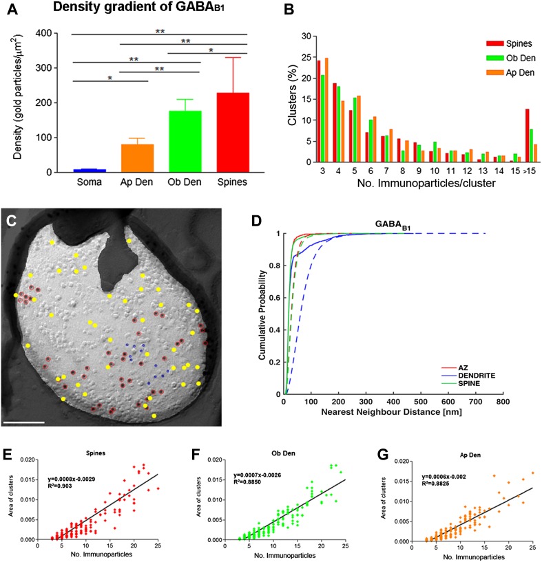
Density gradient and distribution profile of immunoparticles labelling GABAB1 along the surface of PCs. a Density of GABAB1 immunoparticles (including both isolated particles and those within small aggregations) increased from soma to distal dendrites (soma = 8.71 ± 1.43/μm2; apical dendrite = 79.14 ± 18.98/μm2; oblique dendrite = 175.33 ± 34.63/μm2; spines = 227.62 ± 102.18/μm2; Kruskal–Wallis test, pairwise Mann–Whitney U test and Dunn’s method, *p = 0.003; **p < 0.001). b The graph shows the quantification for the size of GABAB1 clusters in the spines, oblique dendrites and apical dendrites. Approximately 75% of clusters consisted of 3–8 immunoparticles. c Example showing random simulation of GABAB1 immunoparticles in a dendritic spine. Red: real GABAB1; Yellow: simulated GABAB1; blue: real GIRK2. Scale bar: 100 nm. d Cumulative probability plots of GABAB1 to GABAB1 NND. Solid and dotted lines show real and simulated GABAB1, respectively. AZ active zone. e–g Positive linear correlation was found between the number of GABAB1 immunoparticles and the area of clusters in the three dendritic compartments [spines, oblique dendrites (Ob Den) and apical dendrites (Ap Den)]
Quantification and analysis of SDS-FRL data
The labelled replicas were examined using a transmission electron microscope (JEOL-1010) and photographed at magnifications of 60,000, 80,000, and 100,000. All antibodies used in this study were visualised by immunoparticles on the protoplasmic face (P-face), consistent with the intracellular location of their epitopes. Non-specific background labelling was measured on E-face surfaces.
Density gradient of GABAB1 along the neuronal surface
Quantitative analysis of immunogold labelling for GABAB1 was performed on three different dendritic compartments of PCs in the inner 1/3 of the ML, in PC somata in the PC layer and the axon terminals establishing synaptic contact with PC spines in the ML. The dendritic compartments analysed were the main apical dendrites, oblique dendrites and dendritic spines. Oblique dendrites were identified based on their small diameter and the presence of at least one emerging spine from the dendritic shaft. Dendritic spines were considered as such if: (1) they emerged from a dendritic shaft, or (2) they opposed an axon terminal recognised by the presence of synaptic vesicles on their cross-fractured portions. Axon terminals were identified based on: (1) the presence of synaptic vesicles in cross-fractures, or (2) the presence of an active zone, recognised by the concave shape of the P-face and the high density of IMPs. Non-specific background labelling was measured on E-face structures surrounding the measured P-faces. Images of the identified PC compartments were selected randomly over the entire dendritic tree of PCs and then captured with an ORIUS SC1000 CCD camera (Gatan, Munich, Germany). The area of the selected profiles and the number of immunoparticles were measured using our GPDQ software (Fig. 1a, b). Immunoparticle densities are presented as mean ± SD between animals. Statistical comparisons were performed with GraphPad Prism 5 software (La Jolla, CA, USA). Digitised images were then modified for brightness and contrast using Adobe PhotoShop CS5 (Mountain View, CA, USA) to optimise them for quantitative analysis.
Analysis of the spatial associations of GABAB1 receptors and GIRK2 or CaV2.1 channels
For each of the molecules, we compared the mean intra-type NND of each image to the mean intra-type NNDs obtained from 500 random simulations generated from the same image. Individual images were considered significantly different from chance, if the real mean NND was within the lowest or highest 2.5% of the simulated mean NNDs, corresponding to a two-tailed test on a significance level of α = 0.05. They were considered associated when mean NND was within the lowest 2.5% and dissociated when mean NND was within highest 2.5% of the simulated mean NNDs. Lack of significant association or dissociation was concluded, when mean NND was within the remaining 95% of simulated mean NNDs. To assess whether a significant association exists for each compartment, we compared the real mean NNDs obtained from each image (n = 19–91) with 500 simulated mean NNDs by two-sided paired t test followed by Holm–Bonferroni correction for multiple testing, with a p < 0.05 being considered significant.
Analysis of colocalization between GABAB1 receptors and GIRK2 or CaV2.1 channels
For each image, inter-type NNDs from 5 nm immunoparticles (GIRK2 or CaV2.1) to10 nm gold particles (GABAB1) were measured using the GPDQ software. The mean NND was compared to 500 mean inter-type NNDs obtained from fitted simulations of 5 nm immunoparticles (GIRK2 or CaV2.1) generated from the same image. Association or dissociation of 10 and 5 nm particles was considered significant in each image if the real mean NND was within the lowest or highest 2.5% of the simulated mean NNDs, corresponding to a two-tailed test on a significance level of α = 0.05. To assess whether a significant interaction exists as a whole for each compartment, we compared the real mean NNDs obtained from each image (n = 19–81) with 500 simulated mean NNDs by paired t test followed by Holm–Bonferroni correction for multiple testing, with a p < 0.05 being considered significant.
Controls
To test method specificity in the procedures for SDS-FRL, antisera against GIRK2 and GIRK3 were tested on cerebellar slices of GIRK2 and GIRK3 knockout mice, respectively. In the replica samples, the immunogold signal disappeared completely in the knockout mouse cerebellum, while a strong signal was present in WT replicas. Furthermore, the primary antibody was either omitted or replaced with 5% (v/v) normal serum of the species of the primary antibody, resulting in total loss of the signal. To test for any cross-reactivity of secondary antibodies when double labelling was used by the SDS-FRL technique, some replicas were incubated with only one primary antibody and the full complement of the secondary antibodies. No cross-labelling was detected that would influence the results. In addition, some replicas were incubated with the two primary antibodies, but we swapped the size of immunogold in the secondary antibodies for the two targets proteins. No differences in distances of the two target proteins were detected that would influence our results. Finally, when double labelling was used, some replicas were incubated with a cocktail of two primary antibodies (GABAB1 and GIRK or Cav) followed by a cocktail of secondary antibodies. Other replicas were incubated with a primary antibody, and then incubated with the second primary antibody, followed by secondary antibodies, and other replicas were incubated with a changed sequence of primary antibodies, applying first primary antibody for GIRK or Cav followed by secondary antibody, and then we applied the second primary antibody (GABAB1) followed by secondary antibody. Under these conditions, we observed similar spatial distribution between two particles, hence that steric hindrance does not seem to affect interparticle distance.
Data analysis
Statistical analyses for morphological data were performed using SigmaStat Pro (Jandel Scientific) and data were presented as mean ± SD) unless indicated otherwise. Statistical significance was defined as p < 0.05. The statistical evaluation of the immunogold densities was performed using the Kruskal–Wallis test, pairwise Mann–Whitney U test and Dunn’s method. Correlations were assessed using Pearson’s correlation test. To assess colocalisation between receptor and ion channels for each compartment, two-sided paired t test followed by Holm–Bonferroni correction for multiple testing was used.
Results
Immunoreactivity for GABAB1 is non-uniform in PCs
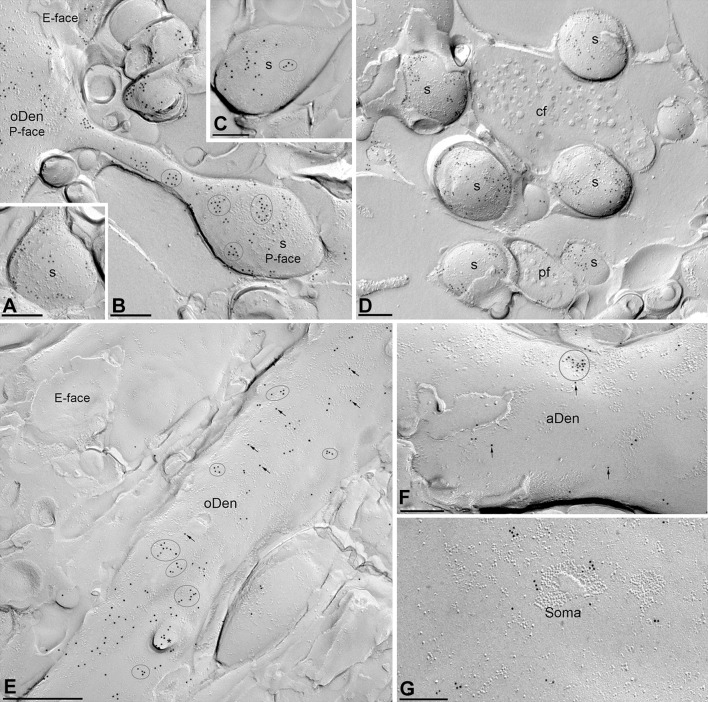
Using the pre-embedding immunogold method, we previously reported that GABAB receptors are widely distributed in developing and adult PCs (Kulik et al. 2002; Luján and Shigemoto 2006). To accurately visualise the two-dimensional distribution of GABAB receptors along somato-dendritic compartments of PCs, and to analyse receptor densities quantitatively, we used highly sensitive immunogold labelling in SDS-FRL (Masugi-Tokita and Shigemoto 2007) in this study. Electron microscopic analysis of the replicas revealed immunoparticles for the GABAB1 subunit on P-faces of PC plasma membranes (Fig. 2). Immunoparticles for GABAB1 were observed throughout the dendritic spines including the spine neck (Fig. 2a–d), dendritic shafts (Fig. 2e, f) and somata (Fig. 2g). The neuronal compartments that showed the highest density of immunoparticles for GABAB1 were dendritic spines, including those establishing synapses with parallel and climbing fibres (Fig. 2a–d). Regardless of the neuronal compartment, immunoparticles for GABAB1 were observed throughout the surface of PCs with two distinct patterns of distribution: scattered and clustered. The clustered pattern consists of aggregation of immunoparticles (> 3 gold particles) and the scattered pattern consists of isolated single immunoparticle detected on dendritic spines and shafts (Fig. 2a–f). Virtually no labelling was observed on the E-face (Fig. 2a–f) or on the cross-fracture of dendrites, spines or axon terminals.
Fig. 2.
Somato-dendritic distribution of GABAB1 in PCs. Representative SDS-FRL electron micrographs of different compartments of PCs. a–d Clusters of GABAB1 immunoparticles (ellipses/circles) associated with the P-face were detected in dendritic spines (s) of PCs, both establishing synaptic contact with parallel fibres (pf) and climbing fibres (cf). Lower density of immunoparticles for GABAB1 was also detected scattered (arrows) outside those clusters. e In oblique dendrites (oDen), both clustered (ellipses/circles) and scattered (arrows) immunoparticles for GABAB1 were detected. Fractured spine necks are indicated with asterisks (*). The E-face is free of any immunolabelling. f In apical dendrites (aDen), we also detected clustered (circles) and scattered (arrows) immunoparticles for GABAB1, though at lower frequency. g A high-magnification image of a PC soma labelled for the GABAB1 subunit. Immunoparticles for GABAB1 in PC soma was low in density and always outside P-face IMP clusters. Scale bars: a–d, f, g 0.2 μm; e 0.5 μm
Next, we performed a quantitative comparison of the GABAB1 densities in different somato-dendritic compartments. A graded increase in the density of GABAB1 immunoparticles was found from the soma to the dendritic spines (Fig. 3a). Dendritic spines showed 26 times higher density of GABAB1 immunoparticles than soma, 3 times higher than apical dendrites and 1.2 times higher than oblique dendrites (Fig. 3a; p < 0.001 for soma vs. dendritic spines; p = 0.003 for dendritic spines vs. oblique dendrites; p < 0.001 for oblique dendrites vs. apical dendrites, Kruskal–Wallis test, pairwise Mann–Whitney U test, and Dunn’s method). We then conducted random simulations (Fig. 3c, d) to investigate whether GABAB1 immunoparticles were clustered. By comparing the NNDs between real and simulated particles from all images (Table 1), we found a highly significant clustering of GABAB1 immunoparticles in spines, dendrites and active zone (p < 0.001 for all compartments). We then asked whether we could detect significant clustering on individual images. We compared the mean NND of each image with the mean NNDs of the simulations, and judged the image to show a significant association if the real mean NND was within the smallest 2.5% of simulated mean NNDs or a significant dissociation if the real mean NND was within the largest 2.5% of simulated mean NNDs. We found that between 79 and 96% of profiles, depending on compartment, showed a significant association of GABAB1 immunoparticles with each other (Table 1), also indicating a clustered distribution of GABAB1. We further analysed the size and immunogold composition of clusters at different dendritic compartments. The size of clusters was similar between spines, oblique and apical dendrites, and quantification of immunoparticles revealed that around 75% of clusters were in the range of 3–8 immunoparticles (Fig. 3b). In these three compartments, the surface area of clusters (defined by the software) and the immunoparticle number showed a strong positive linear correlation (Fig. 3e–g; r = 0.864, 0.884, 0.866 for spines, oblique dendrites, and apical dendrites, respectively), indicating constant density of GABAB1 across clusters.
Table 1.
Clustered distribution of GABAB1, GIRK2 and CaV2.1 in active zones and dendritic shafts and spines
| Molecule | Compartment | Real NND | Simulated NND | p value | Association (%) | N |
|---|---|---|---|---|---|---|
| GABAB1 | Active zone | 22.5 ± 5.6 | 35.8 ± 13.2 | 2.3E−04 | 78.9 | 19 |
| Dendrite | 38.9 ± 17.4 | 83.2 ± 28.6 | 2.8E−10 | 96.0 | 25 | |
| Spine | 27.1 ± 16.5 | 44.9 ± 26.2 | 5.3E−19 | 89.0 | 91 | |
| GIRK2 | Active zone | 19.8 ± 5.2 | 48.7 ± 23.3 | 8.7E−05 | 89.5 | 19 |
| Dendrite | 43.2 ± 29.2 | 204.8 ± 123.9 | 5.3E−06 | 100.0 | 24 | |
| Spine | 30.6 ± 22.2 | 80.6 ± 37.7 | 1.2E−24 | 83.0 | 88 | |
| CaV2.1 | Active zone | 20.0 ± 5.8 | 37.3 ± 24.4 | 3.9E−04 | 97.1 | 35 |
| Dendrite | 36.9 ± 16.9 | 141.8 ± 102.6 | 2.4E−04 | 100.0 | 26 | |
| Spine | 27.6 ± 17.5 | 66.0 ± 33.5 | 1.5E−04 | 84.2 | 19 |
NNDs are reported as mean ± standard deviation of image means, in case of simulations, image means are the means over all 500 simulations of that image. p values were obtained by two-sided paired t test followed by Holm–Bonferroni correction. “Association” shows the percentage of image means within the lowest 2.5% of simulation means. In none of the images did we detect a significant dissociation, i.e. a mean NND within the highest 2.5% of simulated mean NNDs. N indicates the number of images used for analysis
Coupling of GABAB1 receptors with GIRK and CaV2.1 channels in cerebellar membranes
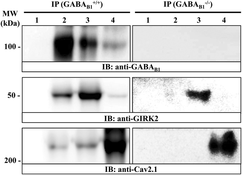
To assess the formation of putative macromolecular complexes containing GABAB1 receptor and its effector molecules, namely GIRK channel and CaV2.1 channel, co-immunoprecipitation experiments were performed. Accordingly, using soluble membrane extracts from mouse cerebellum the anti-GABAB1, the anti-GIRK2 and the anti-CaV2.1 antibodies were able to immunoprecipitate a band of ~ 100 kDa (Fig. 4, lane 2, IP: GABA+/+B1, IB: anti-GABAB1), ~ 50 kDa (Fig. 4, lane 3, IP: GABA+/+B1, IB: anti-GIRK2) and ~ 250 kDa (Fig. 4, lane 4, IP: GABA+/+B1, IB: anti-CaV2.1) which correspond to GABAB1, GIRK2 and CaV2.1 subunits, respectively. Interestingly, the anti-GABAB1 antibody was able to co-immunoprecipitate the GIRK2 channel (Fig. 4: lane 2, IP: GABA+/+B1, IB: anti-GIRK2), as expected (Ciruela et al. 2010), and the CaV2.1 channel (Fig. 4: lane 2, IP: GABA+/+B1, IB: anti-CaV2.1). On the other hand, the anti-GIRK2 antibody co-immunoprecipitated the GABAB1 receptor and the CaV2.1 channel (Fig. 4, lane 3, IP: GABA+/+B1, IB: anti-GABAB1 and IB: anti-CaV2.1, respectively), and the anti-CaV2.1 antibody co-immunoprecipitated the GABAB1 receptor and the GIRK2 channel (Fig. 4, lane 4, IP: GABA+/+B1, IB: anti-GABAB1 and IB: anti-GIRK2, respectively). Importantly, these bands did not appear when an irrelevant rabbit IgG (control IgG) was used for immunoprecipitation (Fig. 4, lane 1), showing that the immunoprecipitation was specific. In addition, when the co-immunoprecipitation experiments were performed using soluble extracts from GABAB1 receptor knockout mouse cerebellum the anti-GABAB1 antibody, unable to immnoprecipitate the GABAB1 receptor (Fig. 4: lane 2, IP: GABA−/−B1, IB: anti-GABAB1), did not co-immunoprecipitate neither the GIRK2 channel nor the CaV2.1 channel (Fig. 4: lane 2, IP: GABA−/−B1, respectively; IB: anti-GIRK2 and IB: anti-CaV2.1, respectively).
Fig. 4.
Co-immunoprecipitation of GABAB1 receptor and GIRK2 and CaV2.1 channels from mouse cerebellum. Solubilised cerebellar membrane extracts from wild type (+/+) and GABAB1 receptor knock-out (−/−) mice were subjected to immunoprecipitation analysis using control rabbit IgG (2 μg, lane 1), rabbit anti-GABAB1 (2 μg, lane 2), rabbit anti-GIRK2 (2 μg, lane 3) and rabbit anti-CaV2.1 (2 μg, lane 4). Immunoprecipitates (IP) were analysed by SDS-PAGE and immunoblotted using a rabbit anti-GABAB1 (1 μg/mL), guinea pig anti-GIRK2 (1 μg/mL) and guinea pig anti-CaV2.1 (1 μg/mL). Immunoreactive bands were detected as described in experimental procedures
In addition, in the very same soluble extract, the anti-GIRK2 and the anti-CaV2.1 antibodies only immunoprecipitated the GIRK2 channel and the CaV2.1 channel, respectively (Fig. 4, lane 3, IP: GABA−/−B1, IB: anti-GIRK2 and lane 4, IP: GABA−/−B1, IB: anti-CaV2.1, respectively). Of note, the absence of orthogonal GIRK2 and CaV2.1 co-immunoprecipitation from GABA−/−B1 cerebellar extracts might reveal an essential role of GABAB receptor nucleating the GIRK2/GABAB/CaV2.1 heterocomplex, yet this contention will need to be further studied in the future. Alternatively, a lack of sensitivity of either the immunoprecipitation and/or immunoblot process would also explain the absence of orthogonal GIRK2 and CaV2.1 co-immunoprecipitation. Overall, these results suggest that in mouse cerebellum, GABAB receptor/GIRK channel, GABAB receptor/CaV2.1 channel might assemble into stable protein–protein complexes resistant to co-immunoprecipitation processing, thus reinforcing the idea that these oligomeric complexes might have physiological relevance in vivo.
Preferential localization of GABAB1 with GIRK channels in PC spines
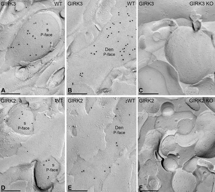
We previously reported the molecular interaction between GABAB receptors and GIRK channels in the cerebellum (Fernández-Alacid et al. 2009; Ciruela et al. 2010). PCs express GIRK1, GIRK2 and GIRK3, although the most predominant subunit is GIRK3 (Aguado et al. 2008; Fernández-Alacid et al. 2009). To provide morphological insights into the GABAB–GIRK interaction, we carried out double-labelling SDS-FRL experiments. Since our anti-GIRK3 antibody was raised in the same species as our anti-GABAB1 antibody, we performed double-labelling SDS-FRL experiments with an anti-GIRK2 antibody only. However, we first compared the subcellular distribution of GIRK2 and GIRK3 in PCs. In single-labelling experiments, immunoparticles for GIRK3 (Fig. 5a, b) and GIRK2 (Fig. 5d, e) were found on the P-face of dendritic shafts and spines. These immunogold labelling patterns were abolished in the GIRK3 KO (Fig. 5c) and GIRK2 KO (Fig. 5f) mice, respectively. Thus, we conclude that GIRK2 and GIRK3 exhibit comparable subcellular distributions in PCs.
Fig. 5.
Subcellular localization for GIRK channel subunits in PCs. Distribution of immunoparticles for the GIRK3 and GIRK2 subunits using the SDS-FRL technique in wild-type (WT) and GIRK knockout (KO) mice. a, b Immunoparticles for the GIRK3 subunit are detected in dendritic spines (s) and dendritic shafts of oblique dendrites (oDen) of PCs. c The antibody specificity was controlled and confirmed in replicas of GIRK3 KO mice that were free of any immunolabelling. d, e Immunoparticles for the GIRK2 subunit are also detected in dendritic spines (s) and dendritic shafts (Den) of PCs, but at lower frequency than GIRK3. f The immunogold labelling for GIRK2 was abolished in the GIRK2 KO mice. Scale bars: a–f 0.2 μm
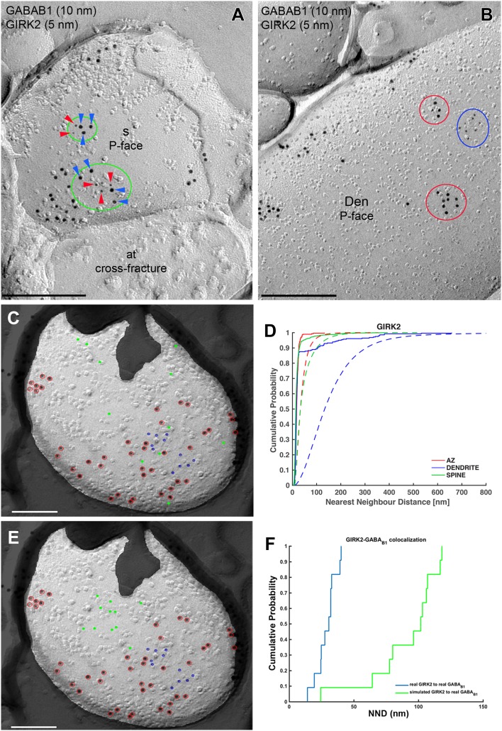
Next, we performed double-labelling experiments for GABAB1 and GIRK2 (Fig. 6). Immunoparticles for GABAB1 appeared co-clustered with those for GIRK2 along the extrasynaptic plasma membrane of dendritic spines (Fig. 6a), but not on dendritic shafts, where clusters of GABAB1 immunoparticles seemed mostly segregated from those of GIRK2 (Fig. 6b). To quantitatively analyse the extent of the spatial relationship between GABAB1 and GIRK2, we first asked whether GIRK2 immunoparticles themselves showed a clustered distribution. We compared NNDs of random simulations of GIRK2 immunoparticles with the real distributions (Fig. 6c, d) and found significant clustering in all compartments (p < 0.001 for all compartments), with 83–100%, depending on compartment, of individual profiles showing a significant association (Table 1). To understand the spatial relationship between GABAB1 and GIRK2, we then conducted fitted simulations (see “Materials and methods”) of GIRK2 immunoparticles (Fig. 6e) to keep the spatial relationship among GIRK2 immunoparticles as close to reality as possible. We then compared NNDs from real and simulated GIRK2 particles to real GABAB1 particles in dendritic shafts and spines (Fig. 6f), and found a significant association between GABAB1 and GIRK2 in dendritic spines (p < 0.001), while we observed a significant dissociation in dendritic shafts (p < 0.01) (Table 2). On the level of individual dendritic spines, we found that 21% showed a significant association, while 1% showed a significant dissociation. For dendritic shafts, we found a significant dissociation in 50% of profiles and a significant association in 4% of profiles. Consistent with these results, we found that inter-cluster distances between GIRK2 and GABAB1 clusters were significantly smaller in dendritic spines compared with dendritic shafts (Table 3).
Fig. 6.
Compartment-dependent co-localization of GABAB1 with GIRK2. Electron micrographs showing double-labelling for GABAB1 (10 nm) and GIRK2 (5 nm) in PCs, as detected using the SDS-FRL technique. a In dendritic spines (s), immunoparticles for GIRK2 (red arrowheads) co-clustered (green ellipses) with those for GABAB1 (blue arrowheads). b In dendritic shafts (Den) of PCs, clusters of GABAB1 immunoparticles (red ellipses) were segregated from clusters of GIRK2 immunoparticles (blue ellipses). Red, green and blue ellipses were drawn manually using Adobe Photoshop for illustration purposes, to show the particles corresponding to clusters. They do not represent the area of clusters as defined using GPDQ software and not all of the clusters detected are shown. c Example showing random simulation of GIRK2 immunoparticles in a dendritic spine. Blue: real GIRK2; red: real GABAB1; green: simulated GIRK2. d Cumulative probability plots of GIRK2 to GIRK2 nearest neighbour distance (NND). Solid and dotted lines show real and simulated GIRK2, respectively. e Example showing fitted simulation of GIRK2 immunoparticles in a dendritic spine. Blue: real GIRK2; red: real GABAB1; green: simulated GIRK2. f Cumulative probability plot showing GIRK2 to GABAB1 NND of the particular simulation shown in e. AZ active zone. Scale bars: a, b 200 nm; c, e 100 nm
Table 2.
Spatial relationship of GABAB1 with GIRK2 and CaV2.1
| Simulated molecule | Compartment | Real NND | Simulated NND | p value | Association (%) | Dissociation (%) | N |
|---|---|---|---|---|---|---|---|
| GIRK2 | Active zone | 42.4 ± 13.8 | 47.0 ± 18.4 | 0.36 | 5.3 | 5.3 | 19 |
| Dendrite | 158.4 ± 78.4 | 119.2 ± 48.8 | 0.0088 | 4.2 | 50.0 | 24 | |
| Spine | 42.4 ± 20.9 | 59.2 ± 22.5 | 4.1E−14 | 21.3 | 1.1 | 89 | |
| CaV2.1 | Active zone | 41.8 ± 11.6 | 43.1 ± 11.4 | 0.77 | 14.3 | 2.9 | 35 |
| Dendrite | 44.0 ± 17.8 | 62.9 ± 13.7 | 3.4E−08 | 46.2 | 0.0 | 26 | |
| Spine | 98.0 ± 37.7 | 96.5 ± 34.5 | 0.83 | 10.0 | 10.0 | 20 |
NNDs are reported as mean ± standard deviation of image means, in case of simulations, image means are the means over all 500 simulations of that image. p values were obtained by two-sided paired t test followed by Holm–Bonferroni correction. “Percent significant” show the percentage of images whose mean NNDs are within the top or bottom 2.5% of simulation means. “Dissociation” and “Association” show the percentage of image means within the top and bottom 2.5%, respectively, of simulation means. N indicates the number of images used for analysis
Table 3.
Spatial relationship between CaV2.1 or GIRK2 and GABAB1 clusters
| Clusters | Compartment | Inter-cluster distance | N |
|---|---|---|---|
| GIRK2 to GABAB1 | Active zone | 84.2 ± 46.1 | 21 |
| Dendrite | 219.6 ± 119.0 | 54 | |
| Spine | 110.7 ± 73.4 | 100 | |
| CaV2.1 to GABAB1 | Active zone | 68.5 ± 45.1 | 44 |
| Dendrite | 124.2 ± 126.7 | 85 | |
| Spine | 154.9 ± 85.9 | 31 |
Inter-cluster distances from CaV2.1 or GIRK2 clusters to the nearest neighbouring GABAB1 clusters are reported as mean ± standard deviation. Inter-cluster distances between GIRK2 and GABAB1 in dendritic shafts differed significantly from both the ones in spines (p < 0.001) and in active zones (p < 0.001), while there was no significant difference between inter-cluster distances in spines and active zones (p = 0.12). Inter-cluster distances between CaV2.1 and GABAB1 were showed significant differences between active zones and dendritic shafts (p = 0.004), active zones and dendritic spines (p < 0.001) as well as dendritic shafts and spines (p = 0.009). p values were obtained by Mann–Whitney U test followed by Holm–Bonferroni correction. N indicates the number of CaV2.1 or GIRK clusters analysed
Preferential localization of GABAB1 with CaV2.1 channels in PC dendrites
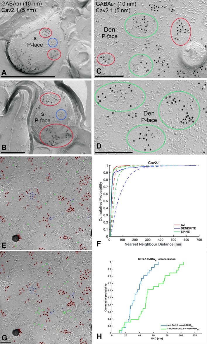
We next performed double-labelling experiments for GABAB1 and CaV2.1 (Fig. 7). Immunoparticles for CaV2.1 were found on the P-face of dendritic shafts and spines, but not on the E-face or on cross-fractures (Fig. 7a–d). Immunoparticles for GABAB1 were close to those for CaV2.1 but seemed not co-clustered along the extrasynaptic plasma membrane of dendritic spines (Fig. 7a, b). In contrast, in dendritic shafts GABAB1 immunoparticles appeared co-clustered with those for CaV2.1 (Fig. 7c, d), although many clusters of GABAB1 immunoparticles were not apparently associated with clusters of CaV2.1 immunoparticles (Fig. 7c). Random simulations of CaV2.1 (Fig. 7e, f) showed a significant clustering of CaV2.1 immunoparticles with themselves in all compartments (p < 0.001), with 84–100%, depending on compartment, of individual profiles showing significant association (Table 1). To quantify their extent of spatial relation, the NNDs between immunoparticles for GABAB1 and CaV2.1 were compared with those between real GABAB1 and simulated CaV2.1 particles (fitted simulations, see “Materials and methods”) (Fig. 7g, h). We found a significant association of GABAB1 with CaV2.1 in dendritic shafts (p < 0.001). In dendritic spines, however, co-clustering occurred only on chance level (p = 0.83) (Table 2). On the level of individual dendritic spines, we found that 10% each showed significant association or dissociation. For dendritic shafts, we found a significant association in 46% of profiles and no profile showing significant dissociation. In line with these results, we found that inter-cluster distances between CaV2.1 and GABAB1 clusters were significantly smaller in dendritic shafts compared with dendritic spines (Table 3).
Fig. 7.
Preferential colocalisation of GABAB1 with CaV2.1 in PC dendritic shafts. Electron micrographs showing double-labelling for GABAB1 (10 nm) and CaV2.1 (5 nm) in PCs, as detected using the SDS-FRL technique. a, b In dendritic spines (s), clusters of GABAB1 immunoparticles (red ellipses) are close to but mostly segregated from clusters of CaV2.1 immunoparticles (blue ellipses). c, d In dendritic shafts (Den), immunoparticles for GABAB1 immunoparticles co-clustered with immunoparticles for CaV2.1 (green ellipses). Clusters of GABAB1 immunoparticles without immunoparticles for CaV2.1 (red ellipses) were also found in dendritic spines of PCs. Red, green and blue ellipses were drawn manually using Adobe Photoshop for illustration purposes, to show the particles corresponding to clusters. They do not represent the area of clusters as defined using GPDQ software and not all of the clusters detected are shown. e Example showing random simulation of CaV2.1 immunoparticles in a dendritic shaft. Blue: real CaV2.1; red: real GABAB1; green: simulated CaV2.1. f Cumulative probability plots of CaV2.1 to CaV2.1 NND. Solid and dotted lines show real and simulated CaV2.1, respectively. g Example showing fitted simulation of CaV2.1 immunoparticles in a dendritic shaft. Blue: real CaV2.1; red: real GABAB1; green: simulated CaV2.1. h Cumulative probability plot showing CaV2.1 to GABAB1 NND of the particular simulation shown in g. AZ active zone. Scale bars: a–d 200 nm; e, g 100 nm
GABAB1 is not co-clustered with but close to GIRK or CaV2.1 channels at presynaptic sites
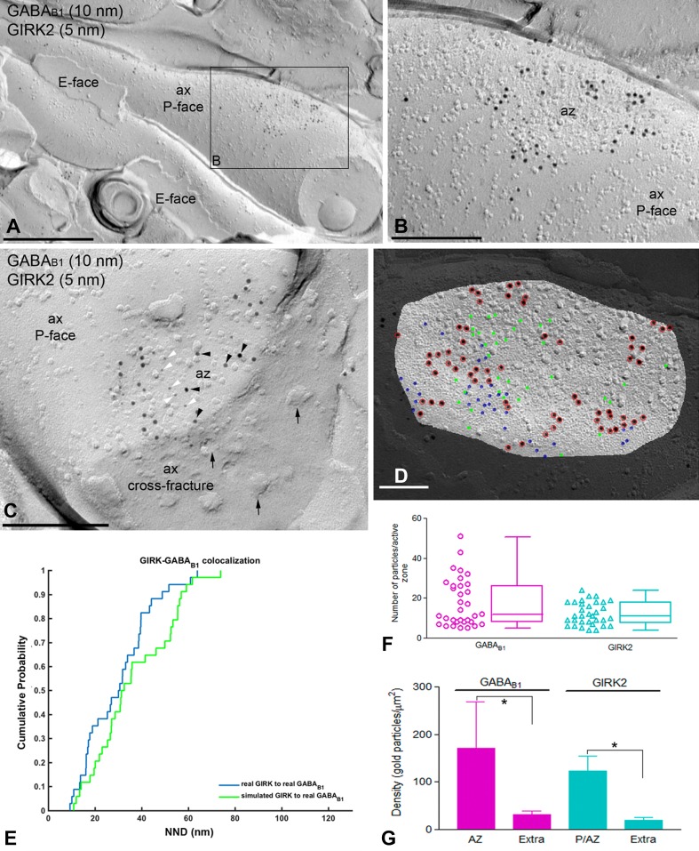
Immunoparticles for GABAB1 were not only confined to somato-dendritic domains of PCs, but also present in axon terminals, as previously reported (Kulik et al. 2002; Luján and Shigemoto 2006). Accordingly, we analysed whether GABAB receptors co-clustered with their effector ion channels at presynaptic sites. First, we examined the co-clustering and spatial relationship of GABAB1 with GIRK2 channels. Most immunoparticles for GIRK2 were detected close to active zones (Fig. 8a–c), and in some cases concentrated in the active zone of axon terminals (Fig. 8c). The NNDs from GIRK2 to GABAB1 immunoparticles in the active zone were as short as that (42.4 nm) found in dendritic spines (Table 1). However, we found no difference (p = 0.36) between real NND and that from simulated GIRK2 to GABAB1 immunoparticles (Fig. 8d, e), indicating absence of significant co-clustering. In active zones, 5% of individual profiles showed association and 5% showed dissociation (Table 2). The inter-cluster distance between GIRK2 and GABAB1 clusters, however, was not significantly different between active zones and dendritic spines, where a significant association had been detected (Table 3). In addition, the number of immunoparticles was highly variable and ranged from 5 to 51 per active zone for GABAB1 (Fig. 8f; mean = 17.8, median = 12, interquartile range = 8–26.25, n = 33 active zone profiles from three animals), and ranged from 4 to 24 per active zone for GIRK2 (Fig. 8f; mean = 12.2, median = 11, interquartile range = 7.75–18, n = 33 active zone profiles from three animals). The density of GABAB1 and GIRK2 at the active zone was 171.66 ± 97.02 and 123.36 ± 31.91 immunogold/μm2, respectively (Fig. 8g). Low densities of GABAB1 (mean = 31.1 immunogold/μm2, median = 34, interquartile range = 26.1–35.9, n = 15 profiles from three animals) and GIRK2 (mean = 19.8 immunogold/μm2, median = 22.3, interquartile range = 18.2–23.7, n = 15 profiles from three animals) were in axon terminals (Fig. 8g), indicating accumulation of both GABAB receptors and GIRK2 in the active zone.
Fig. 8.
Co-distribution of GABAB1 and GIRK2 within the presynaptic active zone of axon terminals. Electron micrographs showing the P-face and cross-fracture of axon terminals (ax) identified by presence of synaptic vesicles (arrows) and active zones (az), recognised by the concave shape of the P-face and the high density of IMPs. a A low-magnification image of an axon terminal (ax) with synaptic contact co-labelled for GABAB1 and the GIRK2. b Higher magnification image of the boxed area shown in a. Small clusters of immunogold particles for GABAB1 (10 nm) were mostly found at the edge of active zones co-distributed with immunogold particles for GIRK2 (5 nm). c In a few axon terminals, immunoparticles for GABAB1 (black arrowheads) co-distributed with immunoparticles for GIRK2 (white arrowheads) in the active zone (az). d Example showing fitted simulation of GIRK2 immunoparticles in an active zone. Blue: real GIRK2; red: real GABAB1; green: simulated GIRK2. e Cumulative probability plot showing GIRK2 to GABAB1 NNDs of the particular simulation shown in d. f High variability of number of GABAB1 immunoparticles (range 5–51) and GIRK2 immunoparticles (range 4–24) was found at the edge and inside of active zones. Box chart shows fifth, 25th, 75th, and 95th percentiles and median (bar). g Densities of GABAB1 and GIRK2 immunoparticles at the active zone and extrasynaptic areas of axons. The density of both GABAB1 and GIRK2 immunoparticles was significantly higher at the edge and inside of active zones (P/AZ; GABAB1 = 171.66 ± 97.03/μm2; GIRK2 = 123.36 ± 31.91/μm2) than at extrasynaptic sites (Extra; GABAB1 = 31.06 ± 8.26/μm2; GIRK2 = 19.83 ± 5.90/μm2) (Kruskal–Wallis test, pairwise Mann–Whitney U test and Dunn’s method, *p < 0.001). Scale bars: a 0.5 μm; b, c 0.2 μm; d 0.1 µm
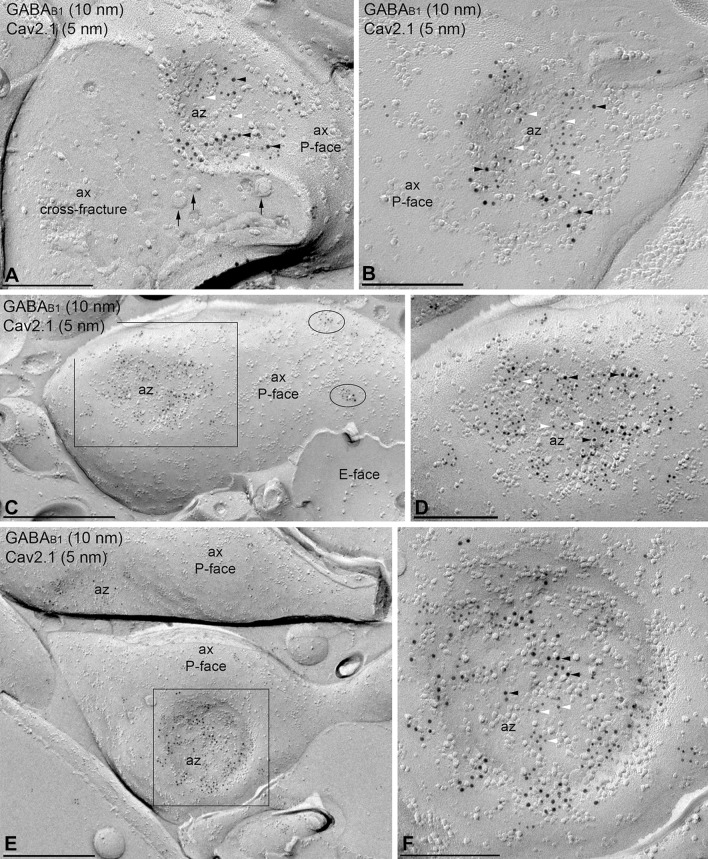
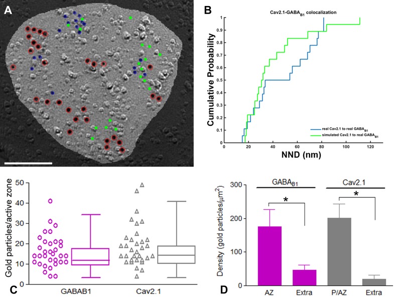
Next, we examined the co-clustering and spatial relation of GABAB1 and CaV2.1 channels. Immunoparticles for CaV2.1 were always concentrated in the presynaptic active zone of axon terminals (Fig. 9a, b), as also described previously (Kulik et al. 2004; Indriati et al. 2013). Within active zones, immunoparticles for both GABAB1 and CaV2.1 were not homogenously distributed, but small aggregations were detected (Fig. 9a–f). To quantify the spatial relation of the two proteins, we measured the NNDs from CaV2.1 (5 nm) to GABAB1 (10 nm) immunoparticles in active zones, and compared it with that of fitted simulations of CaV2.1 particles (Fig. 10a, b). Although the mean NND (42 nm) in active zone was similar to that in dendritic shaft (44 nm), where CaV2.1 and GABAB1 immunoparticles associated more closely than chance, we found no difference between real and simulated NNDs in active zones (Table 2, p = 0.77), indicating absence of significant co-clustering of GABAB1 with CaV2.1 as a whole. On the level of individual active zone profiles, however, we found that 14% showed a significant association while only 2.9% (about chance level which is 2.5%) showed a significant dissociation. This may indicate plastic or dynamic association of GABAB1 with CaV2.1. Interestingly, inter-cluster distance of CaV2.1 and GABAB1 clusters in active zones was significantly smaller than in both dendritic shafts and spines (Table 3). The number of particles was highly variable and ranged from 4 to 41 per active zone for GABAB1 (Fig. 10c; mean = 16.3, median = 14, interquartile range = 11–21, n = 33 active zone profiles from three animals), and ranged from 4 to 49 per active zone for CaV2.1 (Fig. 10c; mean = 19.3, median = 17, interquartile range = 12–22.2, n = 33 active zones three animals). Around 80% of clusters were in the range of 3–11 immunoparticles for GABAB1 and 3–12 immunoparticles for CaV2.1, indicating that the size of clusters was similar between the receptor and the ion channel. The density of GABAB1 and CaV2.1 at the active zones was 175.64 ± 51.07 and 200.35 ± 42.77 immunogold/μm2, respectively (Fig. 10d). Low density of both GABAB1 (mean = 45.96 immunogold/μm2, median = 40.8, interquartile range = 35.7–53.6, n = 15 profiles from three animals) and CaV2.1 (mean = 19.68 immunogold/μm2, median = 17.4, interquartile range = 13–21.9, n = 15 profiles from three animals) were detected outside the active zone, along the extrasynaptic plasma membrane of axon terminals (Fig. 10d).
Fig. 9.
Co-distribution of GABAB1 and CaV2.1 in the presynaptic active zone of axon terminals. a–f Electron micrographs showing the P-face and cross-fracture of axon terminals (ax) identified by presence of synaptic vesicles (arrows) and active zones (az) recognised by the concave shape of the P-face and the high density of IMPs. Black boxes in c and e represent images enlarged in d and f, respectively. Immunoparticles for GABAB1 (10 nm, black arrowhead) were found within the active zone (az) co-distributed, but not co-clustered, with immunoparticles for CaV2.1 (5 nm, white arrowhead). In a few axon terminals (b), immunoparticles for GABAB1 co-distributed with immunoparticles for CaV2.1 only at the edge but not in the central part of the active zone. Few clusters of GABAB1 and CaV2.1 were detected at extrasynaptic sites of axon terminals (black ellipses in c). Scale bars: a, b, d, f 0.2 μm; c, e 0.5 μm
Fig. 10.
Variability and density of GABAB1 and CaV2.1 in active zone and axon terminals. a Example showing fitted simulation of CaV2.1 immunoparticles in an active zone. Blue: real CaV2.1; red: real GABAB1; green: simulated CaV2.1. Scale bar, 100 nm. b Cumulative probability plot showing CaV2.1 to GABAB1 NND of the particular simulation shown in a. c High variability of number of GABAB1 immunoparticles (range 4–56) and CaV2.1 immunoparticles (range 4–54) was found at the edge and inside of active zones. Box chart shows fifth, 25th, 75th, and 95th percentiles and median (bar). d Densities of GABAB1 and CaV2.1 immunoparticles at the active zone and extrasynaptic sites of axon terminals. The density of both GABAB1 and CaV2.1 immunoparticles was significantly larger at the active zones (AZ; GABAB1 = 301.66 ± 124.82/μm2; CaV2.1 = 334.32 ± 130.65/μm2) than at extrasynaptic sites (Extra; GABAB1 = 45.96 ± 15.09/μm2; CaV2.1 = 30.06 ± 20.49/μm2) (Kruskal–Wallis test, pairwise Mann–Whitney U test and Dunn’s method, *p < 0.001)
Discussion
The present study describes the two-dimensional distribution of the GABAB1 subunit of the GABAB receptor and its spatial relationship to the GIRK2 subunit of the GIRK channel and the CaV2.1 subunit of P/Q-type Ca2+ channels along the plasma membrane of PCs using the highly sensitive and quantitative immunogold SDS-FRL technique. Our data reveal novel insights into the subcellular localization of GABAB receptors and the selective coupling of these receptors to effector ion channels. Notably, we have demonstrated that although GABAB1 receptors are distributed along the entire somato-dendritic domain, they are not homogeneously distributed, instead showing a distance-dependent localization along the cell surface of PCs. Moreover, our data suggest that the GABAB–GIRK channel and GABAB–CaV2.1 signalling cascades are present along the dendritic domains of PCs, while showing compartment-specific differences. Indeed, double-labelling studies revealed that GABAB1 and GIRK2 showed a high degree of co-clustering on dendritic spines, whereas GABAB1 and CaV2.1 were mainly co-clustering on dendritic shafts. Finally, our data highlight some differences in the presynaptic co-localization of GABAB receptors with their effector ion channels between distinct domains of the axon terminals.
Distance-dependent increase of GABAB receptors in PC dendrites
GABAB receptors are highly expressed in the cerebellar cortex, and are found at a particularly high density in PCs, consistent with previous in situ hybridization (Kaupmann et al. 1998; Bischoff et al. 1999; Liang et al. 2000) and immunohistochemical (Fritschy et al. 1999; Kulik et al. 2002; Luján and Shigemoto 2006) studies. Previous pre-embedding immunogold data showed that the highest densities of the two GABAB receptor subunits, GABAB1 and GABAB2, are found in PC spines (Kulik et al. 2002; Luján and Shigemoto 2006). Our data obtained using more sensitive approaches show the highest density of GABAB1 immunolabelling in dendritic compartments, particularly around the glutamatergic synapses between PC spines and parallel fibre axon terminals. These results correlate with electrophysiological data showing that GABAB receptors are responsible for mediating major cellular functions in PCs. For example, PCs are hyperpolarized following GABAB receptor activation (Vigot and Batini 1997), and GABAB receptor activation suppresses the rebound potentiation of inhibitory synaptic inputs to PCs (Kawaguchi and Hirano 2000). Moreover, GABAB receptor activation enhances postsynaptic responses mediated by mGlu1 receptors (Hirono et al. 2001), while also enhancing long-term depression of a glutamate-evoked currents, increasing the magnitude of depression (Kamikubo et al. 2007).
While GABAB receptor immunoparticles were present along the entire somato-dendritic axis of PCs, they were distributed in non-uniform fashion. The density of GABAB1 immunoparticles increased from the soma towards dendritic spines, with significant differences observed between the main apical dendrites, oblique dendrites, or dendritic spines at approximately the same distance from the soma. Although understanding how integration of signals in PC dendrites is affected by this uneven distribution of GABAB receptors will require detailed electrophysiological investigations, they could contribute to the activity of the cerebellar microcircuit by amplifying and modifying synaptic signals (Hanson and Smith 2002) or synaptic plasticity (Kohl and Paulsen 2010). In other cell types, we previously reported an exclusive gradient along the somato-dendritic domains for different ion channels, including GIRK and SK channels, in hippocampal pyramidal cells (Fernández-Alacid et al. 2011; Ballesteros-Merino et al. 2012, 2014).
Regardless of the neuronal compartment, our high-resolution immunoelectron microscopic studies revealed two distinct patterns of GABAB1 along the surface of PCs, one consisting of GABAB1 isolated and other consisting of GABAB1 clustered in plasma membrane domains. The formation of clustered and scattered pools of GABAB receptors were also observed in the neuronal plasma membrane of hippocampal pyramidal cells (Kulik et al. 2006; Degro et al. 2015), and as such, this may represent a common organizational principle in different central neurons. Similar distribution patterns have been detected for other receptors and ion channels in central neurons (Kaufmann et al. 2009, 2010; Ballesteros-Merino et al. 2012; Indriati et al. 2013). Most GABAB1 immunoparticles were found forming clusters and this might represent an effective way by which GABAB receptors induce local changes in the electrophysiological properties of the neuronal membrane. We further analysed the size of clusters along the dendritic surface of PCs and found that the area of the clusters, the number of GABAB1 and density of GABAB1 within the clusters were similar among the different dendritic compartments. These data indicate that the somato-dendritic gradient observed in PCs is due to a progressive increase in the number of clusters rather than an increase in the size and/or composition of GABAB1 clusters.
Compartment-dependent co-clustering of postsynaptic GABAB receptors with ion channels
One of the best-characterised downstream effectors modulated by GABAB receptors is the GIRK channel (Lüscher et al. 1997). Activation of postsynaptic GABAB receptors generally causes activation of GIRK channels, thereby hyperpolarizing the postsynaptic plasma membrane (Kaupmann et al. 1998). In the cerebellum, GIRK channels are expressed in a cell type-dependent manner (Aguado et al. 2008), and PCs express GIRK1, GIRK2, and GIRK3 (Karschin et al. 1996). We have demonstrated that GIRK1, GIRK2, and GIRK3 subunits co-precipitated together in the cerebellum (Aguado et al. 2008), and further revealed that GIRK subunits co-immunoprecipitated GABAB receptors in an expression system and solubilised cerebellar membranes (Fernández-Alacid et al. 2009; Ciruela et al. 2010). The association GIRK/GABAB is not supported by proteomic approaches (Schwenk et al. 2016), possibly suggesting that the interaction is via G protein. Furthermore, GABAB receptors preferentially localise to the extrasynaptic plasma membrane of PC spines, where they co-localise with GIRK channels (Fernández-Alacid et al. 2009). Consistent with these data, our immunogold labelling revealed a high degree of co-clustering of GABAB1 and GIRK2 on dendritic spines, whereas on dendritic shafts they are mainly segregated. Similar spine-specific co-clustering of GABAB1 and GIRK3 has been found in hippocampal pyramidal cells (Kulik et al. 2006). In spines, the mean distance between the ion channels to the receptor was 43 nm, and this short distance suggests the existence of preformed macromolecular complexes that would ensure reliable and efficient GABAB–GIRK interaction (Ciruela et al. 2010). However, the observed segregation between GABAB receptors and GIRK channels on dendritic shafts raises the question as to how GIRK channels are activated in this compartment. GIRK channels may be activated by GABAB receptors, as the mean distance between the receptor and ion channel in dendritic shafts (143 nm) may be sufficient for GABAB–GIRK coupling (Karschin 1999). It is also possible that GIRK channels couple to a different GPCR(s), including metabotropic glutamate receptors, adenosine A1 receptors, cannabinoid CB2 receptors; indeed, these receptors have been linked to GIRK channel activation in the cerebellum (North 1989).
If GABAB receptors do not couple to GIRK channels in dendritic shafts, N- or P/Q-type voltage-dependent Ca2+ channels might (Bettler et al. 2004). Although GABAB receptors couple with CaV2.1 channels at presynaptic sites (Huston et al. 1995; Takahashi et al. 1998), there is evidence indicating that they also trigger Ca2+ influx across postsynaptic membranes (Catterall 1998). Light and electron microscopic studies have shown that PCs also express high density of CaV2.1 channels (Kulik et al. 2004; Indriati et al. 2013). Therefore, it seems reasonable to consider that GABAB receptors and CaV2.1 channels are functionally coupled at postsynaptic compartments. Supporting this idea, we demonstrated that CaV2.1 co-immunoprecipitated with GABAB receptors in solubilised cerebellar membranes. This association between CaV2.1 and GABAB may be indirect via KCTD16 (Schwenk et al. 2016; Pin and Bettler 2016). However, the neuronal compartments where these effectors couple to GABAB receptors in PCs should be further elucidated.
Similar to the observed distribution of GABAB receptors, CaV2.1 channels are distributed non-uniformly from soma to distal dendrite with graded increase in density (Indriati et al. 2013). The present study shows that receptor and ion channel distributions are overlapping, but show divergence in their co-clustering pattern. We found that the mean nearest distance of CaV2.1 to GABAB1 was 44 nm in dendritic shafts, and about twice that distance (82.2 nm) in spines. Therefore, our study confirms that GABAB–CaV2.1 complexes are present along the dendritic domains of cerebellar PCs, and suggest a preferential and more efficient coupling in dendritic shafts. This coupling is supported by the results of whole-cell patch recording showing that GABAB receptors inhibit P-type Ca2+ channels through a G protein-mediated mechanism (Mintz and Bean 1993). Regardless of the possible coupling between GABAB receptors and CaV2.1 channels in dendritic spines, their close spatial relationship was not unexpected. First, activation of GABAB receptors by agonists or extracellular Ca2+ enhanced mGlu1-mediated inward currents and Ca2+ signals in PCs, demonstrating a cross-talk between mGlu1 and GABAB receptors (Hirono et al. 2001; Tabata et al. 2004). Second, previous studies showed a direct molecular and functional coupling between CaV2.1 channels and the mGlu1 receptor (Kitano et al. 2003). Therefore, it seems reasonable to expect that CaV2.1 channels distributed in dendritic spines might also be involved in spatiotemporal regulation of intracellular Ca2+ in glutamatergic neurotransmission through both mGlu and GABAB receptors.
Short distance of GABAB receptors from ion channels in axon terminals
The release of neurotransmitter can be modulated by GABAB receptors inhibiting the action of CaV2.1 and CaV2.2 channels (Mintz and Bean 1993; North 1989; Lüscher et al. 1997; Takahashi et al. 1998). We observed a high density of immunoparticles for CaV2.1 in a restricted area of the presynaptic plasma membrane, suggesting a preferential localization of CaV2.1 channels at the active zone of axon terminals, as described (Indriati et al. 2013). Indeed, double‐labelling for CaV2.1 and presynaptic active zone proteins RIM1 and RIM2 provided evidence that these proteins were confined to the same compartment of the presynaptic plasma membrane in the cerebellum (Baur et al. 2015). Although our quantitative analysis showed that CaV2.1 was not significantly associated to GABAB receptor compared with simulated CaV2.1 in the active zone, the mean NND from CaV2.1 to GABAB particles (42 nm) was comparable to that in dendritic shafts (44 nm), where they significantly co-clustered. This is because of the high overall density of both molecules in the active zone, which may allow close interaction between GABAB receptor and CaV2.1 channels in the presynaptic active zone.
We previously reported that the three GIRK channel subunits are localised at presynaptic sites in the cerebellum (Aguado et al. 2008; Fernández-Alacid et al. 2009), as well as in other brain regions (Morishige et al. 1996; Ponce et al. 1996; Koyrakh et al. 2005; Marker et al. 2005). Although electrophysiological studies do not support a role for a pre-synaptic GIRK activation as a primary mechanism by which GABAB receptors modulate neurotransmitter release (Lüscher et al. 1997), using functional assays we reported that GIRK channel-mediated inhibition of glutamate release occurs through GABAB receptors in the cerebral cortex (Ladera et al. 2008) and cerebellum (Fernández-Alacid et al. 2009). The presynaptic coupling in the cerebellum is supported by the up-regulation of GIRK3 and GABAB receptors in parallel fibre terminals after genetic ablation of GABAB1 and GIRK3, respectively (Fernández-Alacid et al. 2009). Using more sensitive immunolocalisation techniques, we not only confirmed the presynaptic distribution of GIRK channels but also revealed short NND (42 nm) to GABAB receptors, which is exactly the same NND as in spine (42 nm). Although we did not find a significant difference between real and simulated inter-NNDs, their proximity to each other suggests an involvement of GABAB–GIRK interaction in the regulation of neurotransmitter release (Ladera et al. 2008; Fernández-Alacid et al. 2009). Altogether, our data clearly suggest that coupling of GABAB receptors to their effector ion channels differs in dendritic spine and shaft domains but may be similar in axon terminal domains.
Altogether, our data clearly demonstrated that both particle and the shortest particle and cluster NNDs for CaV2.1 to GABAB and GIRK to GABAB occurred in the dendritic shaft and dendritic spine, respectively, consistent with the functional associations between the ion channels and the receptor in the respective compartments. The cluster NNDs for CaV2.1/GIRK to GABAB in the active zone were also shorter than those observed in dendrites and spines, suggesting that similar molecular and functional interaction can take place in the active zone despite of no significant difference from the simulated distribution.
Acknowledgements
The authors would like to thank to Mrs. Mercedes Gil for the excellent technical assistance and Dr. Zoltan Nusser for helpful discussions. This work was supported by grants from the Spanish Ministerio de Economía y Competitividad (BFU2015-63769-R) and Junta de Comunidades de Castilla-La Mancha (PPII-2014-005-P) to R.L., the European Union (HBP-Project Ref. 720270) to R.L and R.S., the Spanish Ministry of Education and Science (TIN2013-46638-C3-3-P) to JC and LDLO, the National Institutes of Health (MH061933) to KW, Ministerio de Economía y Competitividad/Instituto de Salud Carlos III (SAF2011-24779 and PIE14/00034), Institució Catalana de Recerca i Estudis Avançats (ICREA Academia-2010) and AgentschapvoorInnovatie door Wetenschap en Technologie (SBO-140028) to FC, and Grants-in-Aid for Scientific Research from the Ministry of Education, Culture, Sports, Science and Technology (MEXT) of Japan (16H04662) to YF. FC belongs to the “Neuropharmacology and Pain” accredited research group (Generalitat de Catalunya, 2014 SGR 1251). DK is a recipient of a DOC Fellowship of the Austrian Academy of Sciences.
Author contribution
All authors had full access to all data in the study and take responsibility for the integrity of the data and the accuracy of the data analysis. RL and YF designed the project; FC performed co-immunoprecipitation analysis; JC, DK and LDLO developed in-house software and performed computational analysis; RL and YF performed SDS-FRL immunoelectron microscopy; MW and RS provided reagents; BB and KW provided knock-out tissues and feedback on the manuscript; RL and CA analysed data; RL wrote the paper.
Compliance with ethical standards
Ethical statement
All co-authors of the present manuscript can certify that it has not been submitted to more than one journal for simultaneous consideration and that the manuscript has not been published previously (partly or in full). The authors also can certify that our main study is not split up into several parts to increase the quantity of submissions, that none of the data presented here have been fabricated or manipulated and that we present our own data/text/theories/ideas. Finally, all co-authors and authorities have explicitly provided their consent to submit the present manuscript and in general we all agree with the ethical responsibilities of authors of the journal.
Conflict of interest
The authors declare that they have no conflict of interest.
References
- Aguado C, Colón J, Ciruela F, Schlaudraff F, Cabañero MJ, Perry C, Watanabe M, Liss B, Wickman K, Luján R. Cell type-specific subunit composition of G protein-gated potassium channels in the cerebellum. J Neurochem. 2008;105:497–511. doi: 10.1111/j.1471-4159.2007.05153.x. [DOI] [PubMed] [Google Scholar]
- Ballesteros-Merino C, Lin M, Wu WW, Ferrandiz-Huertas C, Cabañero MJ, Watanabe M, Fukazawa Y, Shigemoto R, Maylie J, Adelman JP, Luján R. Developmental profile of SK2 channel expression and function in CA1 neurons. Hippocampus. 2012;22:1467–1480. doi: 10.1002/hipo.20986. [DOI] [PMC free article] [PubMed] [Google Scholar]
- Ballesteros-Merino C, Watanabe M, Shigemoto R, Fukazawa Y, Adelman JP, Luján R. Differential subcellular localization of SK3-containing channels in the hippocampus. Eur J Neurosci. 2014;39:883–892. doi: 10.1111/ejn.12474. [DOI] [PubMed] [Google Scholar]
- Batchelor AM, Garthwaite J. GABAB receptors in the parallel fibre pathway of rat cerebellum. Eur J Neurosci. 1992;4:1059–1064. doi: 10.1111/j.1460-9568.1992.tb00132.x. [DOI] [PubMed] [Google Scholar]
- Baur D, Bornschein G, Althof D, Watanabe M, Kulik A, Eilers J, Schmidt H. Developmental tightening of cerebellar cortical synaptic influx-release coupling. J Neurosci. 2015;35:1858–1871. doi: 10.1523/JNEUROSCI.2900-14.2015. [DOI] [PMC free article] [PubMed] [Google Scholar]
- Bettler B, Kaupmann K, Mosbacher J, Gassmann M. Molecular structure and physiological functions of GABA (B) receptors. Physiol Rev. 2004;84:835–867. doi: 10.1152/physrev.00036.2003. [DOI] [PubMed] [Google Scholar]
- Bischoff S, Leonhard S, Reymann N, Schuler V, Shigemoto R, Kaupmann K, Bettler B. Spatial distribution of GABABR1 receptor mRNA and binding sites in the rat brain. J Comp Neurol. 1999;412:1–16. doi: 10.1002/(SICI)1096-9861(19990913)412:1<1::AID-CNE1>3.0.CO;2-D. [DOI] [PubMed] [Google Scholar]
- Bowery NG, Hudson AL, Price GW. GABAA and GABAB receptor site distribution in the rat central nervous system. Neuroscience. 1987;20:365–383. doi: 10.1016/0306-4522(87)90098-4. [DOI] [PubMed] [Google Scholar]
- Burgueño J, Blake DJ, Benson MA, Tinsley CL, Esapa CT, Canela EI, Penela P, Mallol J, Mayor F, Jr, Lluis C, Franco R, Ciruela F. The adenosine A2A receptor interacts with the actin-binding protein alpha-actinin. J Biol Chem. 2003;278:37545–37552. doi: 10.1074/jbc.M302809200. [DOI] [PubMed] [Google Scholar]
- Catterall WA. Structure and function of neuronal Ca2+ channels and their role in neurotransmitter release. Cell Calcium. 1998;24:307–323. doi: 10.1016/S0143-4160(98)90055-0. [DOI] [PubMed] [Google Scholar]
- Chu DCM, Albin RL, Young AB, Penney JB. Distribution and kinetics of GABAB binding sites in rat central nervous system: a quantitative autoradiographic study. Neuroscience. 1990;34:341–357. doi: 10.1016/0306-4522(90)90144-S. [DOI] [PubMed] [Google Scholar]
- Ciruela F, Fernández-Dueñas V, Sahlholm K, Fernández-Alacid L, Nicolau JC, Watanabe M, Luján R. Evidence for oligomerization between GABAB receptors and GIRK channels containing the GIRK1 and GIRK3 subunits. Eur J Neurosci. 2010;32:1265–1277. doi: 10.1111/j.1460-9568.2010.07356.x. [DOI] [PubMed] [Google Scholar]
- Clancy SM, Fowler CE, Finley M, Suen KF, Arrabit C, Berton F, Kosaza T, Casey PJ, Slesinger PA. Pertussis-toxin-sensitive Galpha subunits selectively bind to C-terminal domain of neuronal GIRK channels: evidence for a heterotrimeric G-protein-channel complex. Mol Cell Neurosci. 2005;28:375–389. doi: 10.1016/j.mcn.2004.10.009. [DOI] [PubMed] [Google Scholar]
- David M, Richer M, Mamarbachi AM, Villeneuve LR, Dupre DJ, Hebert TE. Interactions between GABA-B1 receptors and kir 3 inwardly rectifying potassium channels. Cell Signal. 2006;18:2172–2181. doi: 10.1016/j.cellsig.2006.05.014. [DOI] [PubMed] [Google Scholar]
- Degro CE, Kulik A, Booker SA, Vida I. Compartmental distribution of GABAB receptor-mediated currents along the somato-dendritic axis of hippocampal principal cells. Front Synaptic Neurosci. 2015;7:6. doi: 10.3389/fnsyn.2015.00006. [DOI] [PMC free article] [PubMed] [Google Scholar]
- Dittman JS, Regehr WG. Contributions of calcium-dependent and calcium-independent mechanisms to presynaptic inhibition at a cerebellar synapse. J Neurosci. 1996;16:1623–1633. doi: 10.1523/JNEUROSCI.16-05-01623.1996. [DOI] [PMC free article] [PubMed] [Google Scholar]
- Fajardo-Serrano A, Wydeven N, Young D, Watanabe M, Shigemoto R, Martemyanov KA, Wickman K, Luján R. Association of Rgs7/Gβ5 complexes with Girk channels and GABAB receptors in hippocampal CA1 pyramidal neurons. Hippocampus. 2013;23:1231–1245. doi: 10.1002/hipo.22161. [DOI] [PMC free article] [PubMed] [Google Scholar]
- Fernández-Alacid L, Aguado C, Ciruela F, Martín R, Colón J, Cabañero MJ, Gassmann M, Watanabe M, Shigemoto R, Wickman K, Bettler B, Sánchez-Prieto J, Luján R. Subcellular compartment-specific molecular diversity of pre- and post-synaptic GABA-activated GIRK channels in Purkinje cells. J Neurochem. 2009;110:1363–1376. doi: 10.1111/j.1471-4159.2009.06229.x. [DOI] [PMC free article] [PubMed] [Google Scholar]
- Fernández-Alacid L, Watanabe M, Molnár E, Wickman K, Luján R. Developmental regulation of G protein-gated inwardly-rectifying K+ (GIRK/Kir3) channel subunits in the brain. Eur J Neurosci. 2011;34:1724–1736. doi: 10.1111/j.1460-9568.2011.07886.x. [DOI] [PMC free article] [PubMed] [Google Scholar]
- Fowler CE, Aryal P, Suen KF, Slesinger PA. Evidence for association of GABA(B) receptors with Kir3 channels and regulators of G protein signalling (RGS4) proteins. J Physiol. 2007;580:51–65. doi: 10.1113/jphysiol.2006.123216. [DOI] [PMC free article] [PubMed] [Google Scholar]
- Fritschy J-M, Meskenaite V, Weinmann O, Honer M, Benke D, Mohler H. GABAB-receptor splice variants GB1a and GB1b in rat brain: developmental regulation, cellular distribution and extrasynaptic localisation. Eur J Neurosci. 1999;11:761–768. doi: 10.1046/j.1460-9568.1999.00481.x. [DOI] [PubMed] [Google Scholar]
- Fritschy J-M, Sidler C, Parpan F, Gassmann M, Kaupmann K, Bettler B, Benke D. Independent maturation of the GABA (B) receptor subunits GABA (B1) and GABA (B2) during postnatal development in rodent brain. J Comp Neurol. 2004;477:235–252. doi: 10.1002/cne.20188. [DOI] [PubMed] [Google Scholar]
- Fujimoto K. Freeze-fracture replica electron microscopy combined with SDS digestion for cytochemical labeling for integral membrane proteins. Application to the immunogold labeling of intercellular junctional complexes. J Cell Sci. 1995;108:3443–3449. doi: 10.1242/jcs.108.11.3443. [DOI] [PubMed] [Google Scholar]
- Gassmann M, Bettler B. Regulation of neuronal GABA(B) receptor functions by subunit composition. Nat Rev Neurosci. 2012;13:380–394. doi: 10.1038/nrn3249. [DOI] [PubMed] [Google Scholar]
- Gower JC, Ross GJS. Minimum spanning trees and single linkage cluster analysis. J R Stat Soc Ser C. 1969;18:56–64. [Google Scholar]
- Hanson JE, Smith Y. Subcellular distribution of high-voltage-activated calcium channel subtypes in rat globus pallidus neurons. J Comp Neurol. 2002;442:89–98. doi: 10.1002/cne.10075. [DOI] [PubMed] [Google Scholar]
- Hirono M, Yoshioka T, Konishi S. GABAB receptor activation enhances mGluR-mediated responses at cerebellar excitatory synapses. Nat Neurosci. 2001;4:1207–1216. doi: 10.1038/nn764. [DOI] [PubMed] [Google Scholar]
- Huston E, Cullen GP, Burley JR, Dolphin AC. The involvement of multiple calcium channel sub-types in glutamate release from cerebellar granule cells and its modulation by GABAB receptor activation. Neuroscience. 1995;68:465–478. doi: 10.1016/0306-4522(95)00172-F. [DOI] [PubMed] [Google Scholar]
- Indriati DW, Kamasawa N, Matsui K, Meredith AL, Watanabe M, Shigemoto R. Quantitative localization of CaV2.1 (P/Q-type) voltage-dependent calcium channels in Purkinje cells: somato-dendritic gradient and distinct somatic coclustering with calcium-activated potassium channels. J Neurosci. 2013;33:3668–3678. doi: 10.1523/JNEUROSCI.2921-12.2013. [DOI] [PMC free article] [PubMed] [Google Scholar]
- Jaén C, Doupnik CA. RGS3 and RGS4 differentially associate with G protein-coupled receptor-Kir3 channel signaling complexes revealing two modes of RGS modulation. Pre-coupling and collision coupling. J Biol Chem. 2006;281:34549–34560. doi: 10.1074/jbc.M603177200. [DOI] [PubMed] [Google Scholar]
- Kamikubo Y, Tabata T, Kakizawa S, Kawakami D, Watanabe M, Ogura A, Iino M, Kano M. Postsynaptic GABAB receptor signalling enhances LTD in mouse cerebellar Purkinje cells. J Physiol. 2007;585(Pt 2):549–563. doi: 10.1113/jphysiol.2007.141010. [DOI] [PMC free article] [PubMed] [Google Scholar]
- Karschin A. G protein regulation of inwardly rectifying K(+) channels. News Physiol Sci. 1999;14:215–220. doi: 10.1152/physiologyonline.1999.14.5.215. [DOI] [PubMed] [Google Scholar]
- Karschin C, Dissmann E, Stuhmer W, Karschin A. IRK(1–3) and GIRK(1–4) inwardly rectifying K+ channel mRNAs are differentially expressed in the adult rat brain. J Neurosci. 1996;16:3559–3570. doi: 10.1523/JNEUROSCI.16-11-03559.1996. [DOI] [PMC free article] [PubMed] [Google Scholar]
- Kaufmann WA, Ferraguti F, Fukazawa Y, Kasugai Y, Shigemoto R, Laake P, Sexton JA, Ruth P, Wietzorrek G, Knaus HG, Storm JF, Ottersen OP. Large-conductance calcium-activated potassium channels in purkinje cell plasma membranes are clustered at sites of hypolemmal microdomains. J Comp Neurol. 2009;515:215–230. doi: 10.1002/cne.22066. [DOI] [PubMed] [Google Scholar]
- Kaufmann WA, Kasugai Y, Ferraguti F, Storm JF. Two distinct pools of large-conductance calcium-activated potassium channels in the somatic plasma membrane of central principal neurons. Neuroscience. 2010;169:974–986. doi: 10.1016/j.neuroscience.2010.05.070. [DOI] [PMC free article] [PubMed] [Google Scholar]
- Kaupmann K, Huggel K, Heid J, Flor PJ, Bischoff S, Mickel SJ, McMaster G, Angst C, Bittiger H, Froestl W, Bettler B. Expression cloning of GABAB receptors uncovers similarity to metabotropic glutamate receptors. Nature. 1997;386:239–246. doi: 10.1038/386239a0. [DOI] [PubMed] [Google Scholar]
- Kaupmann K, Schuler V, Mosbacher J, Bischoff S, Bittiger H, Heid J, Froestl W, Leonhard S, Pfaff T, Karschin A, Bettler B. Human g-aminobutyric acid type B receptors are differentially expressed and regulate inwardly rectifying K+ channels. Proc Natl Acad Sci USA. 1998;95:14991–14996. doi: 10.1073/pnas.95.25.14991. [DOI] [PMC free article] [PubMed] [Google Scholar]
- Kawaguchi S, Hirano T. Suppression of inhibitory synaptic potentiation by presynaptic activity through postsynaptic GABAB receptors in a Purkinje neuron. Neuron. 2000;27:339–347. doi: 10.1016/S0896-6273(00)00041-6. [DOI] [PubMed] [Google Scholar]
- Kitano J, Nishida M, Itsukaichi Y, Minami I, Ogawa M, Hirano T, Mori Y, Nakanishi S. Direct interaction and functional coupling between metabotropic glutamate receptor subtype 1 and voltage-sensitive CaV2.1 Ca2+ channel. J Biol Chem. 2003;278:25101–25108. doi: 10.1074/jbc.M303266200. [DOI] [PubMed] [Google Scholar]
- Kohl MM, Paulsen O. The roles of GABAB receptors in cortical network activity. Adv Pharmacol. 2010;58:205–229. doi: 10.1016/S1054-3589(10)58009-8. [DOI] [PubMed] [Google Scholar]
- Koyrakh L, Luján R, Colón J, Karschin C, Kurachi Y, Karschin A, Wickman K. Molecular and cellular diversity of neuronal G-protein-gated potassium channels. J Neurosci. 2005;25:11468–11478. doi: 10.1523/JNEUROSCI.3484-05.2005. [DOI] [PMC free article] [PubMed] [Google Scholar]
- Kulik A, Nakadate K, Nyiri G, Notomi T, Malitschek B, Bettler B, Shigemoto R. Distinct localisation of GABAB receptors relative to synaptic sites in the rat cerebellum and ventrobasal thalamus. Eur J Neurosci. 2002;15:291–307. doi: 10.1046/j.0953-816x.2001.01855.x. [DOI] [PubMed] [Google Scholar]
- Kulik A, Nakadate K, Hagiwara A, Fukazawa Y, Luján R, Saito H, Suzuki N, Futatsugi A, Mikoshiba K, Frotscher M, Shigemoto R. Immunocytochemical localization of the α1A subunit of the P/Q-type calcium channel in the rat cerebellum. Eur J Neurosci. 2004;19:2169–2178. doi: 10.1111/j.0953-816X.2004.03319.x. [DOI] [PubMed] [Google Scholar]
- Kulik A, Vida I, Fukazawa Y, Guetg N, Kasugai Y, Marker CL, Rigato F, Bettler B, Wickman K, Frotscher M, Shigemoto R. Compartment dependent colocalization of Kir3.2-containing K+-channels and GABAB receptors in hippocampal pyramidal cells. J Neurosci. 2006;26:4289–4297. doi: 10.1523/JNEUROSCI.4178-05.2006. [DOI] [PMC free article] [PubMed] [Google Scholar]
- Ladera C, Godino MC, Cabañero MJ, Torres M, Watanabe M, Luján R, Sánchez-Prieto J. Presynaptic GABAB receptors inhibit glutamate release through GIRK channels. J Neurochem. 2008;107:1506–1517. doi: 10.1111/j.1471-4159.2008.05712.x. [DOI] [PubMed] [Google Scholar]
- Laviv T, Vertkin I, Berdichevsky Y, Fogel H, Riven I, Bettler B, Slesinger PA, Slutsky I. Compartmentalization of the GABAB receptor signaling complex is required for presynaptic inhibition at hippocampal synapses. J Neurosci. 2011;31:12523–12532. doi: 10.1523/JNEUROSCI.1527-11.2011. [DOI] [PMC free article] [PubMed] [Google Scholar]
- Liang F, Hatanaka Y, Saito H, Yamamori T, Hashikawa T. Differential expression of c-aminobutyric acid type receptor-1a and 1b mRNA variants in GABA and non-GABAergic neurons of the rat brain. J Comp Neurol. 2000;416:475–495. doi: 10.1002/(SICI)1096-9861(20000124)416:4<475::AID-CNE5>3.0.CO;2-V. [DOI] [PubMed] [Google Scholar]
- Luján R, Ciruela F. GABAB receptors-associated proteins: potential drug targets in neurological disorders? Curr Drug Targets. 2012;13:129–144. doi: 10.2174/138945012798868425. [DOI] [PubMed] [Google Scholar]
- Luján R, Shigemoto R. Localization of metabotropic GABA receptor subunits GABAB1 and GABAB2 relative to synaptic sites in the rat developing cerebellum. Eur J Neurosci. 2006;23:1479–1490. doi: 10.1111/j.1460-9568.2006.04669.x. [DOI] [PubMed] [Google Scholar]
- Luján R, Fernandez Marron, de Velasco E, Aguado C, Wickman K. New insights into the therapeutic potential of Girk channels. Trends Neurosci. 2014;37:20–29. doi: 10.1016/j.tins.2013.10.006. [DOI] [PMC free article] [PubMed] [Google Scholar]
- Lüscher C, Jan LY, Stoffel M, Malenka RC, Nicoll RA. G protein-coupled inwardly rectifying K+ channels (GIRKs) mediate postsynaptic but not presynaptic transmitter actions in hippocampal neurons. Neuron. 1997;19:687–695. doi: 10.1016/S0896-6273(00)80381-5. [DOI] [PubMed] [Google Scholar]
- Marker C, Luján R, Loh H, Wickman K. Spinal G protein-gated potassium channels contribute in a dose-dependent manner to the analgesic effect of mu and delta but not kappa opioids. J Neurosci. 2005;25:3551–3559. doi: 10.1523/JNEUROSCI.4899-04.2005. [DOI] [PMC free article] [PubMed] [Google Scholar]
- Masugi-Tokita M, Shigemoto R. High-resolution quantitative visualization of glutamate and GABA receptors at central synapses. Curr Opin Neurobiol. 2007;17:387–393. doi: 10.1016/j.conb.2007.04.012. [DOI] [PubMed] [Google Scholar]
- Minsky M. Steps toward artificial intelligence. Proc IRE. 1961;49:8–30. doi: 10.1109/JRPROC.1961.287775. [DOI] [Google Scholar]
- Mintz IM, Bean BP. GABAB receptor inhibition of P-type Ca2+ channels in central neurons. Neuron. 1993;10:889–898. doi: 10.1016/0896-6273(93)90204-5. [DOI] [PubMed] [Google Scholar]
- Mitchel T. Machine learning. New York: McGraw-Hill; 1997. [Google Scholar]
- Morishige KI, Inanobe A, Takahashi N, Yoshimoto Y, Kurachi H, Miyake A, Tokunaga Y, Maeda T, Kurachi Y. G protein-gated K+ channel (GIRK1) protein is expressed presynaptically in the paraventricular nucleus of the hypothalamus. Biochem Biophys Res Commun. 1996;220:300–305. doi: 10.1006/bbrc.1996.0400. [DOI] [PubMed] [Google Scholar]
- North A. Drug receptors and the inhibition of nerve cells. Br J Pharmacol. 1989;98:13–28. doi: 10.1111/j.1476-5381.1989.tb16855.x. [DOI] [PMC free article] [PubMed] [Google Scholar]
- Pin JP, Bettler B. Organization and functions of mGlu and GABAB receptor complexes. Nature. 2016;540(7631):60–68. doi: 10.1038/nature20566. [DOI] [PubMed] [Google Scholar]
- Ponce A, Bueno E, Kentros C, Vega-Saenz de Miera E, Chow A, Hillman D, Chen S, Zhu L, Wu MB, Wu X, Rudy B, Thornhill WB. G-protein-gated inward rectifier K+ channel proteins (GIRK1) are present in the soma and dendrites as well as in nerve terminals of specific neurons in the brain. J Neurosci. 1996;16:1990–2001. doi: 10.1523/JNEUROSCI.16-06-01990.1996. [DOI] [PMC free article] [PubMed] [Google Scholar]
- Ripley BD. The second-analysis of stationary point processes. J Appl Probab. 1976;13:255–266. doi: 10.2307/3212829. [DOI] [Google Scholar]
- Schuler V, Lüscher C, Blanchet C, Klix K, Sansig G, Klebs K, Schmutz M, Heid J, Gentry C, Urban L, Fox A, Spooren W, Jaton A-L, Vigouret J-M, Pozza M, Kelly PH, Mosbacher J, Froestl W, Bettler B. Epilepsy, hyperalgesia, impaired memory, and loss of pre- and postsynaptic GABAB responses in mice lacking GABAB(1) Neuron. 2001;31:47–58. doi: 10.1016/S0896-6273(01)00345-2. [DOI] [PubMed] [Google Scholar]
- Schwenk J, Pérez-Garci E, Schneider A, Kollewe A, Gauthier-Kemper A, Fritzius T, Raveh A, Dinamarca MC, Hanuschkin A, Bildl W, Klingauf J, Gassmann M, Schulte U, Bettler B, Fakler B. Modular composition and dynamics of native GABAB receptors identified by high-resolution proteomics. Nat Neurosci. 2016;19:233–242. doi: 10.1038/nn.4198. [DOI] [PubMed] [Google Scholar]
- Signorini S, Liao YJ, Duncan SA, Jan LY, Stoffel M. Normal cerebellar development but susceptibility to seizures in mice lacking G protein-coupled, inwardly rectifying K+ channel GIRK2. Proc Natl Acad Sci USA. 1997;94:923–927. doi: 10.1073/pnas.94.3.923. [DOI] [PMC free article] [PubMed] [Google Scholar]
- Tabata T, Araishi K, Hashimoto K, Hashimotodani Y, van der Putten H, Bettler B, Kano M. Ca2+ activity at GABAB receptors constitutively promotes metabotropic glutamate signalling in the absence of GABA. Proc Natl Acad Sci USA. 2004;101:16952–16957. doi: 10.1073/pnas.0405387101. [DOI] [PMC free article] [PubMed] [Google Scholar]
- Takahashi T, Kajikawa Y, Tsujimoto T. G-protein-coupled modulation of presynaptic calcium currents and transmitter release by a GABA-B receptor. J Neurosci. 1998;1(8):3138–3146. doi: 10.1523/JNEUROSCI.18-09-03138.1998. [DOI] [PMC free article] [PubMed] [Google Scholar]
- Torrecilla M, Marker CL, Cintora SC, Stoffel M, Williams JT, Wickman K. G-protein-gated potassium channels containing Kir3.2 and Kir3.3 subunits mediate the acute inhibitory effects of opioids on locus ceruleus neurons. J Neurosci. 2002;22:4328–4334. doi: 10.1523/JNEUROSCI.22-11-04328.2002. [DOI] [PMC free article] [PubMed] [Google Scholar]
- Turgeon SM, Albin RL. Pharmacology, distribution, cellular localisation and development of GABAB binding in rodent cerebellum. Neuroscience. 1993;55:311–323. doi: 10.1016/0306-4522(93)90501-6. [DOI] [PubMed] [Google Scholar]
- Vigot R, Batini C. GABAB receptor activation of Purkinje cells in cerebellar slices. Neurosci Res. 1997;29:151–160. doi: 10.1016/S0168-0102(97)00087-4. [DOI] [PubMed] [Google Scholar]
- Vigot R, Barbieri S, Bräuner-Osborne H, Turecek R, Shigemoto R, Zhang YP, Luján R, Jacobson LH, Biermann B, Fritschy JM, Vacher CM, Müller M, Sansig G, Guetg N, Cryan JF, Kaupmann K, Gassmann M, Oertner TG, Bettler B. Differential compartmentalization and distinct functions of GABAB receptor variants. Neuron. 2006;50:589–601. doi: 10.1016/j.neuron.2006.04.014. [DOI] [PMC free article] [PubMed] [Google Scholar]
- Yuen HK, Princen J, Illingworth J, Kittler J. Comparative study of Hough transform methods for circle finding. Image Vis Comput. 1990;8:71–77. doi: 10.1016/0262-8856(90)90059-E. [DOI] [Google Scholar]
- Zhang J, Tan L, Ren Y, Liang J, Lin R, Feng Q, Zhou J, Hu F, Ren J, Wei C, Yu T, Zhuang Y, Bettler B, Wang F, Luo M. Presynaptic excitation via GABAB receptors in habenula cholinergic neurons regulates fear memory expression. Cell. 2016;166:716–728. doi: 10.1016/j.cell.2016.06.026. [DOI] [PubMed] [Google Scholar]


