Abstract
A 66-year-old man presented at our emergency department with severe intermittent abdominal pain. His history revealed that he had eaten several mochi (rice cakes) without sufficiently chewing them before swallowing. Following computed tomography that showed a high value, he was diagnosed with an obstruction caused by mochi. Although mochi obstruction can sometimes improve with conservative treatment, this case required laparotomy. Medical literature in English on small bowel obstruction due to mochi is rare, but fortunately in this case we were able to collect complete laboratory and imaging data. Furthermore, due to the surgical findings, we could clearly diagnose the pathophysiology of mochi obstruction. Here we describe a case of small bowel obstruction due to mochi, and review the literature to determine the characteristics of intestinal obstruction caused by it.
Keywords: abdomen, acute, bezoars, intestinal obstruction
Mochi (rice cake) is a traditional Japanese food made from kneaded glutinous rice. It is particularly common in dishes eaten at New Year celebrations. In recent years, the incidence of small bowel obstruction due to mochi has increased in Japan.1 While mochi is made from starch, which is good for digestion, it can sometimes cause an obstruction, with patients showing severe symptoms that can suggest a strangulated obstruction.2 The condition also shows a high computed tomography (CT) number, which helps with a diagnosis of mochi obstruction.3 In this report, we describe the case of a patient with a small bowel obstruction due to mochi that required surgical removal. We have determined that a thorough medical history, careful physical examination, and consideration of whether a patient requires CT are essential for diagnosis.
PATIENT REPORT
A 66-year-old male who was a live-in cook at a hotel was not feeling well for 2–3 days prior to a medical consultation at our hospital. He had visited another hospital for dizziness the day before the consultation, and as no abnormalities were observed, he went home. The day before he began to feel unwell, he had eaten “zoni,” which is a Japanese traditional soup dish that usually contains mochi and vegetables. At that time, he ate six mochi that were each 4–5 centimeter in size. The day of the emergency visit, he had again eaten zoni with three mochi at around 8 am. At around 9:30 am on the day of the emergency visit, he felt intermittent abdominal pain around the umbilicus. The pain continued throughout the day and increased towards evening. He visited our hospital at around 5 pm.
At the time of presentation, the patient felt nauseous but did not vomit. His last bowel movement was that morning and showed no abnormalities and he reported normal flatulence. He showed no signs of fever, chills, or diarrhea. His lower back teeth on both sides were absent and he acknowledged that chewing mochi was a bit difficult. He had a past history of several hospitalizations for gastric ulcers, which were treated by medical therapy. He had undergone an appendectomy several decades earlier. He reported no history of mass consumption of persimmons, kelp, or konjac (Amorphophallus) and had not ingested raw or old food. He was not aware of anyone else at his location experiencing similar symptoms.At presentation, the patient’s blood pressure was 125/84 mmHg, heart rate was 85 beats per minute, SpO2 was 97%, and temperature was 37.1 °C.
On physical examination, he showed no signs of anemia in the palpebral conjunctiva, and no signs of jaundice nor congestion in the bulbar conjunctiva. Heart and respiratory sounds were normal, and there was no wheeze or crackle. His abdomen was soft and flat, and his bowel sounds were hyperactive. He showed tenderness around the umbilicus, and there was no rebound and no guarding. There was no leg edema.
Results of blood testing were: total protein 7.5 g/dL; Alb 4.6 g/dL; AST 22 IU/L; ALT 20 IU/L; γ-GTP 79 IU/L; T-Bil 1.1 mg/dL; BUN 18.2 mg/dL; Cr 0.71; AMY 36 IU/L; CK 79 IU/L; Na 139 mEq/L; K 100 mEq/L; glucose 135 mg/dL; CRP 0.14 mg/dL; eGFR 84.8 mL/min/1.73 m2; WBC 13 400/µL; neutrophils 90.5%; RBC 5 530 000/µL; Hb 16.8%; HCT 49.2%; PLT 180 000/µL; PT-INR 0.90; and APTT 29.6 s.
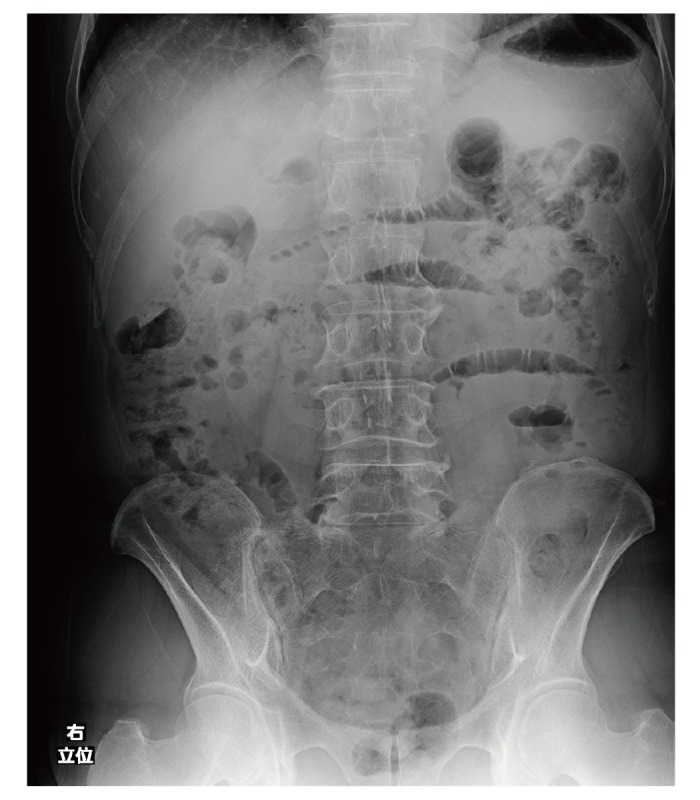
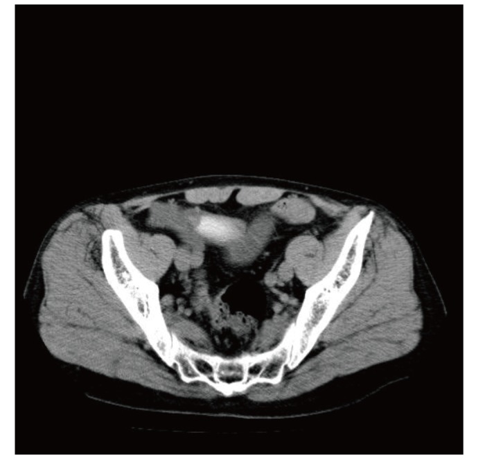
An abdominal echo showed the “keyboard” sign, and the “to-and-fro” sign, but no ascites. On abdominal X-ray, niveau formation was clearly defined (Fig. 1). On CT, there were multiple high-density objects present in his stomach, and one in his small intestine (Fig. 2).
Fig. 1.

Abdominal X-ray. On the day of the first visit, X ray showed multiple niveau formations, and no free air.
Fig. 2.

Abdominal CT of the small intestines showing mochi. On the day of the first visit, abdominal CT showed mochi, which has a high density. At this time, there was no ascites. CT, computed tomography.
Although hospitalization was strongly recommended, the patient was determined to have conservative treatment at home and returned to his residence. He returned to the hospital the following day and was admitted. We initially provided conservative treatment, but the patient experienced severe pain for 3 more days, and gradually developed rebound tenderness and an increase in ascites. On the day of the surgery, blood testing results were: BUN 30.3 mg/dL; Cr 0.92; AMY 21 IU/L; CK 47 IU/L; Na 139 mEq/L; K 4.6 mEq/L; glucose 160 mg/dL; CRP 9.03 mg/dL; eGFR 63.9 mL/min/1.73 m2; WBC 17 300/µL; neutrophils 90.9%; RBC 5 470 000/µL; Hb 16.7%; HCT 48.3%; and PLT 188 000/µL.
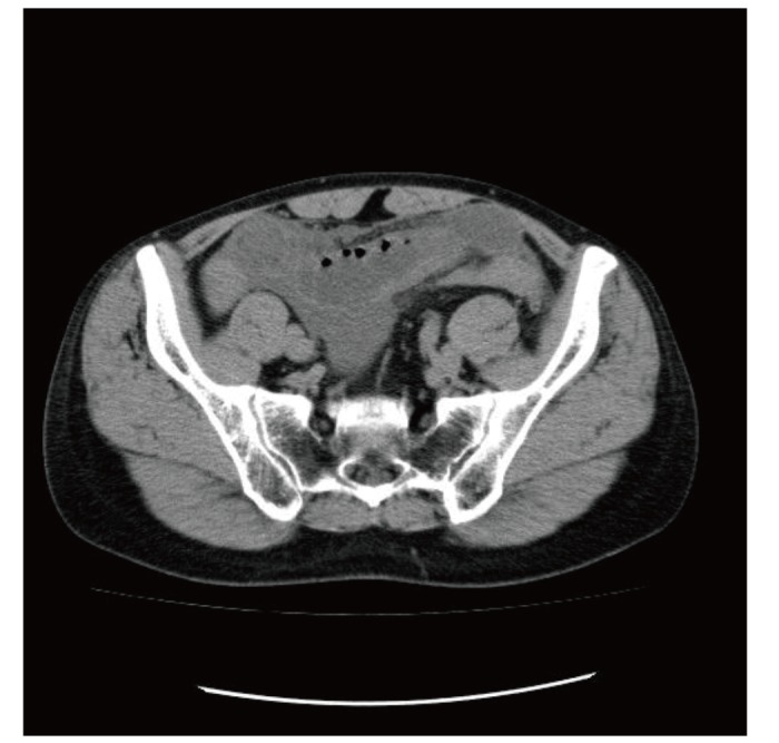
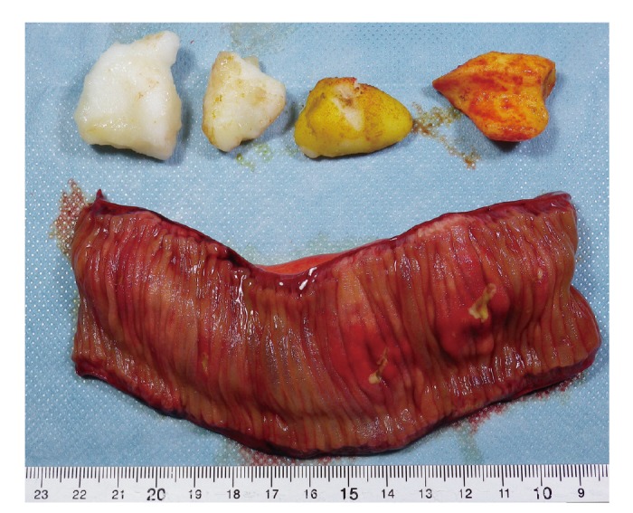
Because of the severe tenderness, signs of peritoneal irritation, and the increase in ascites on CT, we suspected strangulated obstruction (Fig. 3). Emergency surgery was performed the next day, with the removal of a foreign body that involved a small intestinal incision (100 cm from the ligament of Treitz), middle anterior wall incision of the stomach, and small intestine resection (210 cm from the ligament of Treitz). Intestinal resection was performed at the site of the mochi obstruction and macroscopic ulcer due to the risk of necrosis. Several mochi (maximum size: 33 mm) were found in the intestines and stomach, causing an ulcer in the small intestines (Fig. 4). Pathological findings revealed signs of ulcerative change, but no signs of adhesion and necrosis.
Fig. 3.

Abdominal CT clearly showing ascites. Three days after the first visit, abdominal CT showed ascites around intestine. CT, computed tomography.
Fig. 4.

Bezoar from a rice cake. Surgical removal of a section of intestine located 210 cm from the ligament of Treitz revealed a cluster of four mochi. All of the mochi were hard, and the maximum size was 33 mm.
The postoperative course was uneventful. The patient was started on oral food intake from postoperative day 3, and treatment concluded without any problems.
DISCUSSION
We searched MEDLINE and ICHUSHI (Japanese medical bibliography) using the terms “rice cake, obstruction” and “mochi, obstruction.” The MEDLINE search retrieved seven papers in total. The ICHUSHI search (not including reports of proceedings) retrieved 83 papers in total (not including reports of proceedings). Literature related to intestinal obstruction due to rice cake consumption was extracted, and references were examined for other relevant papers. Obstruction due to mochi was reported for the first time in 1968.4 All reports were from Japan and up until 28 May 2017 there were 64 confirmed cases in total.1–18
Regarding the foods causing bowel obstruction, the prevalence of dietary intestinal obstruction is 0.3–5.9%5, 19 for all forms of intestinal obstruction in Japan. Differences in dietary cultures appear to greatly affect the cause of dietary intestinal obstructions. In Japan, the most common intestinal bezoar was from the consumption of persimmon up until the 1960s,4 while konjac, kelp, and wakame (sea mustard) were the most common causes up until the 1990s.20 Konjac (“konnyaku” in Japanese) is a traditional gelatinous food in eastern Asian countries made from the Amorphophallus plant. It contains the plant polysaccharide inulin, which cannot be digested by the human body and is reported to be a cause of intestinal obstruction.21 Recently, many bowel obstructions due to mochi have been reported,1 and similar foods are found in East Asia. Reports of mochi obstruction are increasing, and the ingestion of mochi-type foods may increase globally in the near future with the increase of Japanese food exports.22 However, although bowel obstructions due to konjac and seaweed are not uncommon in Japan, only a few cases have been reported. The proportion of mochi obstructions in all intestinal obstructions due to bezoars in Japan is still unknown.
Mochi is a popular traditional Japanese food made by kneading glutinous rice, and is mainly eaten during New Year celebrations. Therefore, most reports of mochi obstructions occur around January. Oka et al. noted that 59.4% of reported cases of mochi obstructions (including reports of proceedings) occurred in January.3 In the current review of the literature, there were 17 cases reported to have occurred in January, two in February, one in March, one in April, two in May, one in October, two in November and two in December. Among the cases in which the month of occurrence was clear, 60.7% (17/28) occurred in January. While in previous reports intestinal obstructions due to mochi were most common in January around New Year celebrations,1 the current case occurred in May and may have resulted from eating mochi made in winter and stored.
In the current review of the literature, the average age of intestinal obstruction by mochi was 62.7 years, which is much younger than that of suffocation due to mochi. The proportion of males was 57.1%.
Patients with a bezoar intestinal obstruction in Japan characteristically have a history of laparotomy, particularly Billroth II gastrectomy.2, 7, 15, 20, 21 In the current review of the literature, 37.0% (20/54) of cases had a history of abdominal surgery.
Factors that can hinder chewing, such as dentures, tooth defects, or swallowing may also cause mochi obstructions.3, 19 Oka et al. cite two main causes for mochi obstructions: i) problems associated with chewing (tooth defects, dental prosthesis, eating too quickly and swallowing with insufficient chewing); and ii) difficulties associated with chewing and digesting the food itself.3 In the current case, the patient had difficulties chewing as his lower back teeth were absent.
It is generally thought that foods high in fibrous content and substances that cannot be digested can result in gastrointestinal bezoars, causing a bowel obstruction.23 Rice cakes are a readily digestible food, with the main component being starch; however, the preparatory process makes rice cakes difficult to digest physically and chemically. It is observed that mochi becomes hard and sticky when cold, and is not easily dissolved in hot water. This is because the main component of rice cakes is amylopectin, which is insoluble in hot water and the kneading process makes it indigestible.3
Sampei et al. reported that elderly people take more time to swallow mochi than younger healthy adults. This increased swallow time creates more pharyngeal residue with waxy rice mochi compared with waxy wheat mochi22; however, the impact on intestinal obstruction is not clear.
From our search of the literature, the average size of reported bezoars was 32.1 mm (minimum: 18 mm, maximum: 70 mm). In the current case, the maximum size was 33 mm. In most reported cases, the bezoar size was greater than 20 mm; however, it is reported that mochi can expand 5.4 times in the intestinal tract.2
In terms of symptoms and characteristics of intestinal obstruction due to mochi, patients in 71.8% (23/32) of cases extracted from the literature presented with symptoms within 1 day after eating mochi. In such situations, taking a medical history can be easier because little time has passed since mochi ingestion. There is one report in which an intestinal obstruction was thought to be the result of ingestion of mochi 3 months earlier.7 Symptoms of dietary obstruction include severe abdominal pain, with findings reminiscent of strangulated obstruction. Miura et al. reported that although tenderness is present in all cases, guarding was only present in four of 14 cases.6 In the current case, the patient presented with a soft abdomen.
In most of the reviewed cases, CT was used for diagnosis. Only 12.9% (8/62) of reviewed cases were diagnosed without the use of CT, and were diagnosed from the medical history, abdominal echo, and X-ray. Rice cakes show a bright image on CT.3 In the reviewed literature, the average CT number was 149 HU (120–188 HU).
Dietary intestinal obstruction is commonly relieved with conservative treatment, especially those cases associated with the consumption of mochi.6 However, when there are relatively severe symptoms, dietary intestinal obstruction can be difficult to differentiate from strangulated obstruction. Cases of perforation of the small intestines have been reported.3, 7 Therefore, it is suggested that treatment options be considered carefully and cautiously. Although they are very rare, mochi obstructions also cause multiple gastric ulcers16 and perforation of the colon diverticulum,18 and there is a reported case in which a pyloric obstruction was relieved by shredding the mochi into 10-mm pieces using an upper tract endoscope.16
In the current case, although conservative treatment was initiated, the symptoms exacerbated and the patient developed signs of peritoneal irritation with increasing ascites. Clinicians must determine when conservative treatment is no longer effective and when a different course of action is required.
In the current review of the literature, surgical treatment was performed in 45.3% (29/64) of cases. In two cases, laparoscopic surgery was performed.4, 11 The most common site of obstruction was within 100 cm from the end of the ileum in previous reports. This may be because the terminal ileum has low mobility and weak intestinal peristalsis, and food passage is halted by the ileocecal valve.13 In the current case, the site of obstruction was 53.1 cm from the terminal ileum.
In conclusion, considering the risk for intestinal obstruction caused by mochi, especially in situations when a person has a tooth defect (impacting chewing), it is important that mochi be cut into small pieces before eating. It is suggested that conservative treatment may be used initially for a dietary obstruction in certain situations following a careful detailed history and observation, but if pain is severe and difficult to control, CT imaging can help with a diagnosis and surgical intervention should be considered.
The authors declare no conflict of interest.
REFERENCES
- 1. Kunishige T, Ikeda N, Ueno M, Kanamura K, Enomoto K. Rice cake-induced small bowel obstruction treated by laparoscopic-assisted surgery: a case report. JJCS. 2014;39:1127-31. DOI: 10.4030/jjcs.39.1127 Japanese with English Abstract. [DOI] [Google Scholar]
- 2. Yamashita I, Takemori S, Yamashita Y, Tsukada K, Onishi Y, Horikawa N, et al. A case of simple ileus due to mochi that was difficult to distinguish from a strangulation ileus. J Abdom Emerg Med. 1994;14:927-30. DOI: 10.11231/jaem1993.14.927 Japanese with English Abstract. [DOI] [Google Scholar]
- 3. Oka A, Amano Y, Uchida Y, Kagawa K, Takatori K, Kitajima N, et al. Small bowel obstruction and gastric ulceration resulting from rice cake ingestion –computed tomography diagnosis in eight patients-. Nihon Shokakibyo Gakkai Zasshi. 2013;110:1804-13. Japanese with English Abstract. [PubMed] [Google Scholar]
- 4. Koganezawa S. Japanese situation of dietary obstruction. J Jpn Prac Surg Soc. 1968;29:61-70. DOI: 10.3919/ringe1963.29.61 Japanese with English Abstract. [DOI] [Google Scholar]
- 5. Ootaki Y, Matsuda S, Kurisu S, Oyabu H, Hatta K, Koyama T, et al. Ten cases of intestinal obstruction due to foods. J Jpn Prac Surg Soc. 1997;58:606-11. DOI: 10.3919/ringe1963.58.606 Japanese with English Abstract. [DOI] [Google Scholar]
- 6. Miura T, Kimura N, Nakamura J, Yamada S, Miura T, Yanagi M, et al. Rice cake ileus--a rare and ethnic but important disease status in east-southern Asia. Intern Med. 2011;50:2737-9. [DOI] [PubMed] [Google Scholar]
- 7. Yamamoto H, Nara S, Hida K, Yamamoto E, Konishi H, Takeda A, et al. A case of alimentary ileus-like condition due to ‘mochi’ persisted for 3 month with resultant perforation of the small intestine causing generalized peritonitis. J Jpn Surg Assoc. 2003;64:370-4. DOI: 10.3919/jjsa.64.370 Japanese with English Abstract. [DOI] [Google Scholar]
- 8. Yamasaki Y, Yamaoka H, Nishikawa H, Takata M, Nakajima K. Two cases of dietetic ileus caused by rice cakes. J Jpn Surg Assoc. 2004;65:2362-7. DOI: 10.3919/jjsa.65.2362 Japanese with English Abstract. [DOI] [Google Scholar]
- 9. Futamura N, Matsutomo M, Yasumura M, Tateyama K, Tarao M, Sakamoto K, et al. Two Cases of Food-induced Ileus Caused by Rice Cakes. J Abdom Emerg Med. 2004;24:73-7. DOI: 10.11231/jaem1993.24.73 Japanese with English Abstract. [DOI] [Google Scholar]
- 10. Tsunashima H, Kajiyama Y, Kobayashi T, Kikuchi K. Rice cake ileus in diabetic patient. Progress of Digestive Endoscopy. 2013;82:176-7. DOI: 10.11641/pde.82.1_176 Japanese with English Abstract. [DOI] [Google Scholar]
- 11. Nakagawa K, Tukidate H, Fukamachi S. Analysis of food: induced small bowel obstruction. Surgical Therapy. 2011;105:587-90. Japanese with English Abstract. [Google Scholar]
- 12. Tokura M, Moon S, Uesaka K, Seima Y, Sugimoto T. A case of food-induced ileus due to “mochi” rice cakes treat by conservative therapy. J Jpn Surg Assoc. 2009;70:2726-31. DOI: 10.3919/jjsa.70.2726 Japanese with English Abstract. [DOI] [Google Scholar]
- 13. Yamamoto Y, Hiraguchi M, Maeda C, Mizukami Y, Horigome N, Kaneko G, et al. A case report of rice cake treated with conservative treatment. J Jpn Surg Assoc. 2016;77:70-3. DOI: 10.3919/jjsa.77.70 Japanese with English Abstract. [DOI] [Google Scholar]
- 14. Kobayashi S, Matsuyama H, Yoshida M, Hamano M, Masuda H. two cases of dietetic ileus. J Jpn Surg Assoc. 2005;66:393-7. DOI: 10.3919/jjsa.66.393 Japanese with English Abstract. [DOI] [Google Scholar]
- 15. Maeda T, Ikeda Y, Kinoshita K, Ide K. A case ileus in which intestinal foreign body was confirmed by preoperative CT scanning. J Jpn Pract Surg Soc. 1993;54:2116-9. DOI: 10.3919/ringe1963.54.2116 Japanese with English Abstract. [DOI] [Google Scholar]
- 16. Fujii M, Sakashita M, Wakamura K, Horimatsu T, Tanaka S, Obata D, et al. Multiple gastric ulcers caused by a rice cake as an intragastric foreign body. Nihon Shokakibyo Gakkai Zasshi. 2006. March;41:282-3. [DOI] [PubMed] [Google Scholar]
- 17. Baba A, Okuyama Y, Kimura K, Mogami T. Small bowel obstruction due to rice cake (mochi). Intern Med. 2016;55:1933-4. [DOI] [PubMed] [Google Scholar]
- 18. Ichikawa T, Katayama N, Ikeda M, Tsukune Y, Sakai S. Two cases of alimentary tract perforation owing to ingested foreign bodies: diagnosis by CT. Nippon Acta Radiologica. 2001;61:175-6. Japanese with English Abstract. [PubMed] [Google Scholar]
- 19. Matsuzaki H, Akagi D, Takenoue T, Shinkai H, Kobayashi I. A case of preoperatively diagnosed food ileus. J Jpn Surg Assoc. 2011;72:2050-5. DOI: 10.3919/jjsa.72.2050 Japanese with English Abstract. [DOI] [Google Scholar]
- 20. Takami M, Kimura S, Takeushi N, Fujimoto T, Shimizu H, Ota T, et al. Intestinal obstruction due to food boli and gallstones. Progress in Acute Abdominal Medicine. 1987;7:855-60. DOI: 10.11231/jaem1984.7.855 Japanese with English Abstract. [DOI] [Google Scholar]
- 21. Otsuji E, Kikuoka N, Tsujimoto H, Kuwata H, Nakamura T, Suga K, et al. A case of bowel obstruction by “Konnyaku” in the postgastrectomy patient. J Jpn Prac Surg Soc. 1993;54:991-4. DOI: 10.3919/ringe1963.54.991 Japanese with English Abstract. [DOI] [Google Scholar]
- 22. Sanpei R, Tohara H, Fujita S, Yanagimachi M, Abe K, Nakayama E, et al. Video-endoscopic comparison of swallowing waxy rice mochi and waxy wheat mochi: improvement of a traditional Japanese food that presents a choking hazard. Biosci Biotechnol Biochem. 2014;78:472-7. [DOI] [PubMed] [Google Scholar]
- 23. Robles R, Parrilla P, Escamilla C, Lujan JA, Torralba JA, Liron R, et al. Gastrointestinal bezoars. Br J Surg. 1994. July;81:1000-1. [DOI] [PubMed] [Google Scholar]
- 24. Sawai K, Ueda Y, Taguchi S. A case of sigmoid colon perforation due to a rice cake. J Jpn Surg Assoc. 2015;76:1985-8. DOI: 10.3919/jjsa.76.1985 Japanese with English Abstract. [DOI] [Google Scholar]


