Abstract
Mutant serine/threonine protein kinase B-Raf (BRAF) protein is expressed in over half of all melanoma tumors. Although BRAF inhibitors (BRAFi) elicit rapid anti-tumor responses in the majority of patients with mutant BRAF melanoma, the tumors inevitably relapse after a short time. We hypothesized that polyamines are essential for tumor survival in mutant BRAF melanomas. These tumors rely on both polyamine biosynthesis and an upregulated polyamine transport system (PTS) to maintain their high intracellular polyamine levels. We evaluated the effect of a novel arylpolyamine (AP) compound that is cytotoxic upon cellular entry via the increased PTS activity of melanoma cells with different BRAF mutational status. Mutant BRAF melanoma cells demonstrated greater PTS activity and increased sensitivity to AP compared to wild type BRAF (BRAFWT) melanoma cells. Treatment with an inhibitor of polyamine biosynthesis, α-difluoromethylornithine (DFMO), further upregulated PTS activity in mutant BRAF cells and increased their sensitivity to AP. Furthermore, viability assays of 3D spheroid cultures of mutant BRAF melanoma cells demonstrated greater resistance to the BRAFi, PLX4720, compared to 2D monolayer cultures. However, co-treatment with AP restored the sensitivity of melanoma spheroids to PLX4720. These data indicate that mutant BRAF melanoma cells are more dependent on the PTS compared to BRAFWT melanoma cells, resulting in greater sensitivity to the PTS-targeted cytotoxic AP compound.
Keywords: polyamines, α-difluoromethylornithine, polyamine transport system, melanoma, mutant BRAF
1. Introduction
Melanoma is a highly aggressive tumor with poor prognosis in the metastatic stage. Multiple oncogenic mutations (including genes encoding serine/threonine protein kinase B-Raf (BRAF), the neuroblastoma RAS homolog (NRAS), and the proto-oncogene receptor tyrosine protein kinase KIT) drive this highly heterogeneous disease, with mutations in the BRAF gene detected in half of all melanoma tumors [1]. The treatment of metastatic melanoma has been revolutionized over the last decade with the discovery of highly prevalent BRAF mutations, which drive constitutive activation of the RAS-RAF-MEK-ERK pathway and promote uncontrolled proliferation [1]. Ninety percent of reported BRAF mutations result in substitution of glutamic acid for valine at amino acid 600 (the V600E mutation) [2,3]. The subsequent rapid development of selective inhibitors of mutant BRAFV600E proteins (vemurafenib and dabrafenib) demonstrated a major advance in the treatment of melanoma patients harboring the BRAFV600E mutation. However, nearly 100% of the patients exhibit disease progression within seven months after treatment with BRAF inhibitors [4,5,6]. Thus, new ways to overcome the acquired resistance to these inhibitors are urgently needed to increase survival in melanoma patients.
An alternative approach is to target a downstream pathway that is essential for survival of oncogene-addicted tumor cells. While oncogenes indeed drive proliferation, they do so via downstream effector molecules. For example, downstream of extracellular regulated kinase (ERK) signaling is c-MYC (myelocytomatosis viral proto-oncogene homolog), a known regulator of ornithine decarboxylase (ODC) transcription and polyamine biosynthesis [7]. The native polyamines (putrescine, spermidine and spermine) are amino acid-derived polycations that have been implicated in a wide array of biological processes, including cellular proliferation, differentiation, chromatin remodeling, hypusination of the eukaryotic initiation factor-5A (eIF-5A) and apoptosis [8]. Multiple oncogene-encoded proteins, including c-MYC and RAS, are known to upregulate key polyamine biosynthetic enzymes [7,9,10] as well as the cellular uptake of polyamines by activating the polyamine transport system (PTS) [11,12,13,14]. Compared to normal cells, tumor cells have been shown to contain elevated levels of polyamines [15,16,17,18]. These intracellular polyamine levels are maintained via tightly-regulated biosynthetic, catabolic, and uptake and export pathways [19]. Polyamine uptake is upregulated in many tumor types, especially in melanoma tumor cells when compared to normal cells [11,20]. Thus, melanoma tumor cells notoriously replete with multiple oncogenic mutations have a greatly increased need for polyamines compared to normal cells to meet their increased metabolic needs [20].
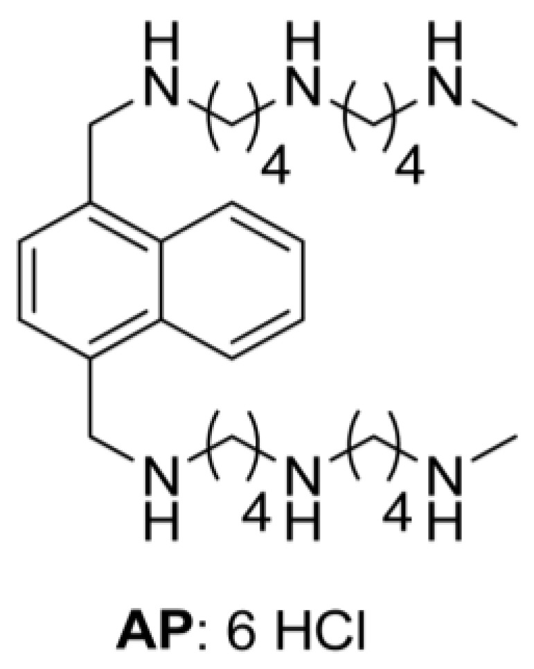
Our objective was to exploit the oncogene-induced polyamine transport activity in melanoma cells by selectively targeting the PTS with a novel arylmethyl-polyamine (AP) compound (Figure 1, [21]). The two-armed design of AP predicated upon a naphthyl core provides PTS hyperselectivity and high potency [21]. Key to our drug design is that both exogenous polyamines and polyamine-based drugs are imported into tumors via a specific uptake system [8,21,22]. Here, we show that polyamine uptake is increased in mutant BRAFV600E melanoma cells, and that AP treatment significantly increases cell death in BRAFV600E melanoma cells compared to BRAFWT melanoma cells. Furthermore, we show that BRAF inhibitor-resistance in melanoma tumor spheroid cultures can be overcome by treatment with AP. These studies provide valuable insights into developing more effective treatment strategies to restore sensitivity of melanoma tumor cells to BRAF inhibitors. In short, the mutant BRAF-driven polyamine addiction can be targeted by cytotoxic polyamine compounds, which selectively target melanoma cells with high polyamine import activity.
Figure 1.

Structure of the arylpolyamine (AP).
2. Materials and Methods
2.1. Cell Lines and Reagents
All human melanoma cell lines including WM983B, WM3734, WM3743, WM989, WM88, WM3451, WM3211, and 1205Lu were obtained as kind gifts from Dr. Meenhard Herlyn (The Wistar Institute, Philadelphia, PA, USA). These cells were maintained in MCDB153 (Sigma-Aldrich, St. Louis, MO, USA) and Leibovitz’s L-15 (Mediatech Inc, Manassas, VA, USA) medium (4:1 ratio) supplemented with 2% fetal calf serum and 2 mmol/L CaCl2. B16F10 cells were obtained from the American Type Culture Collection (Manassas, VA, USA) and maintained in Dulbecco’s Modified Eagle Medium (DMEM) (Invitrogen, Waltham, MA, USA) supplemented with 10% fetal bovine serum and 100 U/mL Penicillin/Streptomycin. The YUMM1.7 cell line (kindly provided by Marcus Bosenburg, Yale University, New Haven, CT, USA) that harbors a BRAFV600E mutation and inactivation of the Phosphatase and tensin homolog (PTEN) gene was maintained in DMEM/F12 (Invitrogen, Waltham, MA, USA) medium supplemented with 10% fetal bovine serum and 100 U/mL Penicillin/Streptomycin.
PLX4720, (S1152; Selleckchem, Houston, TX, USA) a derivative related to PLX4032/Vemurafenib (Plexxikon, Berkeley, CA, USA) was prepared as a 50 mM stock solution in dimethyl sulfoxide and stored at −20 °C. The synthesis of the AP compound has been described previously [21]. The compound was dissolved in phosphate buffered saline (PBS) to provide an initial stock (10 mM), which was filtered through a 0.2 um filter to ensure sterility. Subsequent dilutions were made in PBS to generate the desired stock solutions.
2.2. 3D Spheroid Culture
The nanoscale scaffolding NanoCulture plates (NCP) were purchased from (Organogenix Inc, Woburn, MA, USA). The base of each NCP is constructed with a transparent cyclo-olefin resinous sheet with a nanoscale indented pattern. To form spheroids, 1205Lu human melanoma cells were seeded in a 96-well NCP at 1 × 10⁴ cells/well in MCDB153 (Sigma-Aldrich, St. Louis, MO, USA) and Leibovitz’s L-15 (Mediatech Inc, Manassas, VA, USA) medium (4:1 ratio) supplemented with 2% heat-inactivated fetal calf serum and 2 mM CaCl2 and incubated in a conventional cell incubator at 37 °C in an atmosphere of 5% CO2 and normal O2 levels. When visible spheroids began to form on day 3 after the cells were seeded on the NCPs, treatment with PLX4720 and/or AP was initiated. After drug treatment for 48 h, the spheroid cultures were assayed for cell viability.
2.3. Cell Viability Assay
Cell proliferation assays were conducted in 96-well plates at 25–30% starting confluence to determine the effect of exposure to increasing concentrations of PLX4720 or AP with or without 1 mM α-difluoromethylornithine (DFMO) for 72 h. Cell viability was assessed using the EZQuant Cell Quantifying Kit (Alstem, Richmond, CA, USA) in which the tetrazolium salt WST-8 is reduced by the metabolic activity of live cells to formazan dye. For spheroids treated with PLX4720 and/or AP, viability of the spheroid cells was estimated by quantification of the adenosine triphosphate present using a CellTiter-Glo Luminescent Cell Viability Assay (Promega Co., Madison, WI, USA). The 72 h half maximal inhibitory concentration (IC50) values for AP were calculated using nonlinear regression (sigmoidal dose response) of the plot of percentage inhibition versus the log of inhibitor concentration in GraphPad Prism (v5; GraphPad Software, Inc., La Jolla, CA, USA). The IC50 value is defined as the concentration of the compound required to inhibit 50% cell viability compared to an untreated control.
2.4. Radiolabeled Spermidine Transport Assays
Polyamine transport in tumor cells was evaluated essentially as described previously [23,24]. Radioactive spermidine (Net-522, Spermidine Trihydrochloride, [Terminal Methylenes-3H(N)], specific activity 16.6 Ci/mmol; Perkin Elmer, Boston, MA, USA) was used. Cells were plated in 96 well plates and grown to approximately 80% confluence. Half of the cells were treated with 1 mM DFMO for 40 h. After repeated washing with PBS, 3H-spermidine was added at 0.5 µM and incubated for 60 min at 37 °C. Cells were then washed with cold PBS containing 50 µM spermidine and lysed in 0.1% sodium dodecyl sulfate solution at 37 °C for 30 min with mixing. Cell lysates were then aliquoted for scintillation counting and for protein assay using a microplate Bio-Rad protein assay (Bio-Rad, Hercules, CA, USA). Results were expressed as counts per minute (CPM)/µg protein.
2.5. Statistical Analysis
All in vitro experiments were performed at least in triplicate, and data were compiled from two to three separate experiments. Analyses were done using a one-way analysis of variance with a Tukey test for statistical significance or a Students t-test. In all cases, values of p ≤ 0.05 were regarded as being statistically significant.
3. Results
3.1. Human Mutant BRAFV600E Melanoma Cells Are More Sensitive to Cytotoxic Effects of AP Than BRAFWT Melanoma Cells
A panel of human melanoma cell lines with different BRAF mutational status was screened for their sensitivity to the BRAF inhibitor PLX4720. We confirmed previous findings that mutant BRAFV600E melanoma cells, including WM983B, WM3734, 1205Lu, WM989, and WM88, demonstrated marked sensitivity to PLX4720 (IC50 values ≤ 3.0 µM), whereas BRAFWT melanoma cells, including WM3451, WM3743, and WM3211, demonstrated relative resistance to treatment with PLX4720 (IC50 > 3.0 µM) [25]. This approach allowed us to rank the relative sensitivity of each cell line to the BRAF inhibitor, PLX4720. Thus, the sensitivity of these BRAFV600E melanoma cells to BRAF inhibition with PLX4720 reflected their functional dependence on mutant BRAF signaling to sustain their proliferation and viability.
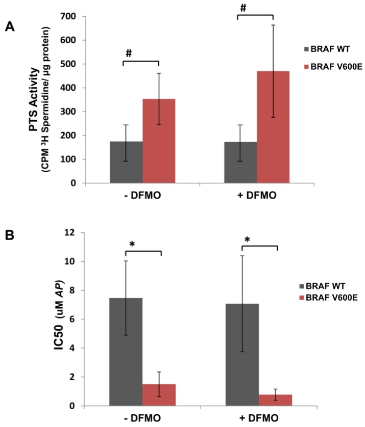
Likewise, we tested whether mutant BRAFV600E cells were more sensitive to increasing concentrations of the cytotoxic polyamine transport ligand, AP (Figure 1), compared to BRAFWT melanoma cells. Table 1 shows that mutant BRAFV600E melanoma cells demonstrated greater sensitivity to AP (IC50 < 2.5 µM) than BRAFWT melanoma cells (IC50 > 4.0 µM). This observation was reflected by the greater polyamine transport activity in BRAFV600E melanoma cells compared to BRAFWT melanoma cells (Figure 2A and Table 1). Since AP accumulated at a faster rate in BRAFV600E human melanoma cells with higher polyamine transport rates compared to BRAFWT human melanoma cells (Figure 2A), BRAFV600E melanoma cells were significantly (p < 0.01) more sensitive to AP exposure than BRAFWT melanoma cells (Figure 2B). In summary, cell lines with high polyamine import activity were more sensitive to the cytotoxic polyamine compound.
Table 1.
Polyamine transport activity and sensitivity to AP in human melanoma cells with different BRAF mutational status cultured ± DFMO a.
| − DFMO | + DFMO | ||||
|---|---|---|---|---|---|
| Cell Line | BRAF Mutational Status |
IC50 (µM AP) |
PTS Activity b
(cpm 3H Spd/µg Protein) |
IC50 (µM AP) |
PTS Activity b
(cpm 3H Spd/µg Protein) |
| WM983B | V600E | 2.6 | 487 ± 64 | 0.9 | 706 ± 78 * |
| WM3734 | V600E | 2.2 | 444 ± 46 | 1.2 | 654 ± 59 * |
| 1205Lu | V600E | 0.7 | 230 ± 22 | 0.6 | 299 ± 54 |
| WM989 | V600E | 1.2 | 304 ± 28 | 1.0 | 348 ± 48 |
| WM88 | V600E | 0.8 | 302 ± 18 | 0.2 | 343 ± 59 |
| WM3451 | WT | 9.0 | 130 ± 21 | 5.1 | 157 ± 34 |
| WM3743 | WT | 8.9 | 117 ± 23 | 10.9 | 110 ± 25 |
| WM3211 | WT | 4.5 | 278 ± 58 | 5.2 | 251 ± 46 |
a The mean values for half maximal inhibitory concentration (IC50) for AP and polyamine transport system (PTS) activity for human melanoma cells expressing mutant BRAFV600E are compared with that for melanoma cells expressing wild type (WT) BRAF under conditions where cells were cultured without added DFMO or with 1 mM α-difluoromethylornithine (DFMO). b PTS activity expressed as counts per minute (CPM) 3H Spermidine (Spd)/µg protein ± standard deviation. PTS assays and cell viability assays were performed at least three times with each cell line. * p ≤ 0.001 when compared to the PTS activity in the absence of DFMO.
Figure 2.
Greater PTS activity and increased sensitivity to AP in mutant BRAFV600E human melanoma cells compared to wild type (WT) BRAFWT cells. (A) BRAFV600E human melanoma cells (WM983B, WM3734, 1205Lu, WM989, and WM88) and BRAFWT human melanoma cells (WM3451, WM3743, and WM3211) were cultured with and without 1 mM DFMO for 40 h and then pulsed with 0.5 µM 3H-spermidine for 60 min at 37 °C. Cell lysates were assayed for CPM 3H-spermidine per mg protein by scintillation counting. The mean PTS activity ± SD for BRAFWT melanoma cells is compared with that of BRAFV600E melanoma cells under conditions where cells were cultured without added DFMO or with 1 mM DFMO. (B) BRAFV600E human melanoma cells (WM983B, WM3734, 1205Lu, WM989, and WM88) and BRAFWT human melanoma cells (WM3451, WM3743, and WM3211) were treated with increasing doses of AP with or without 1 mM DFMO, using 5–6 samples per dose of AP. After 72 h of culture, cell survival was determined via EZQuant Cell Quantifying assay (Alstem, Richmond, CA, USA). AP IC50 values were calculated by GraphPad Prism 6. The mean AP IC50 values ± SD for BRAFWT melanoma cells is compared with that of BRAFV600E melanoma cells under conditions where cells were cultured without added DFMO or with 1 mM DFMO; # p ≤ 0.05; * p < 0.01.
It is well known that lowering intracellular levels of polyamines with inhibitors of polyamine biosynthesis can increase uptake of extracellular polyamines as well as exogenous polyamine analogues [20,26]. WM983B and WM3743 melanoma cells were pretreated for 40 h with 1 mM DFMO, an inhibitor of ODC, the first and rate-limiting enzyme in polyamine biosynthesis, before measuring their polyamine transport activity. DFMO treatment dramatically increased polyamine transport activity in BRAFV600E WM983B melanoma cells, but not in the BRAFWT WM3743 melanoma cells (Supplementary Materials Figure S1A). In general, polyamine depletion with DFMO treatment enhanced polyamine uptake more in the screened human BRAFV600E melanoma cells compared to that seen in BRAFWT melanoma cells (Table 1, Figure 2A). Since DFMO treatment increases polyamine transport activity, we tested whether co-treatment with AP and DFMO will increase the sensitivity of melanoma cells to AP. For instance, BRAFV600E WM983B melanoma cells are significantly (p ≤ 0.0001) more sensitive to AP treatment when co-treated with DFMO (IC50 = 0.7 µM) compared to that with AP alone (IC50 = 2.3 µM) (Supplementary Materials Figure S1C). In contrast, sensitivity to AP was not increased in DFMO-co-treated BRAFWT melanoma cells (Table 1, Figure 2B). These data indicate that human BRAFV600E melanoma cells demonstrate greater polyamine transport activity and increased sensitivity to AP compared to BRAFWT melanoma cells, and their sensitivity can be increased by inhibition of polyamine biosynthesis with DFMO.
3.2. AP Is More Cytotoxic to BRAFV600E Murine Melanoma Cells Than BRAFWT Melanoma Cells
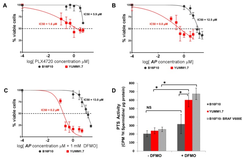
Since human melanoma cells possess multiple oncogenic mutations in addition to BRAFV600E, we compared AP cytotoxicity and PTS activity in the murine B16F10 melanoma cell line that is BRAFWT with BRAFV600E YUMM1.7 cell line that was derived from a melanoma tumor that spontaneously developed in a BRAFV600E/PTENnull transgenic mouse [27]. As expected, YUMM1.7 cells were very sensitive to PLX4720 with a lower IC50 compared to B16F10 cells (Figure 3A). In addition, BRAFV600E YUMM1.7 cells were significantly (p < 0.0001) more sensitive to AP and had a much lower IC50 value for AP compared to BRAFWT B16F10 cells (Figure 3B). In particular, DFMO co-treatment increased the sensitivity of YUMM1.7 cells to AP (IC50 = 0.8 µM AP without DFMO and IC50 = 0.2 µM AP with DFMO co-treatment), and this correlated with a marked DFMO-induction of PTS activity in BRAFV600E YUMM1.7 cells (Figure 3D). In contrast, BRAFWT B16F10 cells demonstrated no significant induction in polyamine uptake following DFMO treatment (Figure 3D). However, B16F10 cells retrovirally infected to express the mutant BRAFV600E protein exhibited a similar PTS activity profile as that seen with YUMM1.7 cells. DFMO treatment was shown to enhance polyamine uptake in the B16F10-BRAFV600E cells as was seen with YUMM1.7 cells (Figure 3D). AP was also more cytotoxic in B16F10-BRAFV600E cells (IC50 = 24.4 µM) compared to control-infected B16F10-pBABE cells that were infected with retrovirus expressing the empty plasmid (IC50 = 36.4 µM). These data suggest that melanoma cells with a mutant BRAFV600E protein are more dependent on the polyamine uptake system compared to cells with a BRAFWT protein, resulting in greater sensitivity to the PTS-targeted cytotoxic AP compound that enters and kills melanoma cells via the polyamine transport system.
Figure 3.
BRAFV600E murine melanoma cells are more sensitive to AP than BRAFWT melanoma cells. (A) Murine BRAFV600E YUMM1.7 melanoma cells and BRAFWT B16F10 melanoma cells were treated with increasing doses of PLX4720. After 72 h of culture, cell survival was determined via EZQuant Cell Quantifying assay. IC50 values were calculated by GraphPad Prism 6; p = 0.0013. (B) Murine BRAFV600E YUMM1.7 melanoma cells and BRAFWT B16F10 melanoma cells were treated with increasing doses of AP. After 72 h of culture, cell survival was determined via EZQuant Cell Quantifying assay. IC50 values were calculated by GraphPad Prism 6; p < 0.0001. (C) Murine BRAFV600E YUMM1.7 melanoma cells and BRAFWT B16F10 melanoma cells were treated with increasing doses of AP ± 1 mM DFMO. After 72 h of culture, cell survival was determined via EZQuant Cell Quantifying assay. IC50 values were calculated by GraphPad Prism 6; p < 0.0001. (D) YUMM1.7 and B16F10 melanoma cells and B16F10 cells retrovirally infected to express the mutant BRAFV600E protein were cultured with and withoutS 1 mM DFMO for 40 h and then pulsed with 0.5 µM 3H-spermidine for 60 min at 37 °C. Cells were washed with cold PBS containing 50 µM spermidine, and cell lysates were assayed for CPM 3H-spermidine per mg protein by scintillation counting; * p < 0.0001; NS: not significant.
3.3. Increased Resistance of Spheroid Melanoma Cells to PLX4720 Is Overcome with AP Co-Treatment
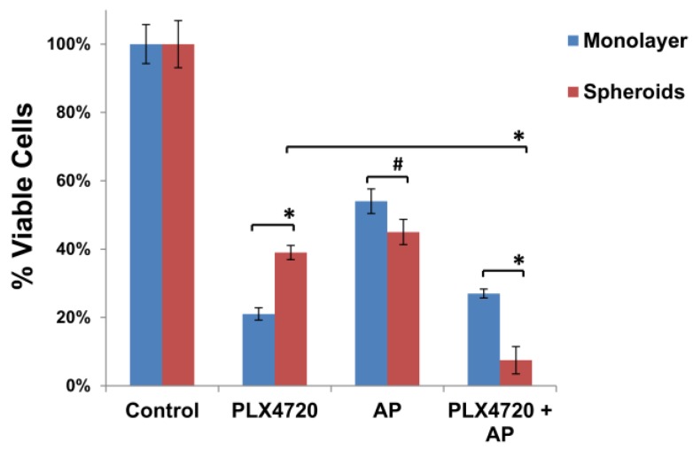
Because growth of cells in a 3D culture system has been found to be more representative of the in vivo microenvironment, we cultured 1205Lu human melanoma cells using a nanoscale, scaffold-based NCP in which tumor cells easily form 3D spheroids [28]. Although these tumor spheroid cultures are grown in ambient air, the spheroid microenvironment closely resembles that in tumors with a hypoxic core and is more relevant for drug sensitivity compared to that seen with monolayer cultures [28,29,30]. Similar to previous reports [31], BRAFV600E mutant 1205Lu melanoma cells grown as spheroids on NCPs were more resistant to 48 h treatment with PLX4720 (25 µM) compared to the same cells grown in 2D monolayer cultures in ambient air (Figure 4). Both spheroid and monolayer cultures were similarly sensitive to 48 h treatment with a high concentration of AP (25 µM) alone. We then tested the effect of AP treatment on the PLX4720-resistant phenotype of the 1205Lu spheroid and monolayer cultures. Co-treatment with both PLX4720 (25 µM) and AP (25 µM) led to a dramatic reduction in cell viability in the spheroid cultures unlike monolayer cultures that showed no further reduction in cell viability when compared to PLX4720 treatment alone (Figure 4). Thus, the increased resistance of the melanoma spheroid cultures to PLX4720 was eliminated with AP co-treatment.
Figure 4.
Increased resistance of spheroid melanoma cells to PLX4720 is overcome with AP co-treatment. BRAFV600E mutant 1205Lu melanoma cells were seeded at 1 × 10⁴ cells in each well of 24-well NanoCulture plates (NCPs). When spheroids were formed on day 3, the 3D cultures of spheroids were treated with PLX4720 (25 µM) and/or AP (25 µM). 2D monolayer cultures of 1205Lu melanoma cells were also treated with PLX4720 (25 µM) and/or AP (25 µM). After drug treatment for 48 h, the viability of spheroids and monolayer cultures was assayed using the CellTiter-Glo Luminescent Cell Viability Assay. The percent cell survival in each treatment group was calculated relative to cells treated with medium only under the same conditions. As controls, the growth of cells without drug treatment under each condition was normalized as 100% separately. The means are presented ± SD; * p < 0.0001; # p = 0.0028.
4. Discussion
Melanoma is challenging to treat due to its genetic heterogeneity, and successful therapy requires targeting multiple molecular vulnerabilities. Our data show that melanoma tumor cells expressing mutant BRAFV600E exhibit a high demand for polyamine growth factors and a greatly upregulated PTS. Utilizing the PTS for drug delivery, the AP compound attacks the melanoma cells via one of its key modes of survival. Indeed, we propose that polyamines are essential for the survival of melanomas. Polyamine levels are dramatically elevated in tumor cells compared to normal cells, often the result of oncogenic induction [11,20]. Previous studies have shown that the c-MYC and RAS can upregulate polyamine biosynthesis [9,10] and increase cellular uptake of polyamines by inducing PTS activity [12,13,14]. Although melanoma cells are notoriously replete with multiple oncogenic mutations, more than half of all melanoma tumors express a mutant BRAF protein [20]. Our data suggest that BRAFV600E melanoma tumors have a greatly increased metabolic need for polyamines compared to normal cells. We have exploited the BRAFV600E-induced PTS activity in metastatic melanoma cells by targeting the PTS with AP.
Putrescine, spermidine, and spermine play key roles in cellular proliferation, signal transduction, gene expression, and autophagic states that contribute to tumor survival [32,33,34,35]. These endogenous polyamines and the polyamine-based AP compete to be imported into tumors via the PTS [21]. However, studies suggest that arylmethyl-polyamines similar to AP have enhanced cytotoxic potency via their multiple electrostatic interactions with DNA [36] and topoisomerase II [37]. Since AP selectively targets tumor cells with high polyamine transport rates, normal cells are significantly less sensitive to AP since they have low PTS activity [21]. Studies have shown that polyamine biosynthesis and cellular uptake are induced in hypoxic regions of tumors and in tumor spheroids [38]. Moreover, depletion of polyamines during hypoxia resulted in increased apoptosis [38], indicating that polyamines play an essential role in the ability of tumor cells to adapt to hypoxic stress and reactive oxygen species. Indeed, polyamines are known to exert anti-oxidant functions [39]. Although the melanoma spheroid cultures in this study were cultured with ambient air, it is well documented that the cells at the center of spheroids are hypoxic, thus modeling the heterogeneous 3D structure of in vivo melanoma tumors that often contain hypoxic regions [28,31]. Solid tumors contain poorly vascularized, hypoxic regions that contribute to tumor progression by activating a hypoxia stress response via hypoxia inducible factor-1α that promotes cell survival, tumor angiogenesis, and metastasis [40,41]. Studies have shown that hypoxic tumor cells and spheroid cultures are more resistant to chemotherapy including BRAF inhibitors [31,42,43]. Likewise, we have found that 3D cultures of 1205Lu melanoma cells grown as spheroids on NCPs are more resistant to PLX4720 treatment compared to 1205Lu cells grown in 2D monolayer culture in ambient air. Knowing that polyamine uptake is induced in hypoxic regions of tumor spheroids, we hypothesized that treatment with the PTS ligand AP would increase the sensitivity of 1205Lu spheroid cells to PLX4720. Indeed, the increased resistance of melanoma spheroids to PLX4720 was overcome with AP co-treatment. In contrast, AP co-treatment had no significant effect on the sensitivity of 2D monolayer cultures of 1205Lu cells to PLX4720.
Accumulating literature shows that treatment with a BRAF inhibitor such as PLX4720 enriches a slow-cycling cancer stem cell-like (CSC) subpopulation of melanoma cells that is characterized by stem cell markers such as Lysine-Specific Demethylase 5B (JARID1B) and spheroid formation [44,45]. It is thought that cancer stem cell populations exist in a hypoxic microenvironment [46,47,48]. Roesch et al. [45] have found that endogenous reactive oxygen species (ROS) levels are increased in slow cycling JARID1Bhigh melanoma cells as a result of increased mitochondrial respiration and oxidative phosphorylation. This high oxygen consumption contributes to hypoxic conditions that have been shown to favor the JARID1Bhigh slow-cycling CSC-like phenotype [44]. Using 1205Lu melanoma cells stably transduced with a JARID1B-promoter-green fluorescent protein (GFP)-reporter construct [44], we found that a short 2-day exposure to PLX4720 led to a 4-fold enrichment of JARID1B-driven GFP expressing 1205Lu melanoma cells grown as spheroids (data not shown). Our data show that PLX4720-resistant melanoma spheroids are made more sensitive to PLX4720 with AP co-treatment, and it is likely that AP is targeting CSC subpopulations that are enriched in the PLX4720-resistant melanoma spheroids.
Polyamines may also contribute to tumor survival by inducing an autophagic state [32,33,34,35]. For instance, spermidine has been shown to induce autophagy in multiple systems including yeast cells, Caenorhabditis elegans, Drosophila melanogaster, and human tumor cells [35,49] and to increase survival of pluripotent stem cells in culture [50]. Autophagy has recently emerged as a common survival process that tumors undergo when assaulted by chemotherapy and radiation [51]. It is induced by cellular stress such as nutrient deprivation, withdrawal of growth factors, and hypoxia [52]. In established tumors, autophagy is also a resistance mechanism to many therapeutic modalities including BRAF inhibitors [53]. Increased polyamine uptake provides a mechanism for BRAFi-resistant melanoma cells to acquire sufficient polyamines to undergo autophagy to survive treatment with BRAF inhibitors.
Previous clinical trials have tested the anti-tumor efficacy of the ODC inhibitor DFMO. However, treatment with DFMO alone demonstrated only moderate success in treating cancer patients [54]. Subsequent studies discovered that DFMO-inhibition of polyamine biosynthesis leads to upregulation of PTS activity with resulting increased uptake of polyamines from the diet and gut flora into the tumor cells [18]. Our findings show that DFMO induces PTS activity and increases AP sensitivity in melanoma cells that harbor a mutated BRAF protein. In summary, treatment with AP, with or without DFMO, offers an exciting potential as adjunct cancer therapy to overcome drug resistance in mutant BRAFV600E melanoma.
Acknowledgments
We thank Meenhard Herlyn (The Wistar Institute, Philadelphia, PA, USA) and Marcus Bosenburg (Yale University, New Haven, CT, USA) for kindly providing melanoma cell lines. This work was supported by the United States Department of Defense grant CA150356 (S.K.G.).
Supplementary Materials
The following are available online at www.mdpi.com/2076-3271/6/1/3/s1, Figure S1: Greater PTS activity and increased sensitivity to AP in BRAFV600E human melanoma cells compared to BRAFWT cells.
Author Contributions
S.K.G. conceived and designed the experiments; M.C.P. and A.M. performed the experiments; S.K.G., M.C.P. and A.M. analyzed the data; O.P. provided materials and edited the paper; and S.K.G. wrote the paper.
Conflicts of Interest
Both the composition of matter and use of the AP with DFMO in the treatment of cancers have been patented by the University of Central Florida (UCF). Commercialization of this intellectual property is being conducted jointly by both UCF and the Lankenau Institute for Medical Research under a collaborative research agreement. The founding sponsors had no role in the design of the study; in the collection, analyses, or interpretation of data; in the writing of the manuscript, and in the decision to publish the results.
References
- 1.Davies H., Bignell G.R., Cox C., Stephens P., Edkins S., Clegg S., Teague J., Woffendin H., Garnett M.J., Bottomley W., et al. Mutations of the BRAF gene in human cancer. Nature. 2002;417:949–954. doi: 10.1038/nature00766. [DOI] [PubMed] [Google Scholar]
- 2.Solit D.B., Rosen N. Resistance to BRAF inhibition in melanomas. N. Engl. J. Med. 2011;364:772–774. doi: 10.1056/NEJMcibr1013704. [DOI] [PubMed] [Google Scholar]
- 3.Haq R., Fisher D.E. Targeting melanoma by small molecules: Challenges ahead. Pigment Cell Melanoma Res. 2013;26:464–469. doi: 10.1111/pcmr.12109. [DOI] [PMC free article] [PubMed] [Google Scholar]
- 4.Flaherty K.T., Puzanov I., Kim K.B., Ribas A., McArthur G.A., Sosman J.A., O’Dwyer P.J., Lee R.J., Grippo J.F., Nolop K., et al. Inhibition of mutated, activated BRAF in metastatic melanoma. N. Engl. J. Med. 2010;363:809–819. doi: 10.1056/NEJMoa1002011. [DOI] [PMC free article] [PubMed] [Google Scholar]
- 5.Sosman J.A., Kim K.B., Schuchter L., Gonzalez R., Pavlick A.C., Weber J.S., McArthur G.A., Hutson T.E., Moschos S.J., Flaherty K.T., et al. Survival in BRAF V600-mutant advanced melanoma treated with vemurafenib. N. Engl. J. Med. 2012;366:707–714. doi: 10.1056/NEJMoa1112302. [DOI] [PMC free article] [PubMed] [Google Scholar]
- 6.Hauschild A., Grob J.J., Demidov L.V., Jouary T., Gutzmer R., Millward M., Rutkowski P., Blank C.U., Miller W.H., Jr., Kaempgen E., et al. Dabrafenib in BRAF-mutated metastatic melanoma: A multicentre, open-label, phase 3 randomised controlled trial. Lancet. 2012;380:358–365. doi: 10.1016/S0140-6736(12)60868-X. [DOI] [PubMed] [Google Scholar]
- 7.Bello-Fernandez C., Packham G., Cleveland J.L. The ornithine decarboxylase gene is a transcriptional target of c-Myc. Proc. Natl. Acad. Sci. USA. 1993;90:7804–7808. doi: 10.1073/pnas.90.16.7804. [DOI] [PMC free article] [PubMed] [Google Scholar]
- 8.Casero R.A., Jr., Marton L.J. Targeting polyamine metabolism and function in cancer and other hyperproliferative diseases. Nat. Rev. Drug Discov. 2007;6:373–390. doi: 10.1038/nrd2243. [DOI] [PubMed] [Google Scholar]
- 9.Forshell T.P., Rimpi S., Nilsson J.A. Chemoprevention of B-cell lymphomas by inhibition of the Myc target spermidine synthase. Cancer Prev. Res. 2010;3:140–147. doi: 10.1158/1940-6207.CAPR-09-0166. [DOI] [PubMed] [Google Scholar]
- 10.Origanti S., Shantz L.M. Ras transformation of RIE-1 cells activates cap-independent translation of ornithine decarboxylase: Regulation by the Raf/MEK/ERK and phosphatidylinositol 3-kinase pathways. Cancer Res. 2007;67:4834–4842. doi: 10.1158/0008-5472.CAN-06-4627. [DOI] [PubMed] [Google Scholar]
- 11.Poulin R., Casero R.A., Soulet D. Recent advances in the molecular biology of metazoan polyamine transport. Amino Acids. 2012;42:711–723. doi: 10.1007/s00726-011-0987-y. [DOI] [PMC free article] [PubMed] [Google Scholar]
- 12.Bachrach U., Seiler N. Formation of acetylpolyamines and putrescine from spermidine by normal and transformed chick embryo fibroblasts. Cancer Res. 1981;41:1205–1208. [PubMed] [Google Scholar]
- 13.Chang B.K., Libby P.R., Bergeron R.J., Porter C.W. Modulation of polyamine biosynthesis and transport by oncogene transfection. Biochem. Biophys. Res. Commun. 1988;157:264–270. doi: 10.1016/S0006-291X(88)80042-1. [DOI] [PubMed] [Google Scholar]
- 14.Roy U.K., Rial N.S., Kachel K.L., Gerner E.W. Activated K-RAS increases polyamine uptake in human colon cancer cells through modulation of caveolar endocytosis. Mol. Carcinog. 2008;47:538–553. doi: 10.1002/mc.20414. [DOI] [PMC free article] [PubMed] [Google Scholar]
- 15.Pegg A.E. Polyamine metabolism and its importance in neoplastic growth as a target for chemotherapy. Cancer Res. 1988;48:759–774. [PubMed] [Google Scholar]
- 16.Tabor C.W., Tabor H. Polyamines. Ann. Rev. Biochem. 1984;53:749–790. doi: 10.1146/annurev.bi.53.070184.003533. [DOI] [PubMed] [Google Scholar]
- 17.Pegg A.E. Recent advances in the biochemistry of polyamines in eukaryotes. Biochem. J. 1986;234:249–262. doi: 10.1042/bj2340249. [DOI] [PMC free article] [PubMed] [Google Scholar]
- 18.Gerner E.W., Meyskens F.L., Jr. Polyamines and cancer: Old molecules, new understanding. Nat. Rev. Cancer. 2004;4:781–792. doi: 10.1038/nrc1454. [DOI] [PubMed] [Google Scholar]
- 19.Wallace H.M., Fraser A.V., Hughes A. A perspective of polyamine metabolism. Biochem. J. 2003;376:1–14. doi: 10.1042/bj20031327. [DOI] [PMC free article] [PubMed] [Google Scholar]
- 20.Seiler N., Delcros J.G., Moulinoux J.P. Polyamine transport in mammalian cells. An update. Int. J. Biochem. Cell Biol. 1996;28:843–861. doi: 10.1016/1357-2725(96)00021-0. [DOI] [PubMed] [Google Scholar]
- 21.Muth A., Kamel J., Kaur N., Shicora A.C., Ayene I.S., Gilmour S.K., Phanstiel O. Development of polyamine transport ligands with improved metabolic stability and selectivity against specific human cancers. J. Med. Chem. 2013;56:5819–5828. doi: 10.1021/jm400496a. [DOI] [PubMed] [Google Scholar]
- 22.Phanstiel O., Kaur N., Delcros J.G. Structure-activity investigations of polyamine-anthracene conjugates and their uptake via the polyamine transporter. Amino Acids. 2007;33:305–313. doi: 10.1007/s00726-007-0527-y. [DOI] [PubMed] [Google Scholar]
- 23.Kramer D.L., Miller J.T., Bergeron R.J., Khomutov R., Khomutov A., Porter C.W. Regulation of polyamine transport by polyamines and polyamine analogs. J. Cell. Physiol. 1993;155:399–407. doi: 10.1002/jcp.1041550222. [DOI] [PubMed] [Google Scholar]
- 24.Nilsson J.A., Keller U.B., Baudino T.A., Yang C., Norton S., Old J.A., Nilsson L.M., Neale G., Kramer D.L., Porter C.W., et al. Targeting ornithine decarboxylase in Myc-induced lymphomagenesis prevents tumor formation. Cancer Cell. 2005;7:433–444. doi: 10.1016/j.ccr.2005.03.036. [DOI] [PubMed] [Google Scholar]
- 25.Schayowitz A., Bertenshaw G., Jeffries E., Schatz T., Cotton J., Villanueva J., Herlyn M., Krepler C., Vultur A., Xu W. Functional profiling of live melanoma samples using a novel automated platform. PLoS ONE. 2012;7:e52760. doi: 10.1371/journal.pone.0052760. [DOI] [PMC free article] [PubMed] [Google Scholar]
- 26.Alhonen-Hongisto L., Seppanen P., Janne J. Intracellular putrescine and spermidine deprivation induces increased uptake of the natural polyamines and methylglyoxal bis(guanylhydrazone) Biochem. J. 1980;192:941–945. doi: 10.1042/bj1920941. [DOI] [PMC free article] [PubMed] [Google Scholar]
- 27.Obenauf A.C., Zou Y., Ji A.L., Vanharanta S., Shu W., Shi H., Kong X., Bosenberg M.C., Wiesner T., Rosen N., et al. Therapy-induced tumour secretomes promote resistance and tumour progression. Nature. 2015;520:368–372. doi: 10.1038/nature14336. [DOI] [PMC free article] [PubMed] [Google Scholar]
- 28.Yoshii Y., Waki A., Yoshida K., Kakezuka A., Kobayashi M., Namiki H., Kuroda Y., Kiyono Y., Yoshii H., Furukawa T. The use of nanoimprinted scaffolds as 3D culture models to facilitate spontaneous tumor cell migration and well-regulated spheroid formation. Biomaterials. 2011;32:6052–6058. doi: 10.1016/j.biomaterials.2011.04.076. [DOI] [PubMed] [Google Scholar]
- 29.Yamada K.M., Cukierman E. Modeling tissue morphogenesis and cancer in 3D. Cell. 2007;130:601–610. doi: 10.1016/j.cell.2007.08.006. [DOI] [PubMed] [Google Scholar]
- 30.Haycock J.W. 3D Cell Culture: Methods and Protocols. Springer; Berlin, Germany: 2011. 3D cell culture: A review of current approaches and techniques; pp. 1–15. [DOI] [PubMed] [Google Scholar]
- 31.Qin Y., Roszik J., Chattopadhyay C., Hashimoto Y., Liu C., Cooper Z.A., Wargo J.A., Hwu P., Ekmekcioglu S., Grimm E.A. Hypoxia-driven mechanism of vemurafenib resistance in melanoma. Mol. Cancer Ther. 2016;15:2442–2454. doi: 10.1158/1535-7163.MCT-15-0963. [DOI] [PMC free article] [PubMed] [Google Scholar]
- 32.Cufi S., Vazquez-Martin A., Oliveras-Ferraros C., Martin-Castillo B., Vellon L., Menendez J.A. Autophagy positively regulates the CD44+ CD24-/low breast cancer stem-like phenotype. Cell Cycle. 2011;10:3871–3885. doi: 10.4161/cc.10.22.17976. [DOI] [PubMed] [Google Scholar]
- 33.Mirzoeva O.K., Hann B., Hom Y.K., Debnath J., Aftab D., Shokat K., Korn W.M. Autophagy suppression promotes apoptotic cell death in response to inhibition of the PI3K—mTOR pathway in pancreatic adenocarcinoma. J. Mol. Med. 2011;89:877–889. doi: 10.1007/s00109-011-0774-y. [DOI] [PubMed] [Google Scholar]
- 34.Morselli E., Galluzzi L., Kepp O., Marino G., Michaud M., Vitale I., Maiuri M.C., Kroemer G. Oncosuppressive functions of autophagy. Antioxid. Redox Signal. 2011;14:2251–2269. doi: 10.1089/ars.2010.3478. [DOI] [PubMed] [Google Scholar]
- 35.Morselli E., Marino G., Bennetzen M.V., Eisenberg T., Megalou E., Schroeder S., Cabrera S., Benit P., Rustin P., Criollo A., et al. Spermidine and resveratrol induce autophagy by distinct pathways converging on the acetylproteome. J. Cell Biol. 2011;192:615–629. doi: 10.1083/jcb.201008167. [DOI] [PMC free article] [PubMed] [Google Scholar]
- 36.Dallavalle S., Giannini G., Alloatti D., Casati A., Marastoni E., Musso L., Merlini L., Morini G., Penco S., Pisano C., et al. Synthesis and cytotoxic activity of polyamine analogues of camptothecin. J. Med. Chem. 2006;49:5177–5186. doi: 10.1021/jm060285b. [DOI] [PubMed] [Google Scholar]
- 37.Wang H., Davis A., Yu S., Ahmed K. Response of cancer cells to molecular interruption of the CK2 signal. Mol. Cell Biochem. 2001;227:167–174. doi: 10.1023/A:1013112908734. [DOI] [PubMed] [Google Scholar]
- 38.Svensson K.J., Welch J.E., Kucharzewska P., Bengtson P., Bjurberg M., Pahlman S., Ten Dam G.B., Persson L., Belting M. Hypoxia-mediated induction of the polyamine system provides opportunities for tumor growth inhibition by combined targeting of vascular endothelial growth factor and ornithine decarboxylase. Cancer Res. 2008;68:9291–9301. doi: 10.1158/0008-5472.CAN-08-2340. [DOI] [PubMed] [Google Scholar]
- 39.Mozdzan M., Szemraj J., Rysz J., Stolarek R.A., Nowak D. Anti-oxidant activity of spermine and spermidine re-evaluated with oxidizing systems involving iron and copper ions. Int. J. Biochem. Cell Biol. 2006;38:69–81. doi: 10.1016/j.biocel.2005.07.004. [DOI] [PubMed] [Google Scholar]
- 40.Pouyssegur J., Dayan F., Mazure N.M. Hypoxia signalling in cancer and approaches to enforce tumour regression. Nature. 2006;441:437–443. doi: 10.1038/nature04871. [DOI] [PubMed] [Google Scholar]
- 41.Keith B., Simon M.C. Hypoxia-inducible factors, stem cells, and cancer. Cell. 2007;129:465–472. doi: 10.1016/j.cell.2007.04.019. [DOI] [PMC free article] [PubMed] [Google Scholar]
- 42.O’Connell M.P., Marchbank K., Webster M.R., Valiga A.A., Kaur A., Vultur A., Li L., Herlyn M., Villanueva J., Liu Q. Hypoxia induces phenotypic plasticity and therapy resistance in melanoma via the tyrosine kinase receptors ROR1 and ROR2. Cancer Discov. 2013;3:1378–1393. doi: 10.1158/2159-8290.CD-13-0005. [DOI] [PMC free article] [PubMed] [Google Scholar]
- 43.Pucciarelli D., Lengger N., Takáčová M., Csaderova L., Bartosova M., Breiteneder H., Pastorekova S., Hafner C. Hypoxia increases the heterogeneity of melanoma cell populations and affects the response to vemurafenib. Mol. Med. Rep. 2016;13:3281–3288. doi: 10.3892/mmr.2016.4888. [DOI] [PubMed] [Google Scholar]
- 44.Roesch A., Fukunaga-Kalabis M., Schmidt E.C., Zabierowski S.E., Brafford P.A., Vultur A., Basu D., Gimotty P., Vogt T., Herlyn M. A temporarily distinct subpopulation of slow-cycling melanoma cells is required for continuous tumor growth. Cell. 2010;141:583–594. doi: 10.1016/j.cell.2010.04.020. [DOI] [PMC free article] [PubMed] [Google Scholar]
- 45.Roesch A., Vultur A., Bogeski I., Wang H., Zimmermann K.M., Speicher D., Korbel C., Laschke M.W., Gimotty P.A., Philipp S.E., et al. Overcoming intrinsic multidrug resistance in melanoma by blocking the mitochondrial respiratory chain of slow-cycling JARID1Bhigh cells. Cancer Cell. 2013;23:811–825. doi: 10.1016/j.ccr.2013.05.003. [DOI] [PMC free article] [PubMed] [Google Scholar]
- 46.Schwab L.P., Peacock D.L., Majumdar D., Ingels J.F., Jensen L.C., Smith K.D., Cushing R.C., Seagroves T.N. Hypoxia inducible factor-1α promotes primary tumor growth and tumor-initiating cell activity in breast cancer. Breast Cancer Res. 2012;14:R6. doi: 10.1186/bcr3087. [DOI] [PMC free article] [PubMed] [Google Scholar]
- 47.Mathieu J., Zhang Z., Zhou W., Wang A.J., Heddleston J.M., Pinna C.M., Hubaud A., Stadler B., Choi M., Bar M. HIF induces human embryonic stem cell markers in cancer cells. Cancer Res. 2011;71:4640–4652. doi: 10.1158/0008-5472.CAN-10-3320. [DOI] [PMC free article] [PubMed] [Google Scholar]
- 48.Mohyeldin A., Garzon-Muvdi T., Quinones-Hinojosa A. Oxygen in stem cell biology: A critical component of the stem cell niche. Cell Stem Cell. 2010;7:150–161. doi: 10.1016/j.stem.2010.07.007. [DOI] [PubMed] [Google Scholar]
- 49.Eisenberg T., Knauer H., Schauer A., Buttner S., Ruckenstuhl C., Carmona-Gutierrez D., Ring J., Schroeder S., Magnes C., Antonacci L., et al. Induction of autophagy by spermidine promotes longevity. Nat. Cell Biol. 2009;11:1305–1314. doi: 10.1038/ncb1975. [DOI] [PubMed] [Google Scholar]
- 50.Chen T., Shen L., Yu J., Wan H., Guo A., Chen J., Long Y., Zhao J., Pei G. Rapamycin and other longevity-promoting compounds enhance the generation of mouse induced pluripotent stem cells. Aging Cell. 2011;10:908–911. doi: 10.1111/j.1474-9726.2011.00722.x. [DOI] [PubMed] [Google Scholar]
- 51.Strohecker A.M., White E. Targeting mitochondrial metabolism by inhibiting autophagy in BRAF-driven cancers. Cancer Discov. 2014;4:766–772. doi: 10.1158/2159-8290.CD-14-0196. [DOI] [PMC free article] [PubMed] [Google Scholar]
- 52.Kroemer G., Marino G., Levine B. Autophagy and the integrated stress response. Mol. Cell. 2010;40:280–293. doi: 10.1016/j.molcel.2010.09.023. [DOI] [PMC free article] [PubMed] [Google Scholar]
- 53.Ma X.H., Piao S.F., Dey S., McAfee Q., Karakousis G., Villanueva J., Hart L.S., Levi S., Hu J., Zhang G., et al. Targeting ER stress-induced autophagy overcomes BRAF inhibitor resistance in melanoma. J. Clin. Investig. 2014;124:1406–1417. doi: 10.1172/JCI70454. [DOI] [PMC free article] [PubMed] [Google Scholar]
- 54.Seiler N. Thirty years of polyamine-related approaches to cancer therapy. Retrospect and prospect. Part 1. Selective enzyme inhibitors. Curr. Drug Targets. 2003;4:537–564. doi: 10.2174/1389450033490885. [DOI] [PubMed] [Google Scholar]
Associated Data
This section collects any data citations, data availability statements, or supplementary materials included in this article.


