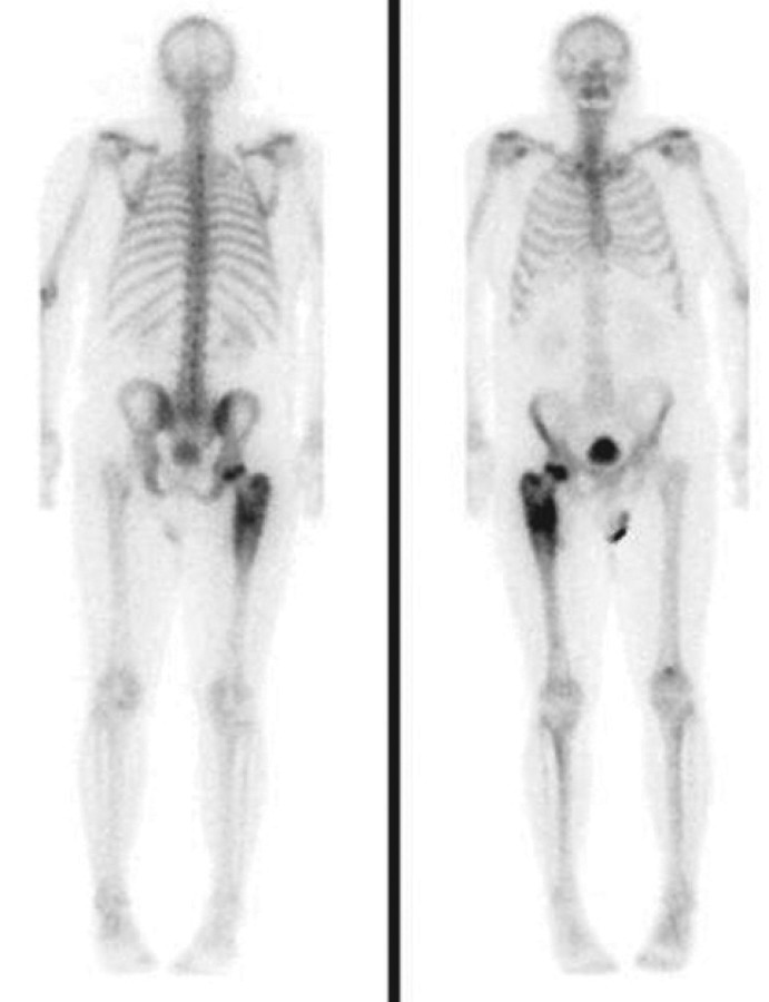
A 58-year-old Caucasian man presented to his general practitioner with over 1 year of severe back pain and was discovered to have very low vitamin D3 levels (<10 nmol/l). Alkaline phosphatase was elevated at 160 U/l, calcium levels decreased to a low of 2.06 mmol/l and phosphate to 0.76 mmol/l. He was prescribed vitamin D supplements and levels began to normalise, but developed increasing pain in his right femur; an HDP Tc-99m bone scan revealed marked increased activity in the right proximal femur, indicating an aggressive osteoblastic process, in keeping with an osteosarcoma. Computed tomography (CT) and magnetic resonance imaging (MRI) scans confirmed a large tumour emanating from the right femoral head. The entire femur was resected and replaced with a titanium implant.
Oncogenic osteomalacia, also known as tumour-induced osteomalacia (TIO) is a rare paraneoplastic syndrome of abnormal phosphate and vitamin D metabolism, caused by typically small endocrine tumours that secrete fibroblast growth factor 23 -(FGF-23), a phosphatonin.1 Typical features include hypophosphataemia due to renal phosphate wasting, inappropriately low to normal vitamin D levels and normal to elevated FGF-23. Patients suffer years of symptoms before diagnosis, leading to bone pain, fractures, depression and muscle weakness. fig 2
Fig 1.
Tc99m bone scan.
Fig 2.
MRI scan of anterior thigh. MRI = magnetic resonance imaging.
Reference
- 1.Masi L. The Phosphatonins: New Hormones the Cause of Numerous Congenital Bone Disorders. Clin Cases Miner Bone Metab. 2010;7:174. [PMC free article] [PubMed] [Google Scholar]


