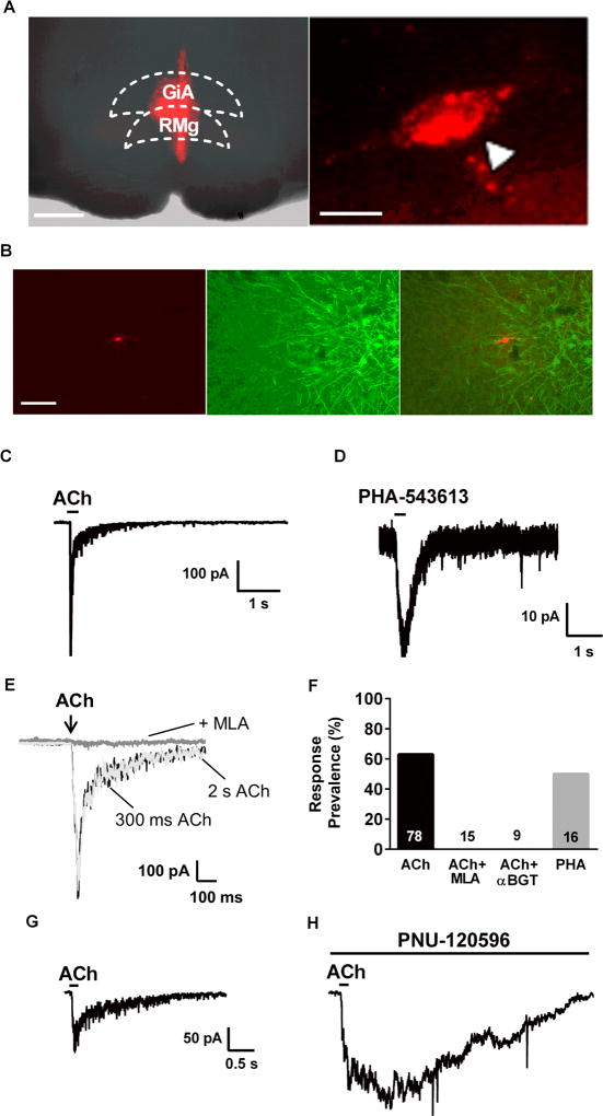
Figure 1. A subpopulation of PAG-RVM projection neurons expresses α7 nAChRs.
A. Left: Fluorescence photomicrograph illustrating a sample RVM injection site (−10.5 mm from Bregma). Gigantocellular Reticular Nucleus (GiA) and Raphe Magnus Nucleus (RMg) are part of the RVM. (Scale: 500 um). Right: Example vlPAG backlabeled neuron. (Scale: 20 µm). B. Representative fluorescence images of a backlabeled vlPAG neuron that projects to the RVM (left), antibody staining for tryptophan hydroxylase (TPH), a marker of serotonergic neurons (center), and image overlay showing no co-localization of the fluorescent signals (right; scale = 100 µm). We saw no overlap with TPH immunofluorescence in 77 backlabeled cells. C. Inward current response to a brief focal application of acetylcholine (ACh; 3 mM; 300 msec duration). Similar responses were seen in 49 of 78 backlabeled neurons tested. D. Inward current response to focal application of the selective α7 agonist, PHA-543613 (100 µM; 300 msec). Similar responses were seen in 8/16 neurons tested. E. Focal ACh application (3mM) induced an inward current with similar kinetics whether the application was 300 msec (black trace) or 2 sec (light grey trace) in duration. Bath application of MLA (10 nM) completely blocked the ACh-induced current in the same neuron. Complete blockade by MLA was seen in all responsive cells tested (n=15). F. Summary of response prevalence of vlPAG neurons to ACh alone and in combination with MLA (10 nM; tested only on responsive cells) or αBGT (50 nM; pretreated for >15 min prior to recording); n values are presented within or above each bar. G. Inward current due to focal application of ACh (3 mM). H. Inward current response to ACh (in the same neuron as G.) after treatment with the α7 positive allosteric modulator, PNU-120596 for15 min. The prolonged inward current is consistent with loss of α7 nAChR desensitization due to PNU-120596.

