

Abstract
PURPOSE:
There is a need for cervical flexion and even cervical hyperflexion for the use of technological devices, especially mobile phones. We investigated the effect of this use on the cervical lordosis angle.
MATERIAL AND METHODS:
A group of 156 patients who applied with only neck pain between 2013–2016 and had no additional problems were included. Patients are specifically questioned about mobile phone, tablet, and other devices usage. The value obtained by multiplying the year of usage and the average usage (hour) in daily life was determined as the total usage value (an average hour per day x year: hy). Cervical lordosis angles were statistically compared with the total time of use.
RESULTS:
In the general ROC analysis, the cut-off value was found to be 20.5 hy. When the cut-off value is tested, the overall accuracy is very good with 72.4%. The true estimate of true risk and non-risk is quite high. The ROC analysis is statistically significant.
CONCLUSION:
The use of computing devices, especially mobile telephones, and the increase in the flexion of the cervical spine indicate that cervical vertebral problems will increase even in younger people in future. Also, to using with attention at this point, ergonomic devices must also be developed.
Keywords: Cervical flexion, Cervical lordosis, Cervical pain, Mobile phone usage, Technological devices
Introduction
Over the years, the use of tablets, mobile phones and interactive computing devices has increased. Social and business life is now almost a part of our lives, and after this point, our life will be more affected. We do not know the damages that the mobile phones give to the spine, especially cervical region, which makes our work easier and is a part of our daily life. In the age of we live, our posture of cervical flexion is increased. Many people use their phones for hours during the day with the increasing use of social media, and this has been taking place in our lives for almost 15-20 years.
Also, the use of tablets and PC’s is much higher. There is a need for flexion of the cervical region to use these devices, especially mobile phones. Increased cervical flexion has a negative effect on cervical lordosis.
We tried to investigate the relationship between the duration of use and cervical lordosis. This is the first study to show how technological devices affect spinal alignment regarding usage time.
Material and Methods
Patients with a complaint of neck pain in the young age population who applied to our polyclinic between 2013 and 2016 and did not have any additional problems were included in this study. The age range of patients in the study is 25-42. Patients are specifically questioned about mobile phone, tablet, and other devices (as gaming consoles) usage. They were asked how many years they have been using and how many hours in a day they spent in the flexion /hyperflexion posture. The value obtained by multiplying the year of usage and the average usage (hour) in daily life was determined as the total usage value. Criteria that we applied when we included patients in the study;
1 - Desk workers and people working in the cervical flexure posture were excluded from the study.
2 - Patients with the condition that could affect the spine were excluded from study (rheumatic diseases, ankylosing spondylitis, deformities, connective tissue disease, previous spine surgery, and significant cervical trauma)
3 - Patients who read books regularly in flexion posture were excluded from the study.
Patients were not grouped because there was not a risky value for cervical lordosis accepted in the literature, and the cut-off was taken as median value firstly. And after general ROC analysis was performed and the sensitivity and specificity of the cut off value obtained here were compared. The cervical lordosis angles were measured by the tangent method in the position of lateral neutral standing (Figure 1). Cervical lordosis angles were statistically compared with total time (year x daily hour) of usage.
Figure 1.
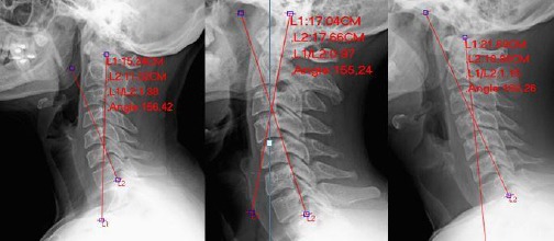
It is shown the tangent method for measure cervical lordosis in our some case
Results
The cut-off value for risk differentiation was set at 12.5 degrees because of the median value of 12.5 degrees for lordosis. According to this, patients with lordosis angle < 12.5 degrees were considered “risky” lordoz angle, patients with lordozis > 12.5 degrees were considered “non-risky” lordosis angle. In this case, 50 % of the patients are in the lordosis risk group, and 50 % are not in the lordosis risk group (Table 1).
Table 1.
Descriptive Statistics
| Frequency | Percent | ||
|---|---|---|---|
| Gender | Female | 93 | 59.6 |
| Male | 63 | 40.4 | |
| Lordosis Risk Group | Non-Risky | 78 | 50.0 |
| Risky | 78 | 50.0 | |
| Mean | SD | ||
| Lordosis Angle | 13.24 | 12.21 | |
| Age | 33.70 | 5.51 | |
| Usage Time | 21.38 | 13.60 | |
Lordosis Risk Group is divided by the median value of 12.5 degrees.
The average age of the patients was 33.7, the mean duration of use was calculated as 21.38 he and the mean lordosis was 13.24 degrees. 59.6% of the patients were female, and 40.4% were male (Table 1). In the general ROC analysis, the cut off value was found to be 20.5 hy. When the cut off value is tested, the overall accuracy is very good with 72.4%. The true estimate of true risk is 71.8 %, and the true estimate of the true non-risk estimate is 73.1% The ROC analysis is statistically significant (Table 2).
Table 2.
ROC Analysis Results
| Total Usage Time | Female Usage Time | Male Usage Time | |
|---|---|---|---|
| Cut-Off* | 20.5 | 17.5 | 20.5 |
| Sensitivity | 71.8% | 63.6% | 91.3% |
| Specificty | 73,1% | 78.9% | 67.5% |
| PPV | 49.4% | 46.2% | 54.0% |
| NPV | 50.6% | 53.8% | 46.0% |
| AUC | 0.779 | 0.794 | 0.819 |
| Accuracy | 72.4% | 69.9% | 76.2% |
Usage times are shown as hy (hour x year); PPV: Positive predictive value; NPV: Negative predictive value; AUC: Area under the curve.
In the ROC analysis for women, the cut off value was found to be 17.5 hy. When cut off value is tested, total accuracy is good with 69.9 %. While the true estimate of true risk is 63.6 %, the true non-risk estimate is 78.9 % The ROC analysis for women is statistically significant (Table 2).
In the ROC analysis for men, the cut off value was found to be 20.5 hy. The best estimate compared to total and women was realised in men ROC analysis. When cut off value is tested, total accuracy is good with 76.2%. While the true estimate of true risk is 61.3%, the true non-risk estimate is 67.5%. The ROC analysis for men is statistically significant (Table 2).
The relationship between the lordosis risky group and gender has been examined. While 59.1% of the women were in the risky group, the rate of risky lordosis group in males was calculated as 36.5%. The difference was statistically significant (Table 3).
Table 3.
The relationship between the Lordosis Risk Group and Gender was examined
| Gender * Lordosis Risk Group Crosstabulation | ||||||
|---|---|---|---|---|---|---|
| Lordosis Risk NonRisky |
Group Risky | p | ||||
| Gender | Female | Count | 38 | 55 | ||
| % within Gender | 40.9% | 59.1% | ||||
| % within Lordosis Risk Group | 48.7% | 70.5% | ||||
| Male | Count | 40 | 23 | < 0.005 | ||
| % within Gender | 63.5% | 36.5% | ||||
| % within Lordosis Risk Group | 51.3% | 29.5% | ||||
| Total | Count | 78 | 78 | |||
| % within Gender | 50.0% | 50.0% | ||||
| % within Lordosis Risk Group | 100.0% | 100.0% | ||||
Fischer’s Exact Test; Lordosis Risk Group is divided by the median value of 12.5 degrees.
The lordosis risk group and mean of usage were compared. The mean duration of total usage was 27.69 hy ± 12.67 hy in the group with lordosis < 12.5 as we said not a risky population, whereas it was calculated as 15.06 ± 11.44 hy in the group with lordosis > 12.5 as we said risky population. The difference was statistically significant (p < 0.0001) (Table 4).
Table 4.
The relationship between the lordosis risk group and the average of usage time was compared Lordosis Risk Group and Total Usage Time
| Usage Time | p | |||
|---|---|---|---|---|
| Mean | SD | |||
| Lordosis Risk Group | Non-Risky | 15.06 | 11.44 | < 0.0001 |
| Risky | 27.69 | 12.67 | ||
Lordosis Risk Group is divided by the median value of 12.5 degrees.
Discussion
The cervical lordosis is the curve of the cervical vertebrae. Cervical lordosis provides horizontal gaze. Cervical lordosis helps to keep the spine in balance, provides it by turning to lordosis from kyphosis in the junction at the cervicothoracic region.
There is no clear study of the physiological curve in the cervical region. It has been stated that there may be differences in many people. The average accepted lordozis angle 20-35 [1] [2] [3] [4] [5]. However, there is no consensus on under which angle of the cervical lordosis can cause problems.
Many conditions affect cervical lordosis. Trauma, working postures in the job, and degenerative diseases affect cervical lordosis. Increased flexion/hyperflexion in the cervical region causes changes in the cervical region. Because of the prolonged tendency to flexion, the weight effect created by the weight of the head and the strain of the neck muscles will cause degeneration of the cervical vertebrae and discs, loss of lordosis and kyphosis. Cervical muscles are strained, ligamentous structures are deteriorating. These are the most important causes of loss of cervical lordosis. As a result of this, cervical disc degenerations, kyphotic cervical stenosis, cord tension, radicular symptoms are encountered [6] [7] [8].
Cervical colon separated to the 3 division by Luis. The anterior column is the disc and the vertebral body while the posteriors are defined as facet joints. The primary loading segments of the cervical column will be affected by the loss of cervical lordosis, which will accelerate the degeneration and the developing sagittal imbalance [9].
Deviations from this curvature, such as a loss of lordosis or the development of cervical kyphosis, are associated with pain and disability [2] [10] [11] [12] [14] [14].
There have been many publications on cervical lordosis and neck pain. While some studies found no correlation between cervical lordosis and neck pain, and some studies found strong correlations between neck pain and cervical lordosis [15][16][17][18][19].
In 1994, Helliwell stated that in patients with chronic neck pain, the cervical lordosis angle is more straight or less [20].
The normal lordotic angle has been shown to be different in many studies because the cervical spinal region is the most mobile segment of the spinal column [2] [12].
In the study of Mc Aviney et al. in 2005, cervical X-rays and angular measurements and examining neck pain, they noted that neck pain was statistically high in cervical curvature less than 20 degrees and could be considered clinically normal at above 30 degrees [13].
Mobile phones and technological devices have begun to be used more and more in our lives in the last 20-30 years. For using these devices, especially mobile phones, the cervical region’s flexion and hyperflexion are essential. The flexion posture of the neck and the weight of head are disturbing the balance of the spine. Also, the use of technological devices and the duration of use during the day increases. The load on the cervical spine while using mobile phones in the cervical region has already been published before [6].
According to the degree of flexion, the stress in the cervical region increases, loads on discs increase, loss of lordosis and degenerative processes accelerate. This can be the source of pain. However, loss of lordosis will lead to greater pathological problems in the later period. It was pointed out in an issue in an article published in 2014. It has been tried to take attention to the danger waiting for us by giving numerical data. In the article, it was estimated that the average time spent in college with reading books and mobile devices in the flexion posture was 5000 hours [21].
The effect of mobile devices on our lives was studied, and the most common symptom was found to be neck pain even in young patients in the study of Azaria et al. [22]. They reported a problem as high as 72.1 %. And the increase in the intensity of use has also increased the symptom of neck pain, which is very short compared to the work we do [22].
The existence of neck symptoms in association with cell phone usage has been previously described, but it is very rare. The association between the degree of cervical lordosis and the usage time of mobile devices has never been mentioned before. We studied the effect of total time of usage on cervical lordosis in our study.
We considered the usage duration as a numerical value and assessed whether it correlated with the cervical lordosis regardless of pain score. Although we think that neck pain is related to cervical lordosis in general, we did not include the pain score in this study. Patients may be able to live pain periodically at different grades, so we made only an angular evaluation to be more objective.
We were meticulous when choosing patient populations. We worked on a young patient population. Our goal here is that this population is relatively remote from the expected degenerative processes with age, and the duration of usage is higher in this population. For this reason, we think the correlation will be more meaningful.
There are two different main methods of measuring cervical lordosis. Cobb and Harrison posterior tangent methods for cervical lordosis measurements have high reliability in evaluating cervical lordosis and are the most commonly used methods in practice [23] [24].
However, due to the degenerative changes in the cervical region, the use of the inferior vertebral endplates in the Cobb method showed that this method is lower in diagnostic accuracy than the tangent method, which uses the posterior edges of the vertebral corpus. So we chose Harrison posterior tangent method.
The relation of the Harrison posterior tangent method to the effective lordosis was found to be higher than the Cobb method, and also the measurement technique is more practical [24].
As a result of our study, cervical lordosis decreases in the correlation between total duration of usage. Especially the values of 20.5 hy were accepted as a critical threshold in patients.
So, according to the statistical analysis, approximately a value of 20 hy is lowered cervical lordosis under 12.5 degrees nearly 70 % in the population. An average of 2 hours per day during a 10-year period creates a risk of loss of cervical lordosis. There are some rehabilitation aimed at the loss of cervical lordosis, and it was written that they are useful. Anterior head weighting procedures were applied for loss of cervical lordosis in a study and improvement of cervical lordosis has been seen in patients [25].
We are all at risk in this age. Maybe we all need cervical rehabilitation. Or using such mobile devices without cervical flexion will relatively protect our cervical spine. Otherwise, cervical spine pathologies may be a problem that we will all encounter.
In conclusion, cervical vertebrae problems that will develop with the necessity of flexion of the cervical region and lifestyle in this way will also force our lives in the future. As a result of our study, cervical lordosis decreases in the correlation between total duration of usage. Especially the values of 20.5 hy were accepted as a critical threshold in patients. This situation is likely to lead to the loss of cervical lordosis in the following years and to cause serious problems associated with it.
Footnotes
Funding: This research did not receive any financial support
Competing Interests: The authors have declared that no competing interests exist
References
- 1.Borden AG, Rechtman AM, Gershon-Cohen J. The normal cervical lordosis. Radiology. 1960;74:806–09. doi: 10.1148/74.5.806. https://doi.org/10.1148/74.5.806. PMid:13802725. [DOI] [PubMed] [Google Scholar]
- 2.Gore DR, Sepic SB, Gardner GM. Roentgenographic findings of the cervical spine in asymptomatic people. Spine. 1986;11(6):521–24. doi: 10.1097/00007632-198607000-00003. https://doi.org/10.1097/00007632-198607000-00003. PMid:3787320. [DOI] [PubMed] [Google Scholar]
- 3.Hald HJ, Danz B, Schwab R, Burmeister K, Bähren W. Radiographically demonstrable spinal changes in asymptomatic young men. Rofo. 1995;163(1):4–8. doi: 10.1055/s-2007-1015936. https://doi.org/10.1055/s-2007-1015936. PMid:7626752. [DOI] [PubMed] [Google Scholar]
- 4.Juhl JH, Miller SM, Roberts GW. Roentgenographic variations in the normal cervical spine. Radiology. 1962;78:591–97. https://doi.org/10.1148/78.4.591. [Google Scholar]
- 5.Nojiri K, Matsumoto M, Chiba K, Maruiwa H, Nakamura M, Nishizawa T, et al. Relationship between the alignment of upper and lower cervical spine in asymptomatic individuals. J Neurosurg (Suppl) 2003;1:80–3. doi: 10.3171/spi.2003.99.1.0080. https://doi.org/10.3171/spi.2003.99.1.0080. [DOI] [PubMed] [Google Scholar]
- 6.Hansraj KK. Assessment of stresses in the cervical spine caused by posture and position of the head. Surg Technol Int. 2014;25:277–79. PMid:25393825. [PubMed] [Google Scholar]
- 7.Scheer JK, Tang JA, Smith JS, et al. Cervical spine alignment, sagittal deformity, and clinical implications: a review. Journal of Neurosurgery Spine. 2013;19(2):141–59. doi: 10.3171/2013.4.SPINE12838. https://doi.org/10.3171/2013.4.SPINE12838. PMid:23768023. [DOI] [PubMed] [Google Scholar]
- 8.Shimizu K, Nakamura M, Nishikawa Y, et al. Spinal kyphosis causes demyelination and neuronal loss in the spinal cord: a new model of kyphotic deformity using juvenile Japanese small game fowls. Spine (Phila Pa 1976) 2005;30:2388–92. doi: 10.1097/01.brs.0000184378.67465.5c. https://doi.org/10.1097/01.brs.0000184378.67465.5c. [DOI] [PubMed] [Google Scholar]
- 9.Louis R. Spinal stability as defined by the three-column spine concept. Anat Clin. 1985;7:33–42. doi: 10.1007/BF01654627. https://doi.org/10.1007/BF01654627. PMid:3994851. [DOI] [PubMed] [Google Scholar]
- 10.Ames CP, Smith JS, Scheer JK, et al. Impact of spinopelvic alignment on decision making in deformity surgery in adults. A review. J Neurosurg Spine. 2012;16:547–64. doi: 10.3171/2012.2.SPINE11320. https://doi.org/10.3171/2012.2.SPINE11320. PMid:22443546. [DOI] [PubMed] [Google Scholar]
- 11.Gay RE. The curve of the cervical spine: variations and significance. J Manipulative Physiol Ther. 1993;16:591–94. PMid:8133194. [PubMed] [Google Scholar]
- 12.Hardacker JW, Shuford RF, Capicotto PN, Pryor PW. Radiographic standing cervical segmental alignment in adult volunteers without neck symptoms. Spine (Phila Pa 1976) 1997;22:1472–80. doi: 10.1097/00007632-199707010-00009. https://doi.org/10.1097/00007632-199707010-00009. [DOI] [PubMed] [Google Scholar]
- 13.McAviney J, Schulz D, Bock R, Harrison DE, Holland B. Determining the relationship between cervical lordosis and neck complaints. J Manipulative Physiol Ther. 2005;28:187–93. doi: 10.1016/j.jmpt.2005.02.015. https://doi.org/10.1016/j.jmpt.2005.02.015. PMid:15855907. [DOI] [PubMed] [Google Scholar]
- 14.Tang JA, Scheer JK, Smith JS, et al. The impact of standing regional cervical sagittal alignment on outcomes in posterior cervical fusion surgery. Neurosurgery. 2015;76:14–21. doi: 10.1227/01.neu.0000462074.66077.2b. https://doi.org/10.1227/01.neu.0000462074.66077.2b. PMid:25692364. [DOI] [PubMed] [Google Scholar]
- 15.Grob D, Frauenfelder H, Mannion AF. The association between cervical spine curvature and neck pain. Eur Spine J. 2007;16:669–78. doi: 10.1007/s00586-006-0254-1. https://doi.org/10.1007/s00586-006-0254-1. PMid:17115202. PMCid:PMC2213543. [DOI] [PMC free article] [PubMed] [Google Scholar]
- 16.Harrison DD, Harrison DE, Janik TJ, et al. Modeling of the sagittal cervical spine as a method to discriminate hypolordosis: results of elliptical and circular modeling in 72 asymptomatic subjects, 52 acute neck pain subjects, and 70 chronic neck pain subjects. Spine. 2004;29:2485–92. doi: 10.1097/01.brs.0000144449.90741.7c. https://doi.org/10.1097/01.brs.0000144449.90741.7c. PMid:15543059. [DOI] [PubMed] [Google Scholar]
- 17.Harrison DE, Harrison DD, Janik TJ, et al. Comparison of axial and flexural stresses in lordosis and three buckled configurations of the cervical spine. Clin Biomech. 2001;16:276–84. doi: 10.1016/s0268-0033(01)00006-7. https://doi.org/10.1016/S0268-0033(01)00006-7. [DOI] [PubMed] [Google Scholar]
- 18.Harrison DE, Jones EW, Janik TJ, Harrison DD. Evaluation of axial and flexural stresses in the vertebral body cortex and trabecular bone in lordosis and two sagittal cervical translation configurations with an elliptical shell model. J Manipulative Physiol Ther. 2002;25:391–401. doi: 10.1067/mmt.2002.126128. https://doi.org/10.1067/mmt.2002.126128. PMid:12183697. [DOI] [PubMed] [Google Scholar]
- 19.Kumagai G, Ono A, Numasawa T, et al. Association between roentgenographic findings of the cervical spine and neck symptoms in a Japanese community population. Journal of Orthopaedic Science. 2014;19(3):390–97. doi: 10.1007/s00776-014-0549-8. https://doi.org/10.1007/s00776-014-0549-8. PMid:24570299. PMCid:PMC4033794. [DOI] [PMC free article] [PubMed] [Google Scholar]
- 20.Helliwell PS, Evans PF, Wright V. The straight cervical spine: does it indicate muscle spasm? Bone Joint J. 1994;76(1):103–06. https://doi.org/10.1302/0301-620X.76B1.8300650. [PubMed] [Google Scholar]
- 21.Harvard Editorial Board: How to soothe a sore neck. The essentials are icing and heat, gentle therapeutic exercise, and good posture. Harv Mens Health Watch. 2014;18(11):5. [PubMed] [Google Scholar]
- 22.AlZarea BK, Santosh RP. Mobile Phone Head and Neck Pain Syndrome: Proposal of a New Entity. Oral Health Dent Manag. 2015;14(5):313–17. [Google Scholar]
- 23.Harrison DE, Harrison DD, Cailliet R, et al. Cobb method or Harrison posterior tangent method: which to choose for lateral cervical radiographic analysis. Spine (Phila Pa 1976) 2000;25:2072–78. doi: 10.1097/00007632-200008150-00011. https://doi.org/10.1097/00007632-200008150-00011. [DOI] [PubMed] [Google Scholar]
- 24.Soylu FN, Özkan FU, Erdem S, et al. Evaluation of cervical lordosis and its relation to cervical pain. (Servikal lordoz açılarıve boyun ağrısıilişkisinin değerlendirilmesi) Marmara Medical. 2014;27:112–15. [Google Scholar]
- 25.Saunders ES, Woggon D, Cohen C, Robinson DH. Improvement of Cervical Lordosis and Reduction of Forward Head Posture with Anterior Head Weighting and Proprioceptive Balancing Protocols. J. Vertebral Subluxation Res. 2003;1:1–5. [Google Scholar]