ABSTRACT
Foot-and-mouth disease virus (FMDV) has a positive-sense single-stranded RNA (ssRNA) genome that includes a single, large open reading frame encoding a polyprotein. The cotranslational “cleavage” of this polyprotein at the 2A/2B junction is mediated by the 2A peptide (18 residues in length) using a nonproteolytic mechanism termed “ribosome skipping” or “StopGo.” Multiple variants of the 2A polypeptide with this property among the picornaviruses share a conserved C-terminal motif [D(V/I)E(S/T)NPG↓P]. The impact of 2A modifications within this motif on FMDV protein synthesis, polyprotein processing, and virus viability were investigated. Amino acid substitutions are tolerated at residues E14, S15, and N16 within the 2A sequences of infectious FMDVs despite their reported “cleavage” efficiencies at the 2A/2B junction of only ca. 30 to 50% compared to that of the wild type (wt). In contrast, no viruses containing substitutions at residue P17, G18, or P19, which displayed little or no “cleavage” activity in vitro, were rescued, but wt revertants were obtained. The 2A substitutions impaired the replication of an FMDV replicon. Using transient-expression assays, it was shown that certain amino acid substitutions at residues E14, S15, N16, and P19 resulted in partial “cleavage” of a protease-free polyprotein, indicating that these specific residues are not essential for cotranslational “cleavage.” Immunofluorescence studies, using full-length FMDV RNA transcripts encoding mutant 2A peptides, indicated that the 2A peptide remained attached to adjacent proteins, presumably 2B. These results show that efficient “cleavage” at the 2A/2B junction is required for optimal virus replication. However, maximal StopGo activity does not appear to be essential for the viability of FMDV.
IMPORTANCE Foot-and-mouth disease virus (FMDV) causes one of the most economically important diseases of farm animals. Cotranslational “cleavage” of the FMDV polyprotein precursor at the 2A/2B junction, termed StopGo, is mediated by the short 2A peptide through a nonproteolytic mechanism which leads to release of the nascent protein and continued translation of the downstream sequence. Improved understanding of this process will not only give a better insight into how this peptide influences the FMDV replication cycle but may also assist the application of this sequence in biotechnology for the production of multiple proteins from a single mRNA. Our data show that single amino acid substitutions in the 2A peptide can have a major influence on viral protein synthesis, virus viability, and polyprotein processing. They also indicate that efficient “cleavage” at the 2A/2B junction is required for optimal virus replication. However, maximal StopGo activity is not essential for the viability of FMDV.
KEYWORDS: picornavirus, replicon, FMDV, ribosomal skipping, protease, StopGo, foot-and-mouth disease virus, proteases
INTRODUCTION
Foot-and-mouth disease virus (FMDV) is the causative agent of foot-and-mouth disease, a highly contagious disease of domestic and wild cloven-hooved animal species. The virus has been successfully eradicated from Europe but is still endemic in many regions of the world (in Asia, Africa, and the Middle East) and can potentially cause major outbreaks in domestic livestock elsewhere, with severe economic losses (reviewed in reference 1). FMDV is the prototypic member of the Aphthovirus genus within the family Picornaviridae. These viruses are small (ca. 25 to 30 nm) and have a positive-sense single-stranded RNA (ssRNA) genome (2). The FMDV genome is ∼8.5 kb in length and includes a single, large open reading frame (ORF) encoding a long polyprotein of over 2,300 residues (3). However, this polyprotein is never observed within infected cells due to rapid co- and posttranslational processing to produce, initially, the mature leader protein (Lpro) and the precursor proteins P1-2A, P2, and P3. The Lpro is a papain-like protease and cleaves the polyprotein at its own C terminus; that is the junction between Lpro and the capsid precursor P1-2A (4, 5). The 3C protease (3Cpro) is responsible for proteolytic cleavage of P1-2A to produce the structural proteins VP0, VP3, and VP1 plus the 2A peptide. The P2 and P3 precursors are also processed by 3Cpro to generate the nonstructural proteins, which are required for the replication of the viral genome. The processing of VP0 to VP4 and VP2 occurs during encapsidation of the viral RNA, although it can also occur on assembly of empty capsid particles (6, 7).
The separation of the P1-2A precursor from 2B (within P2) is achieved by yet another mechanism. There is considerable heterogeneity among the picornaviruses with respect to the 2A peptide/protein that is located on the C-terminal side of the capsid protein precursor. Both the size and the function of the different 2A species vary between different picornavirus genera (8). The entero- and rhinovirus 2A proteins, termed 2Apro, are thiol proteinases of ∼150 amino acids which catalyze the proteolytic cleavage of the junction between the P1 and P2 precursors at their own N termini, i.e., at the P1/2A junction (9, 10). In contrast, in the aphthoviruses and cardioviruses (e.g., encephalomyocarditis virus [EMCV] and Theiler's murine encephalitis virus [TMEV]), the separation of the capsid precursor (P1-2A) from P2 (2BC) occurs at the 2A/2B junction, i.e., at the C terminus of 2A. In FMDV, the 2A peptide is only 18 amino acids long and lacks any characteristic protease motifs (11–13). Earlier studies have demonstrated that the “cleavage” at the 2A/2B junction is not dependent on the FMDV protease Lpro or 3Cpro or on host cell proteases (12, 14, 15). The FMDV 2A peptide contains a highly conserved amino acid sequence at its C terminus, D12(V/I)E(S/T)NPG2A↓P192B. This sequence induces a cotranslational “cleavage” event that is referred to as “ribosomal skipping” (16) or, alternatively, “stop-carry on” or “StopGo” (17, 18). The first residue of 2B (Pro or P) is referred to as P19 as it is a key part of the “cleavage” site. This motif, together with upstream amino acids that form an α-helix over most of its length, is believed to interact with the exit tunnel of the translating ribosome and prevent the formation of a peptide bond between the C-terminal amino acid, glycine (Gly or G), of 2A and the first residue, proline (P), of 2B (16, 19). This produces a break in the growing amino acid chain, but the process of protein synthesis continues, without the requirement for a new translation initiation event. The same conserved motif is also present at the C terminus of the cardiovirus 2A proteins, which are substantially larger; the FMDV 2A sequence appears to be the minimal functional entity to break the growing polypeptide chain (for convenience this process is simply called cleavage below), and other functions have been assigned to the cardiovirus 2A protein as well (20).
The majority of studies on the function of the FMDV 2A peptide have been conducted using in vitro experiments with mRNAs encoding artificial polyproteins comprising two reporter proteins linked via the 2A peptide, thus generating two separate translation products (see, e.g., references 16, 21, and 22). These previous studies have shown that specific amino acid substitutions within the FMDV 2A sequence, and especially within the highly conserved D12(V/I)E(S/T)NPG2A↓P192B motif, drastically reduce the apparent cleavage efficiency and can even block it entirely. These results indicate that these amino acid residues are critical for optimal ribosomal skipping (16, 22). Furthermore, in the context of a synthetic reporter polyprotein, assayed within CHO cells, four different synonymous codons for residue G18 of the 2A peptide were shown to function with very similar apparent cleavage efficiencies at the 2A/2B junction, but the cleavage efficiency was not optimal (only 88 to 89% complete) in this system (23). These results were interpreted as showing that it is this amino acid residue rather than the nucleotide sequence which is critical for achieving cleavage (23). The 2A peptide has been shown to mediate cleavage in all eukaryotic translation systems tested, whereas a number of artificial polyproteins containing this sequence have been examined in prokaryotic systems and no detectable cleavage products were observed (22).
The less conserved part of the 2A sequence, located upstream of the D(V/I)E(S/T)NPG2A↓P2B motif, has also been shown to be important for optimal 2A function. Chimeric FMDV, TMEV, and EMCV 2A peptides were generated by replacing the N- or C-terminal portions with another 2A variant and then assayed within artificial polyprotein systems, where they showed little or no activity (24). In addition, when the FMDV 2A, in an artificial polyprotein system, was elongated by the addition of up to 30 amino acids from the upstream VP1, its apparent cleavage activity was enhanced (16, 22, 25). Thus, the context of the 2A sequence is important. The 2A peptides from other picornaviruses exhibited similar increases in activity when elongated with 30 amino acids from their respective polyprotein precursors (8). Moreover, an extensive alanine (A), glycine (G), and proline (P) scanning mutagenesis of the entire FMDV 2A peptide showed a decrease in apparent cleavage activity for all mutants (24). This supports the view that the specific identity of the amino acid at nearly all positions within the 2A peptide is important for activity and that 2A peptides are fine-tuned to function as a single unit within their natural polyprotein.
In studies by Loughran et al. (26), a number of mutations in the 2A-coding sequences within the full-length TMEV and FMDV genomes were tested for their effects on virus viability and polyprotein processing. Modification of the SNPG↓P2B sequence to SNPL↓V2B at the 2A/2B junction blocked polyprotein cleavage. However, this modification had no significant effect on the growth of Theiler's murine encephalomyelitis virus (TMEV), whereas it was detrimental for the replication of mengovirus (another cardiovirus) and apparently lethal for FMDV. Thus, it was concluded that the 2A/P2 cleavage event is not essential for virus viability for certain cardioviruses but is critical for FMDV.
In this study, we have reinvestigated the effect of 2A modifications in the context of the native FMDV polyprotein and its effect on virus protein synthesis and replication, virus viability, and polyprotein processing. In contrast to earlier studies, mutant infectious FMDVs having certain amino substitutions within the 2A peptide have been obtained but such changes do adversely affect virus replication and polyprotein processing to some degree.
RESULTS
Effect of single amino acid substitutions in 2A on FMDV viability.
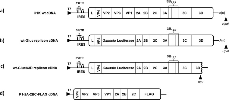
Several studies using artificial polyprotein systems have demonstrated that nearly all positions of the 2A peptide are important for the “StopGo” activity and that modifications can severely impair cleavage (22, 24). To establish whether the StopGo activity plays a crucial role in FMDV viability, this study set out to investigate the constraints on the 2A sequence within the context of the full-length FMDV genome (Fig. 1a).
FIG 1.
Structures of the plasmids used in this study. (a) Full-length FMDV O1K cDNA; (b) Gluc replicon cDNA; (c) RNA polymerase-defective Gluc replicon cDNA. The plasmids were linearized using HpaI or BlpI prior to in vitro transcription. (d) Schematic representation of the P1-2A-2BC-FLAG cDNA cassette expressed in transient-expression assays (as described in Materials and Methods).
To determine the viability of FMDVs with single-amino-acid substitutions within the 2A peptide, selected modifications that were previously found to impair, to different extents, the StopGo activity in artificial polyprotein systems (22, 24) were introduced into the plasmid pT7S3, which contains the full-length FMDV cDNA (27), using site-directed mutagenesis (see Materials and Methods). The resultant plasmids were linearized, and RNA transcripts, prepared in vitro, were introduced into BHK cells by electroporation. Unexpectedly, all of the FMDV 2A mutants (Table 1) produced viable progeny viruses, with full cytopathic effect (CPE) detectable after the second passage. The rescued viruses were then sequenced to identify possible adaptations or reversions (Table 1). After three passages of the 2A mutant viruses in cells, the viruses rescued from the transcripts encoding the N16H, E14Q, S15F, and S15I modifications had each retained the plasmid-derived amino acid substitutions. In contrast, the rescued viruses derived from the N16A, P17A, G18A, P19G, and P19A mutant transcripts all matched the wild-type (wt) sequence (i.e., the rescued viruses were not mutant), even when two nucleotide changes were required to achieve this (e.g., see Fig. 2 for the P19A mutant). To determine whether this reflected reversion to the wt sequence or some form of contamination/carryover of the wt sequence, three synonymous substitutions were inserted ca. 20 nucleotides downstream of the 2A/2B junction in the wt and the N16A, P17A, G18A, and P19A mutant plasmids (Fig. 2), after which the “marked” RNA transcripts were introduced into BHK cells. After three passages, the rescued mutant viruses had lost the 2A modification in each case but had, like the “marked” wt virus, each retained the three synonymous substitutions in the 2B-coding region (Fig. 2); this provides strong evidence that the presence of the wt sequence in the rescued viruses reflects reversion.
TABLE 1.
Amino acid sequences in the encoded 2A peptide within rescued viruses following three passages in BHK cells
FIG 2.
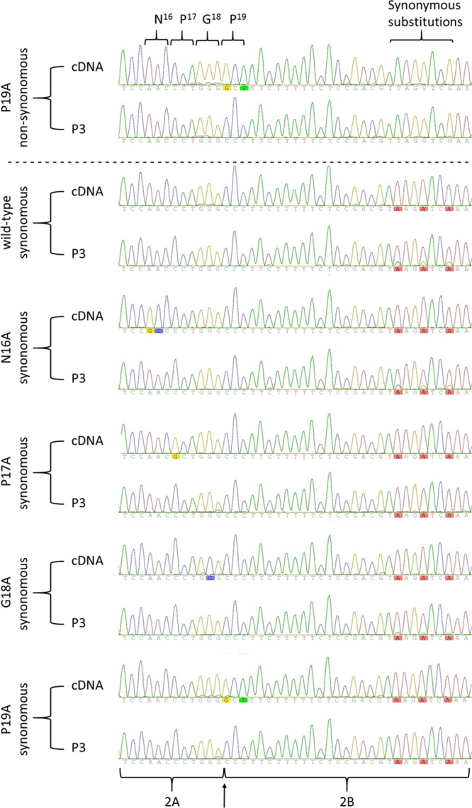
FMDVs rescued from the N16A, P17A, G18A, and P19A mutants have reverted to the wt sequence. Three synonymous mutations downstream of the 2A/2B junction were introduced into the wt and mutant N16A, P17A, G18A, and P19A plasmids. The resultant RNA transcripts were introduced into BHK cells. The rescued viruses were analyzed after 3 passages in BHK cells. The region of the FMDV genome including that encoding the 2A peptide was amplified by RT-PCR, and the PCR products were sequenced. The chromatograms are shown. Note the retained synonymous mutations ca. 20 nucleotides downstream of the 2A/2B junction, which had been introduced as a marker.
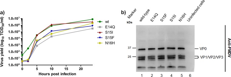
The growth characteristics of the wt and the viable 2A mutant viruses in BHK cells were examined in more detail by determining growth curves using a multiplicity of infection (MOI) of 0.1. Surprisingly, the wt and the viable 2A mutants grew with similar kinetics (Fig. 3a). Analysis of the FMDV capsid proteins within cells infected with the wt and the viable 2A mutant viruses, as determined by immunoblotting using anti-FMDV antibodies, is shown in Fig. 3b. As expected, the production of the capsid proteins was similar for the wt and the 2A mutants in each of the infected-cell extracts (Fig. 3b, lanes 1 to 5).
FIG 3.
Growth curves and assessment of the production of FMDV capsid proteins in BHK cells infected with wt and viable 2A mutant viruses. (a) BHK cells were infected with the wt and the indicated 2A mutants at an MOI of 0.1, and virus was harvested by freezing at 0, 2, 5, 10, and 24 h postinfection. Virus yields were determined as TCID50 by titration in BHK cells. (b) Uninfected or FMDV-infected (MOI, 0.1) BHK cell lysates were analyzed by SDS-PAGE and immunoblotting with antibodies specific for FMDV capsid proteins (anti-FMDV sera). Uninfected cells were used as a negative control. Molecular mass markers (kDa) are indicated on the left.
Requirements for efficient 2A/2B cleavage in its native context.
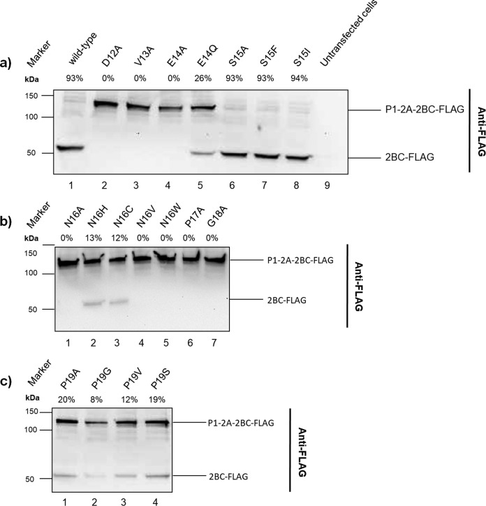
To examine the effects of the 2A mutants on the StopGo cleavage at the 2A/2B junction in its natural context and in cells, a plasmid encoding a truncated FMDV polyprotein, termed the P1-2A-2BC-FLAG protein, with a FLAG epitope at its C terminus was generated (Fig. 1d). The transient expression of this truncated viral polyprotein (without any proteases) was designed to permit the simultaneous assessment of the production of the uncleaved P1-2A-2BC-FLAG (ca. 135 kDa) and of the “cleavage” product 2BC-FLAG (ca. 54 kDa). The coding sequences for the wt or mutant P1-2A-2BC-FLAG products were under the control of the T7 promoter. The plasmids were transfected into BHK cells that had been infected with the recombinant vaccinia virus vTF7-3 (28), which expresses the T7 RNA polymerase. The expression and processing of the proteins generated from these plasmids were visualized in immunoblots using anti-FLAG antibodies. Expression of the wt cassette led to apparently complete cleavage of the P1-2A-2BC-FLAG polyprotein, as expected (Fig. 4a, lane 1), and thus only the 2BC-FLAG product was observed. In contrast, the E14Q mutant generated a mixture of both uncleaved and cleaved products (Fig. 4a, lane 5). Unexpectedly (c.f. references 22 and 24), in the system used here, the S15A, S15F, and S15I mutant proteins were each efficiently cleaved (Fig. 4a, lanes 6 to 8). The N16C, N16H, P19A, P19G, P19V, and P19S mutants all produced a mixture of cleaved and uncleaved products (Fig. 4b, lanes 2 and 3, and c, lanes 1 to 4). However, the D12A, V13A, E12A, N16A, N16V, N16W, P17A, and G18A substitutions resulted in the production of only the uncleaved product, and hence these mutant 2A peptides were all inactive in this system (Fig. 4a, lanes 2, 3, and 4, and b, lanes 1, 4, 5, 6, and 7). Overall, there is partial agreement between the results described here, using assays of the 2A in its near-native context within cells, and those described previously (22, 24). The main discrepancies concern the S15A, S15F, and S15I mutants, which resulted in essentially complete cleavage (≥90%) here but gave rather suboptimal cleavage (42 and 39% of the wt level, respectively) in vitro (22), while the P19A, P19G, P19V, and P19S mutants resulted in detectable, but low-level, cleavage (8 to 20%) here but completely abrogated cleavage in vitro (22). The same cell lysates were also analyzed using an anti-FMDV capsid protein antibody to detect the intact polyprotein and the P1-2A product (data not shown). The pattern of results was fully consistent with those obtained using the anti-FLAG antibodies to detect the intact polyprotein and the 2BC-FLAG product. Thus, it seems that the efficiency of cleavage detected in this assay system is higher than that observed using cell-free translation systems in vitro.
FIG 4.
Transient-expression assays to determine 2A/2B cleavage induced by the wt and mutant FMDV cDNAs. The indicated plasmids were transfected into vTF7-3 infected BHK cells as described in Materials and Methods. After 24 h, cell extracts were prepared and analyzed by SDS-PAGE and immunoblotting using an anti-FLAG antibody. The uncleaved P1-2A-2BC-FLAG and the cleavage product (2BC-FLAG) are marked. Molecular mass markers (kDa) are indicated on the left. The cleavage activities (percentage of cleaved product) of the wt and each 2A mutant were determined by quantifying the intensity of the signal for the FMDV capsid proteins using ImageJ (v1.50) and are indicated above each lane.
Influence of the amino acid substitutions in FMDV 2A on FMDV RNA replication efficiency assessed using a replicon that expresses the Gaussia luciferase.
To evaluate the impact of the 2A mutants on the replication of viral RNA, nine different substitutions within the 2A-coding sequence were introduced into an FMDV replicon (Fig. 1b). In this replicon, the coding sequences for the FMDV structural proteins (VP1 to VP3) have been replaced by the sequence encoding the Gaussia luciferase (Gluc) reporter protein, thus allowing replication to be readily monitored via measurement of Gluc expression. RNA transcripts were produced in vitro from the linearized plasmids and introduced into BHK cells using electroporation. As a negative control, a derivative of the wt-Gluc replicon which lacks a portion of the coding sequence for 3Dpol (the RNA-dependent RNA polymerase) was produced and is termed wt-GlucΔ3D (Fig. 1c). Lysates were prepared from cells at various times after electroporation with the different transcripts and assayed for Gluc activity (Fig. 5). The wt-GlucΔ3D transcript produced Gluc initially, which was already detectable at 1 h postelectroporation, but no further increase in luciferase activity was observed after 2 h. This expression presumably represents the translation of the input RNA. In contrast, the replication-competent wt-Gluc, while generating an initially similar level of Gluc activity at 2 h, showed a sustained increase in expression at later time points. Interestingly, all of the 2A mutants expressed low levels of Gluc activity initially, almost 10-fold less than the wt-GlucΔ3D at 2 h. However, the expression increased to some degree at later time points; the level of Gluc expression first surpassed that of the polymerase knockout mutant after 6 h. It is noteworthy that the mutant transcripts with the E14Q, S15F, S15I, and N16H changes, which were retained in the rescued viruses, did not have better RNA replication efficiencies than the other 2A mutants. This may reflect, to some degree, suboptimal cleavage at the 2A/2B junction due to the absence of the upstream VP1-coding sequences in these replicons (25).
FIG 5.
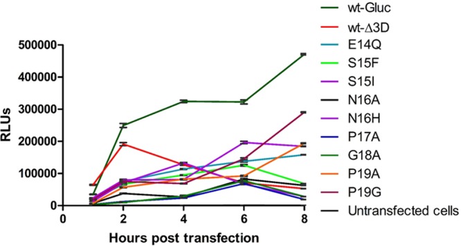
Expression of the luciferase reporter protein, Gluc, by an FMDV replicon. BHK cells were electroporated with wt or mutant RNA transcripts derived from the indicated cDNAs, and at the indicated times, cell lysates were prepared and assayed for Gluc activity. RLUs, relative light units. Data are presented as mean ± standard deviation (SD) from samples (n = 3) harvested at the indicated times.
Influence of the StopGo function on the correct processing of the FMDV P1-2A precursor.
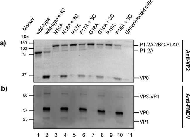
Hahn and Palmenberg (29) demonstrated that amino acid substitutions within the conserved D(V/I)E(S/T)NPG2A↓P2B motif at the C terminus of the 2A protein of EMCV not only severely reduced or abrogated the StopGo function but also impaired the subsequent cleavage of L-P1-2A by 3Cpro. The effects of substitutions in 2A on the FMDV P1-2A processing in cells has now been assayed using the truncated FMDV polyprotein termed P1-2A-2BC-FLAG (as described above) (Fig. 1d) which was coexpressed with the FMDV 3Cpro. The wt and mutant P1-2A-2BC-FLAG plasmids encoding the N16A, P17A, G18A, and P19A substitutions (shown in Fig. 4 to abrogate or impair [P19A] cleavage) were transfected, alone or together with a plasmid that expresses 3Cpro (as described in reference 7), into vTF7-3-infected BHK cells. Analysis of the FMDV P1-2A processing was done by immunoblotting using anti-VP2 antibodies (Fig. 6a). Expression of the wt plasmid alone led to complete cleavage at the 2A/2B junction of the P1-2A-2BC-FLAG polyprotein, to yield P1-2A, as expected (Fig. 6a, lane 1). Furthermore, coexpression of the wt product with 3Cpro produced VP0 (from P1-2A), also as expected (Fig. 6a, lane 2). When the mutant plasmids with defective cleavage at the 2A/2B junction were expressed alone, then the larger, intact P1-2A-2BC-FLAG product was detected (Fig. 6a, lanes 3, 5, 7, and 9), as described above (see Fig. 3). In the presence of 3Cpro, the production of VP0, derived from P1-2A (both detected with an anti-VP2 monoclonal antibody), was still readily apparent in each case (Fig. 6a, lanes 4, 6, 8, and 10). These results were confirmed by immunoblotting using anti-FMDV antibodies (Fig. 6b). Coexpression of the wt and mutant plasmids with 3Cpro produced a very similar pattern of detectable capsid proteins in each case (Fig. 6b, lanes 2, 4, 6, 8, and 10). Thus, abrogating cleavage at the 2A/2B junction did not block the processing of the capsid precursor by 3Cpro in this system. It should be noted that this is in contrast to some earlier studies (14), which showed that a truncated version of FMDV P1-2A (lacking the C terminus of VP1) could not be processed at all by 3Cpro.
FIG 6.
Transient-expression assays to determine the influence of 2A substitutions on the processing of the FMDV capsid precursor P1-2A. The wt and mutant P1-2A-2BC-FLAG plasmids were transfected alone or with pSKRH3C (40) (which expresses FMDV 3Cpro), as indicated, into vTF7-3-infected BHK cells as described in Materials and Methods. After 24 h, cell extracts were prepared and analyzed by SDS-PAGE and immunoblotting using anti-FMDV VP2 (a) and anti-FMDV (to detect all FMDV capsid proteins) (b) antisera as indicated. Molecular mass markers (kDa) are indicated on the left.
Detection of a novel FMDV 2A-2B fusion protein using immunofluorescence.
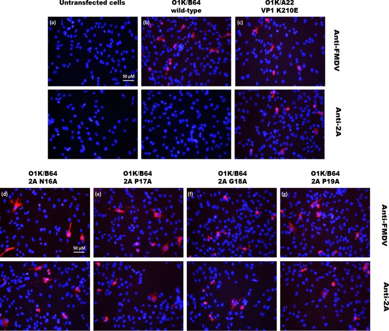
The FMDV capsid protein precursor, P1-2A, is normally processed by 3Cpro to VP0, VP3, VP1, and 2A. In previous studies, it has been shown that when the cleavage of the VP1/2A junction is impaired, then the presence of FMDV 2A (still attached to VP1, as VP1-2A) can be detected in BHK cells by immunofluorescence using anti-2A antibodies (7, 30). When the 2A is released from the VP1, then the 2A is no longer detectable (presumably it is either degraded or not fixed in the procedure). Thus, it seemed possible that substitutions within the 2A peptide that impair the 2A/2B cleavage activity (and prevent formation of viable mutant viruses), would result in the formation of detectable 2A-2B fusion proteins. Full-length FMDV RNA transcripts, with or without modifications in 2A, were introduced into BHK cells by transfection, and after 8 h, the cells were stained with either anti-2A antibodies or anti-FMDV capsid protein antibodies. The FMDV VP1 K210E mutant, as previously described (7), which produces an uncleaved VP1-2A protein, was included as a positive control for the detection of 2A attached to an adjacent protein. FMDV capsid proteins could be detected in cells transfected with each of the RNA transcripts, as expected (Fig. 7b to g). In contrast, no signal for the 2A peptide was observed in cells transfected with the wt O1K RNA (Fig. 7b) or in untransfected cells (Fig. 7a). However, the presence of FMDV 2A (still attached to VP1) was detected in cells transfected with the VP1 K210E mutant RNA (Fig. 7c), consistent with previous results (7, 30). Furthermore, using the transcripts with the mutant 2A/2B junctions, the presence of FMDV 2A, presumably attached to 2B (and maybe VP1), could be detected in the transfected cells (Fig. 7d to g). It should be noted that it is not possible to detect the free 2A peptide by immunoblotting due to its small size (ca. 2 kDa), and attempts to identify the presence of the 2A fused to other proteins in extracts from these RNA-transfected cells were unsuccessful (c.f. detection of VP1-2A within cells infected with the VP1 K210E mutant virus [7, 30]), presumably because the 2A could be attached to a number of different proteins, e.g., within 2A-2B, 2A-2BC, VP1-2A-2B, and VP1-2A-2BC, and not all cells take up and replicate the RNA transcripts.
FIG 7.
Detection of FMDV 2A fusion proteins by immunofluorescence staining within cells. BHK cells were untreated or transfected with wt or mutant FMDV RNA transcripts. At 8 h posttransfection, the cells were fixed. FMDV capsid proteins or the FMDV 2A peptide was detected using anti-FMDV O1K polyclonal antibodies (upper panels) or anti-2A antibodies (lower panels), respectively, plus a secondary antibody labeled with Alexa Fluor 568 (red). The 2A substitutions are indicated. Untransfected cells were used as a negative control, whereas the O1K VP1 K210E mutant, described previously (7), in which the 2A remains joined to VP1, served as a positive control. The cellular nuclei were visualized with DAPI (blue). Bars, 50 μm.
DISCUSSION
The 2A peptide plays a significant role in the FMDV life cycle, as it is required for the cotranslational cleavage of the growing polyprotein into two separate entities at the junction between 2A and 2B. Related 2A peptide sequences are found in a variety of other members of the picornavirus family; this suggests that they contribute significantly to the correct production and function of the viral proteins.
Using artificial polyprotein systems, it has been well documented (18, 22, 24, 26) that point mutations in the highly conserved D12(V/I)E(S/T)NPG2A↓P192B motif, located at the C terminus of FMDV 2A, can either severely reduce or completely abrogate cleavage activity. In this study, we have extended these observations and investigated the effects of single amino acid substitutions in 2A on FMDV RNA replication, on virus viability, and on polyprotein processing in its natural context within cells. The results presented here clearly demonstrate that certain 2A mutants previously found to greatly impair the StopGo activity in artificial polyprotein systems (22, 24) were still able to produce infectious viruses and thus that the wt sequence and maximal cleavage activity are not essential for virus viability. It was anticipated that some mutations might have resulted in lethal phenotypes, since earlier mutagenesis studies using FMDV and EMCV did not produce any viable progeny when the C-terminal 2A sequence was changed from SNPG↓P2B to SNPL↓V2B even after several passages (26). Interestingly, we were able to rescue viruses from all of the RNA transcripts. When the apparent cleavage activity of the mutant 2A was low (<31% of wt activity), then it was found that reversions to the wild-type sequence had occurred. This indicates that some RNA replication must have occurred (to allow the formation of wt revertants) despite the presence of a defective 2A peptide. In contrast, mutants with a higher level of cleavage activity (≥31% of the wt level) retained, in each case, the introduced amino acid substitutions in the rescued viruses. These results clearly indicate that efficient 2A-mediated cleavage activity is advantageous for the virus but that optimal efficiency is not essential. This raises the question of why the separation of the capsid proteins from the nonstructural proteins is so advantageous for some picornaviruses. It seems necessary for these viruses to have a 3C-independent mechanism to break the polyprotein. Some members of the picornavirus family (e.g., enteroviruses) possess a 2A protease to achieve the separation of the capsid protein precursor from the rest of the polyprotein, and the StopGo mechanism that occurs at the 2A/2B junction is clearly a distinct mechanism but one that is used by many members (e.g., aphthoviruses, cardioviruses, sapeloviruses, and teschoviruses) of this virus family (8).
It has been speculated (13) that 2A can act as a translational regulator to modify the amounts of the different parts of the polyprotein that are produced. In FMDV, the 2A peptide is located at the boundary between the upstream capsid proteins and the nonstructural proteins involved in RNA replication. There could be two distinct functions for the 2A peptide. One primary function of 2A could be to achieve the cleavage of the polyprotein, but it may also downregulate downstream translation. Potentially, this could prove to be beneficial to the virus, as the assembly of the FMDV capsid requires up to 60 copies of each of the four structural proteins, whereas fewer copies of the proteins involved in the replication process are required. On the other hand, it could be considered that in the early stages of the virus infection, it would seem advantageous to produce more of the proteins required for replication and processing than of the capsid proteins. It is also noteworthy that most members of the picornavirus family that use a different mechanism for separation of the capsid proteins from the nonstructural proteins do not apparently have any mechanism for modifying the ratio of proteins produced, and thus the need for such a mechanism within the picornaviruses, in general, is not established. However, recently, Napthine et al. (31) have demonstrated that in EMCV a programmed −1 ribosomal frameshift occurs within the 2B-coding region, just downstream of the 2A-coding region. This frameshift results in the production of a distinct protein, termed 2B*, and then termination of translation. The level of ribosomal frameshifting increases dramatically late in infection, and thus the production of the nonstructural proteins involved in virus replication is reduced at this time. The process requires the interaction of the EMCV 2A protein (ca. 16 kDa) with a stem-loop structure some 14 nucleotides downstream of a “slip site” (GGUUUUU) within the 2B-coding region. Although a U-rich motif (UUCUUUUUCU) is present just downstream of the coding region for the 2A/2B junction in the FMDV genome, certain other elements of this process appear to be absent. As indicated above, the FMDV 2A is only 18 residues long, and it lacks the cluster of basic residues (R95 to R97) that appear to be important for the interaction of the EMCV 2A protein with the stem-loop structure that is critical for the high frameshift efficiency. Thus, currently, there is no evidence for such a process within FMDV.
Assessment of the RNA replication efficiency, using a replicon system, demonstrated that alterations in the 2A peptide have a clear, negative effect either on the replication of the viral RNA or on the translation of the polyprotein. Clearly, the processes of translation and replication are linked, since when translation of the polyprotein is reduced, then the levels of protein available to replicate the RNA are also reduced, resulting in a lower level of RNA replication. As indicated above, it may be that the detrimental effect of the changes in 2A were accentuated by the absence of the VP1 coding sequence in the replicons. In the context of the full-length viral polyprotein, it was shown (Fig. 7) that blocking the cleavage at the FMDV 2A/2B junction produced fusion proteins containing 2A (presumably as 2A-2B or possibly VP1-2A-2B, before or after the cleavage of the VP1/2A junction by 3Cpro). However, the addition of just 18 amino acids to the N terminus of the 2B protein may be considered to be unlikely to cause this decrease in replication efficiency (indeed, it has been shown that leaving the 2A peptide fused to the C terminus of VP1 has no apparent effect on virus viability [7, 30]). It should be remembered, however, that the VP1/2A cleavage is the slowest of the 3C-mediated processing events within P1-2A (7, 14, 30). Previously it has been found that cleavage at the VP1/2A junction in poliovirus appears to a have a role in processing of the capsid precursors, since amino acid substitutions that prevented cleavage resulted in a P1 capsid precursor which was resistant to 3Cpro processing (10). Furthermore, Hahn and Palmenberg demonstrated, using in vitro translation assays, that a mutation in the EMCV 2A impaired the processing of the L-P1-2A precursor by 3Cpro (29). This may suggest a critical role for the 2A cleavage to allow proper folding of the (L)-P1-(2A) precursor to permit efficient cleavage by 3Cpro. However, in our studies, blocking the cleavage at the 2A/2B junction did not block the processing of P1-2A by 3Cpro (Fig. 6). It was also observed with TMEV that normal capsid protein processing occurred in mutant viruses in which the 2A/2B processing was blocked (26).
The Gluc replicon, as used here, lacks the coding sequences for the structural proteins except for VP4; however, the replication/translation is still impaired in the 2A mutants compared to the wild type (Fig. 5). It is therefore conceivable that the possible cleavage restrictions that could govern the processing of the structural proteins also apply to the nonstructural proteins. This may mean that correct processing of these proteins, which are required for RNA replication, is impaired, thereby resulting in lower RNA synthesis. Although the processing of the FMDV P1-2A by 3Cpro appears to be unaffected when the 2A peptide is mutated (Fig. 6), this does not rule out the possibility of a negative effect on the 2B-2C (or P3) processing. Surprisingly, there was relatively little difference in the growth characteristics between the viable 2A mutant viruses (E14Q, S15F, S15I, and N16H) and the wt (Fig. 3), which contrasts with the decrease in replication efficiency observed in the context of an FMDV replicon. This could suggest that the changes in the 2A peptide influence the initial rate of viral RNA replication but not the final virus yield.
Investigation of the effect of 2A mutations on the StopGo mechanism revealed that certain amino acid substitutions are severely detrimental for the proper function of the 2A, whereas others only moderately impair the cleavage, resulting in a mixture of products (some cleaved and others not [Fig. 4]). Previous studies have suggested that the 2A geometry is the determining factor for its function (19, 22). The current hypothesis is that the N-terminal portion of 2A (in an α-helical conformation) interacts with the ribosomal exit tunnel to confer specific constraints required for the turn motif (ESNPG) to be in a position to influence events within the peptidyl transferase center of the ribosome. Some amino acid substitutions could severely change the conformation of 2A, thereby preventing the disruption of the peptide bond formation between the G and P residues, and hence result in an uncleaved polyprotein. The substitutions N16C and N16H were found to result in cleavage, although with decreased efficiency (both cleaved and uncleaved products were observed [Fig. 4]). The function of residue N16 within 2A has not yet been determined; however, it has been suggested that N16 forms a hydrogen bond with E14 to stabilize the right turn (22). The substitutions S15A, S15I, and S15F were found to result in essentially complete cleavage, in contrast to earlier studies (22) that reported a reduction in the cleavage activity. Comparison of the 2A sequences from different picornavirus species has shown that a variety of amino acids are allowed at this position within the C terminus of 2A, suggesting that this particular amino acid is of low importance for the StopGo function. However, Sharma et al. (24) demonstrated that replacement of S15 by glycine (G) (in the FMDV sequence), which influenced the peptide secondary structure, impaired function more significantly than Ala or Pro substitutions, suggesting that increased backbone flexibility imposed by the Gly residue at this position was especially detrimental (24).
Interestingly, the substitutions P19A, P19G, P19V, and P19S greatly reduced the level of cleavage but did not abolish it (Fig. 4). This is in contrast to previous studies (22) which have reported that these amino acid substitutions resulted in no apparent cleavage activity in an artificial polyprotein system. A model for the mechanism of 2A-mediated cleavage developed by Donnelly et al. (16) suggests that the P19 residue (at the N terminus of 2B) is an absolute requirement for cleavage, as a poor nucleophilic character in this position is an integral part of the proposed mechanism. However, our data clearly show that Ala (A), Ser (S), and Val (V) residues are also functional at this position, albeit with reduced activity. Rychlík et al. (32) demonstrated that A, S, and V are, in fact, also poor nucleophiles in the context of ribosomal peptidyl transferase activity, but not to the same extent as P and G. This could explain why these amino acids are able to support the cleavage activity to some degree, although not at a level compatible with virus viability. However, this does not account for the reduced cleavage activity observed for the P19G mutants, suggesting that another, not yet identified, characteristic of residue 19 must apply.
The study by Gao et al. (23) found that the codon usage for the NPGP motif is conserved among the seven FMDV serotypes. Through the use of mRNAs encoding artificial polyproteins comprising two reporter proteins, assayed within CHO cells, the study investigated the role of synonymous codons for G18. It was concluded that the different synonymous codon usage for G18 did not influence the cleavage efficiency in that system. However, in separate studies, we have provided evidence that a clear codon bias operates to encode the NPG/P motif at the 2A/2B junction within FMDV-infected cells (33). This raises the interesting possibility that the RNA sequence itself contributes to the cleavage event at the 2A/2B junction.
MATERIALS AND METHODS
Construction of plasmids containing full-length mutant FMDV cDNAs.
The plasmid pT7S3 (27) contains the full-length cDNA for the O1Kaufbeuren (O1K) B64 strain of FMDV. Modification of the coding sequence around the 2A/2B junction was achieved by a 2-step site-directed mutagenesis procedure, a variation of the QuikChange protocol (Stratagene), using Phusion high-fidelity DNA polymerase (Thermo Fisher Scientific). The first round of PCR, using forward mutagenic 2A PCR primers (Table 2) with a single reverse primer 10PPN10 (Table 2) and the plasmid pT7S3 as the template, generated an amplicon (ca. 450 bp) specifying particular amino acid substitutions within 2A. The primary PCR products were gel purified (GeneJET gel extraction kit; Thermo Fisher Scientific) and used as primers for a second round of PCR with plasmid pT7S3 as the template. The DpnI resistant full-length products were selected in chemically competent Escherichia coli TOP10 cells (Thermo Fisher Scientific) and amplified, and then the plasmid DNA was purified (Midiprep kit; Qiagen) and verified by sequencing of the 2A-coding region with a BigDye Terminator v. 3.1 cycle sequencing kit and a 3500 genetic analyzer (Applied Biosystems).
TABLE 2.
PCR primers used to create and sequence mutant FMDV cDNAs
| Primer name | Sequence (5′→3′)a |
|---|---|
| Fwd_2A_D12A | AAGTTGGCGGGAGCCGTCGAGTCCAACCCTGG |
| Fwd_2A_V13A | AAGTTGGCGGGAGACGCCGAGTCCAACCCTGG |
| Fwd_2A_E14A | AAGTTGGCGGGAGACGTCGCGTCCAACCCTG |
| Fwd_2A_E14Q | GATGTCCAGTCCAACCCTGG |
| Fwd_2A_S15A | TTGGCGGGAGACGTCGAGGCCAACCCTG |
| Fwd_2A_S15F | GATGTCGAGTTTAACCCTGC |
| Fwd_2A_S15I | GATGTCGAGATTAACCCTGG |
| Fwd_2A_N16A | GTCCGCCCCTGGGCCCTTC |
| Fwd_2A_N16C | CGAGTCCTGCCCTGGGCCCTTCTTTTTCTCCGA |
| Fwd_2A_N16H | CGAGTCCCACCCTGGGCCCTTCTTTTTCTCCGA |
| Fwd_2A_N16V | CGAGTCCGTCCCTGGGCCCTTCTTTTTCTCCGA |
| Fwd_2A_N16W | CGAGTCCTGGCCTGGGCCCTTCTTTTTCTCCGA |
| Fwd_2A_P17A | GTCCAACGCTGGGCCCTTC |
| Fwd_2A_G18A | GTCCAACCCTGCGCCCTT C |
| Fwd_2A_P19A | CAACCCTGGGGCTTTCTT |
| Fwd_2A_P19G | CAACCCTGGGGGCTTCTT |
| Fwd_2A_P19S | CGAGTCCAACCCTGGGTCCTTCTTTTTCTCCGA |
| Fwd_2A_P19V | CGAGTCCAACCCTGGGGTCTTCTTTTTCTCCGA |
| 8APN203 | CTCCTTCAACTACGGTGCC |
| 8APN206 | CACCCGAAGACCTTGAGAG |
| 10PPN10 | CTTTGACCAACCCGGCCA |
| 13APN1 | CCGGGCCCAGGGTTGGACTCGAC |
| 13APN4 | CCGGATCCGCTAGCCATGGGAGTCAAAGTTCTGTTTGC |
| ATG_P1_fwd | ATGAATACTGGCAGCATAATAAACAACTAC |
| 2C_FLAG_Stop_rev | CTATTACTTGTCGTCATCGTCTTTGTAGTCCTGCTTGAAGATCGGGTGACTCGACAC |
| 2A_Synonomous_Fwd | TCTCCGACGTAAGATCAAACTTCTCCA |
Mutagenic nucleotides are underlined.
The generation of plasmids with three synonymous mutations ca. 20 bp downstream of the modified 2A/2B junction was achieved essentially as described above. The first round of PCRs used the forward mutagenic 2A_Synonymous_Fwd primer (Table 2) with the single reverse primer 10PPN10 (Table 2) and plasmid pT7S3 as the template. The primary PCR products were gel purified (GeneJET gel extraction kit; Thermo Fisher Scientific) and used as primers for a second round of PCR with modified versions of pT7S3, with the codons for N16, P17, G18, or P19 changed to encode an alanine (A) residue in each case, as templates.
Construction of plasmids containing an FMDV replicon containing Gaussia luciferase.
The Gaussia luciferase (Gluc) FMDV replicon was constructed by replacement of the coding region for VP2, VP3, VP1, and 2A from pT7S3-NheI (34) with the coding region for Gluc fused to FMDV 2A (as used previously [35]). The Gluc-2A sequence was amplified by PCR using primers 13APN1 and 13APN4 (Table 2) with the rPad2GL bacterial artificial chromosome (BAC) (35) as the template. The amplicon was inserted into the vector pCR-XL-TOPO (Invitrogen), and the NheI-ApaI fragment was excised and inserted between the same sites within the ca. 5-kb XbaI fragment from pT7S3-NheI (essentially as described previously [34]). The modified XbaI fragment (now containing the Gluc-2A sequence) was reconstructed into the backbone of the O1K FMDV cDNA within the XbaI-digested pT7S3 (27), and the orientation was established by restriction digestion (using EcoRI and HpaI). The Gluc FMDV replicon was termed wt-Gluc.
The replication-defective Gluc FMDV replicon, termed wt-GlucΔ3D, was prepared by digesting the wt-Gluc plasmid with BamHI and HpaI to liberate a fragment of ca. 770 bp corresponding to the 3′ terminus of the FMDV genome (including part of the 3Dpol-coding region [Fig. 1]). The large residual fragment was gel purified, blunt ended, self-ligated, and transformed into E. coli. The wt-GlucΔ3D plasmid DNA was purified (Midiprep kit; Qiagen) and verified by sequencing of the 3Dpol-coding region, as described above.
Construction of plasmids containing FMDV P1-2A-2BC-FLAG cDNA cassettes.
The FMDV cDNA cassette, in the plasmid pP1-2A-2BC-FLAG, was prepared by PCR using Phusion high-fidelity DNA Polymerase (Thermo Fisher Scientific). Briefly, the coding region for P1-2A-2BC from O1K FMDV cDNA (as in pT7S3 [27]) was amplified with the forward primer ATG_P1_fwd, which incorporates an initiation codon, and the reverse primer 2C_FLAG_Stop_rev, which includes the sequence for a FLAG epitope tag followed by a termination codon (Table 2). The blunt-end amplicon (ca. 3,670 bp) was ligated into the pJET1.2 vector (Thermo Fisher Scientific) according to the manufacturer's instructions. Sequencing revealed an unwanted initiation codon between the T7 promoter and the insert, which was then removed. A 2-step site-directed mutagenesis PCR using Phusion high-fidelity DNA polymerase as described above with mutagenic PCR 2A primers (Table 2) and 10PPN10 was used to produce the following plasmids encoding the indicated single-amino-acid substitutions within 2A: pP1-2C-FLAG D12A, V13A, E14A, E14Q, S15A, S15F, S15I, N16A, N16C, N16W, P17A, G18A, P19A, P19G, P19V, and P19S. All plasmids were propagated in E. coli TOP10 cells (Thermo Fisher Scientific), purified (Midiprep kit; Qiagen), and verified by sequencing.
In vitro transcription.
Briefly, 5 μg of replicon plasmid or full-length FMDV plasmid was linearized by digestion with HpaI (Thermo Fisher Scientific), purified (GeneJET PCR purification kit; Thermo Fisher Scientific), and eluted in RNase-free water. Both replicon and full-length FMDV RNA transcripts were prepared using the MEGAscript T7 kit (Ambion). Reaction mixtures were incubated at 37°C for 4 h and treated with 2 units of Turbo DNase for 30 min, after which the RNA was purified using the MEGAclear transcription clean-up kit according to the manufacturer's instructions. RNA integrity was assessed by electrophoresis using an ethidium bromide-stained agarose gel (1%), in Tris-borate-EDTA (TBE) buffer, and quantified by spectrophotometry (NanoDrop 1000; Thermo Fisher Scientific).
Rescue of virus from full-length cDNA plasmids.
For rescue and passage of infectious FMDV, 5 μg full-length FMDV RNA was introduced into BHK cells by electroporation (as described previously [36]). The cells were then transferred to one well of a 6-well plate and incubated for 1 to 3 days at 37°C, after which the viruses were harvested by freezing. The rescued viruses were then amplified using additional passages (P2 and P3) with fresh BHK cells. After the third passage (P3), viral RNA was extracted (RNeasy minikit; Qiagen) and converted to cDNA using Ready-To-Go You-Prime first-strand beads (GE Healthcare Life Sciences) with random hexamer primers. Amplicons (ca. 660 bp), including the 2A coding region, were amplified by PCR (AmpliTaq Gold DNA polymerase; Thermo Fischer Scientific) using the primers 8APN206 and 8APN203 (Table 2). Control reactions, without reverse transcriptase (RT), were used to ensure that the analyzed products were derived from RNA and not from the presence of carryover plasmid DNA template. The amplicons were visualized in 1% agarose gels, purified (GeneJET gel extraction kit; Thermo Fischer Scientific), and sequenced as described above. Sequences were analyzed using Geneious 7.2 (Biomatters, Auckland, New Zealand).
Gaussia luciferase assay.
BHK cells suspended in cold phosphate-buffered saline (PBS) were transferred to a 4-mm cuvette, after which 2 μg replicon RNA was added and briefly mixed, and the cells were electroporated (25 ms and 240 V; one pulse) on a Gene Pulser X-Cell (Bio-Rad). Following incubation for 10 min at room temperature, the cells were transferred to 5 wells of a 24-well plate (140 μl per well with 500 μl Dulbecco modified Eagle medium [DMEM] containing 5% fetal calf serum [FCS]). Following incubation at 37°C for the required time, the medium was removed and the BHK cells were lysed by adding 100 μl of Renilla luciferase assay lysis buffer (Promega) to the cells in each well (24-plate well) and incubated at room temperature for 15 min. The luciferase activity was quantified in a luminometer (Titertek-Berthold) by addition of this lysate (20 μl) to Renilla luciferase assay reagent (100 μl) according to the manufacturer's instructions.
Virus growth kinetics.
Virus titers for the wt and the 2A mutant viruses, E14Q, S15I, S15F, and N16H, were determined in BHK cells as 50% tissue culture infective dose (TCID50) per milliliter, as described previously (37). Monolayers of BHK cells grown in 96-well plates were infected with either wt or mutant FMDV at an MOI of 0.1 at 37°C. At 0, 2, 5, 10, and 24 h postinfection, the infected cells were harvested by freezing (at −80°C) to determine the virus yield as TCID50 per milliliter.
Transient-expression assays.
BHK cells (in 35-mm wells) were grown to 90% confluence and infected with vTF7-3, a recombinant vaccinia virus that expresses the T7 RNA polymerase (28), as described previously (38). Briefly, following the infection, plasmid DNA (pP1-2A-2BC-FLAG and its derivatives, 2 μg) was transfected alone or, when indicated, with pSKRH3C (50 ng) (39), which expresses FMDV 3Cpro, using FuGene6 (Roche), into the infected BHK cells and incubated overnight at 37°C.
Western blotting.
Cell lysates for immunoblotting were prepared by addition of cold buffer C (0.125 M NaCl, 20 mM Tris-HCl [pH 8.0], 0.5% NP-40) to the cells. After incubation (on ice, for at least 5 min), the cell extracts were clarified by centrifugation (20,000 × g for 10 min), and Laemmli sample buffer (with 25 mM dithiothreitol [DTT]) was added (as described previously [40]). Following heating to 98°C for 5 min, samples were resolved by SDS-PAGE (4 to 15% polyacrylamide), transferred to a polyvinylidene difluoride (PVDF) membrane (Bio-Rad), and blocked for 1 h in 5% PBS–Tween (PBS, 0.1% Tween) with 5% nonfat milk. The membranes were incubated overnight at 4°C with either goat anti-FLAG antibodies (Abcam), guinea pig anti-FMDV O1 Manisa serum (to detect FMDV capsid proteins), or mouse anti-FMDV VP2 (4B2) monoclonal antibody (41), as used previously (7). The membranes were washed 3 times with PBS-Tween and incubated for 3 h at room temperature with either horseradish peroxidase (HRP)-conjugated anti-goat IgG (Dako), HRP-conjugated anti-guinea pig IgG (Dako), or HRP-conjugated anti-mouse IgG (Dako). The membranes were then washed 3 times with PBS-Tween, and bound proteins were detected using a chemiluminescence detection kit (ECL Prime; Amersham) with a Chemi-Doc XRS system (Bio-Rad). The intensities of the signals for the FLAG-tagged polyproteins were, when necessary, quantitated using ImageJ software (v1.50).
Immunofluorescence assay.
Monolayers of BHK cells were grown on glass coverslips in 6-well plates, and immediately prior to transfection, cells were washed briefly in PBS and the medium replaced with DMEM without serum. FMDV RNA transcripts were introduced into BHK cells using Lipofectin transfection reagent (Thermo Fisher Scientific) according to the manufacturer's instructions. After 8 h, the cells were fixed, stained, and mounted as described previously (7, 30) using rabbit anti-FMDV O serum or rabbit anti-2A (ABS31; Merck) followed by a donkey Alexa Fluor 568-labeled anti-rabbit IgG (A10042; Life Technologies). The slides were washed in PBS, after which they were mounted with Vectashield (Vector Laboratories) containing DAPI (4′,6′-diamidino-2-phenylindole), and images were captured using an epifluorescence microscope.
ACKNOWLEDGMENTS
We thank Li Yu (Chinese Academy of Agricultural Sciences, China) for providing us with the anti-FMDV VP2 antibody. We also acknowledge the excellent technical assistance of Preben Normann and helpful advice from Thea Kristensen.
REFERENCES
- 1.Jamal SM, Belsham GJ. 2013. Foot-and-mouth disease: past, present and future. Vet Res 44:116–129. doi: 10.1186/1297-9716-44-116. [DOI] [PMC free article] [PubMed] [Google Scholar]
- 2.Martinez-Salas E, Belsham GJ. 2017. Genome organisation, translation and replication of foot-and-mouth disease virus RNA, p 13–42. In Sobrino F, Domingo E (ed), Foot-and-mouth disease: current research and emerging trends. Caister Academic Press, Poole, United Kingdom. [Google Scholar]
- 3.Belsham GJ. 2005. Translation and replication of FMDV RNA. Curr Top Microbiol Immunol 288:43–70. [DOI] [PubMed] [Google Scholar]
- 4.Medina M, Domingo E, Brangwyn JK, Belsham GJ. 1993. The two species of the foot-and-mouth disease virus leader protein, expressed individually, exhibit the same activities. Virology 194:355–359. doi: 10.1006/viro.1993.1267. [DOI] [PubMed] [Google Scholar]
- 5.Strebel K, Beck E. 1986. A second protease of foot-and-mouth disease virus. J Virol 58:893–899. [DOI] [PMC free article] [PubMed] [Google Scholar]
- 6.Curry S, Fry E, Blakemore W, Abu-Ghazaleh R, Jackson T, King A, Lea S, Newman J, Stuart D. 1997. Dissecting the roles of VP0 cleavage and RNA packaging in picornavirus capsid stabilization: the structure of empty capsids of foot-and-mouth disease virus. J Virol 71:9743–9752. [DOI] [PMC free article] [PubMed] [Google Scholar]
- 7.Gullberg M, Polacek C, Bøtner A, Belsham GJ. 2013. Processing of the VP1/2A junction is not necessary for production of foot-and-mouth disease virus empty capsids and infectious viruses: characterization of “self-tagged” particles. J Virol 87:11591–11603. doi: 10.1128/JVI.01863-13. [DOI] [PMC free article] [PubMed] [Google Scholar]
- 8.Luke GA, de Felipe P, Lukashev A, Kallioinen SE, Bruno EA, Ryan MD. 2008. Occurrence, function and evolutionary origins of “2A-like” sequences in virus genomes. J Gen Virol 89:1036–1042. doi: 10.1099/vir.0.83428-0. [DOI] [PMC free article] [PubMed] [Google Scholar]
- 9.Sommergruber W, Zorn M, Blaas D, Fessl F, Volkmann P, Maurer-Fogy I, Pallai P, Merluzzi V, Matteo M, Skern T, Kuechler E. 1989. Polypeptide 2A of human rhinovirus type 2: identification as a protease and characterization by mutational analysis. Virology 169:68–77. doi: 10.1016/0042-6822(89)90042-1. [DOI] [PubMed] [Google Scholar]
- 10.Toyoda H, Nicklin MJH, Murray MG, Anderson CW, Dunn JJ, Studier FW, Wimmer E. 1986. A second virus-encoded proteinase involved in proteolytic processing of poliovirus polyprotein. Cell 45:761–770. doi: 10.1016/0092-8674(86)90790-7. [DOI] [PubMed] [Google Scholar]
- 11.Belsham GJ. 1993. Distinctive features of foot-and-mouth disease virus, a member of the picornavirus family; aspects of virus protein synthesis, protein processing and structure. Prog Biophys Mol Biol 60:241–260. doi: 10.1016/0079-6107(93)90016-D. [DOI] [PMC free article] [PubMed] [Google Scholar]
- 12.Palmenberg AC, Parks GD, Hall D, Ingraham RH, Seng TW, Pallal PV. 1992. Proteolytic processing of the cardioviral cleavage in clone-derived P2 region: primary 2A/2B precursors. Virology 190:754–762. doi: 10.1016/0042-6822(92)90913-A. [DOI] [PubMed] [Google Scholar]
- 13.Tulloch F, Luke GA, Ryan MD. 2017. Foot-and-mouth disease virus proteinases and polyprotein processing, p 43–59. In Sobrino F, Domingo E (ed), Foot-and-mouth disease: current research and emerging trends. Caister Academic Press, Poole, United Kingdom. [Google Scholar]
- 14.Ryan MD, Belsham GJ, King AMQ. 1989. Specificity of enzyme-substrate interactions in foot-and-mouth disease virus polyprotein processing. Virology 173:35–45. doi: 10.1016/0042-6822(89)90219-5. [DOI] [PubMed] [Google Scholar]
- 15.Ryan MD, King AMQ, Thomas GP. 1991. Cleavage of foot-and-mouth disease virus polyprotein is mediated by residues located within a 19 amino acid sequence. J Gen Virol 72:2727–2732. doi: 10.1099/0022-1317-72-11-2727. [DOI] [PubMed] [Google Scholar]
- 16.Donnelly M, Luke G, Mehrotra A, Li X, Hughes LE, Gani D, Ryan MD. 2001. Analysis of the aphthovirus 2A/2B polyprotein “cleavage” mechanism indicates not a proteolytic reaction, but a novel translational effect: a putative ribosomal “skip.” J Gen Virol 82:1013–1025. [DOI] [PubMed] [Google Scholar]
- 17.Atkins JF, Wills NM, Loughran G, Wu C-Y, Parsawar K, Ryan MD, Wang C-H, Nelson CC. 2007. A case for “StopGo”: reprogramming translation to augment codon meaning of GGN by promoting unconventional termination (Stop) after addition of glycine and then allowing continued translation (Go). RNA 13:803–810. doi: 10.1261/rna.487907. [DOI] [PMC free article] [PubMed] [Google Scholar]
- 18.Doronina VA, Wu C, de Felipe P, Sachs MS, Ryan MD, Brown JD. 2008. Site-specific release of nascent chains from ribosomes at a sense codon. Mol Cell Biol 28:4227–4239. doi: 10.1128/MCB.00421-08. [DOI] [PMC free article] [PubMed] [Google Scholar]
- 19.Ryan MD, Donnelly M, Lewis A, Mehrotra AP, Wilkie J, Gani D. 1999. A model for nonstoichiometric, cotranslational protein scission in eukaryotic ribosomes. Bioorg Chem 27:55–79. doi: 10.1006/bioo.1998.1119. [DOI] [Google Scholar]
- 20.Groppo R, Palmenberg AC. 2007. Cardiovirus 2A protein associates with 40S but not 80S ribosome subunits during infection. J Virol 81:13067–13074. doi: 10.1128/JVI.00185-07. [DOI] [PMC free article] [PubMed] [Google Scholar]
- 21.Donnelly M, Gani D, Flint M, Monaghan S, Ryan MD. 1997. The cleavage activities of aphthovirus and cardiovirus 2A proteins. J Gen Virol 78:13–21. doi: 10.1099/0022-1317-78-1-13. [DOI] [PubMed] [Google Scholar]
- 22.Donnelly MLL, Luke GA, Hughes LE, Luke G, Mendoza H, Dam E, Gani D, Ryan MD. 2001. The “cleavage” activities of foot-and-mouth disease virus 2A site-directed mutants and naturally occurring “2A-like” sequences. J Gen Virol 82:1027–1041. doi: 10.1099/0022-1317-82-5-1027. [DOI] [PubMed] [Google Scholar]
- 23.Gao Z, Zhou J, Zhang J, Ding Y, Liu Y. 2014. The silent point mutations at the cleavage site of 2A/2B have no effect on the self-cleavage activity of 2A of foot-and-mouth disease virus. Infect Genet Evol 28:101–106. doi: 10.1016/j.meegid.2014.08.006. [DOI] [PubMed] [Google Scholar]
- 24.Sharma P, Yan F, Doronina VA, Escuin-Ordinas H, Ryan MD, Brown JD. 2012. 2A peptides provide distinct solutions to driving stop-carry on translational recoding. Nucleic Acids Res 40:3143–3151. doi: 10.1093/nar/gkr1176. [DOI] [PMC free article] [PubMed] [Google Scholar]
- 25.Minskaia E, Nicholson J, Ryan MD. 2013. Optimisation of the foot-and-mouth disease virus 2A co-expression system for biomedical applications. BMC Biotechnol 13:67. doi: 10.1186/1472-6750-13-67. [DOI] [PMC free article] [PubMed] [Google Scholar]
- 26.Loughran G, Libbey JE, Uddowla S, Scallan MF, Ryan MD, Fujinami RS, Rieder E, Atkins JF. 2013. Theiler's murine encephalomyelitis virus contrasts with encephalomyocarditis and foot-and-mouth disease viruses in its functional utilization of the StopGo non-standard translation mechanism. J Gen Virol 94:348–353. doi: 10.1099/vir.0.047571-0. [DOI] [PMC free article] [PubMed] [Google Scholar]
- 27.Ellard FM, Drew J, Blakemore WE, Stuart DI, King AMQ. 1999. Evidence for the role of His 142 of protein 1C in the acid induced disassembly of foot and mouth disease virus capsids. J Gen Virol 80:1911–1918. doi: 10.1099/0022-1317-80-8-1911. [DOI] [PubMed] [Google Scholar]
- 28.Fuerst TR, Niles EG, Studier FW, Moss B. 1986. Eukaryotic transient-expression system based on recombinant vaccinia virus that synthesizes bacteriophage T7 RNA polymerase. Proc Natl Acad Sci U S A 83:8122–8126. [DOI] [PMC free article] [PubMed] [Google Scholar]
- 29.Hahn H, Palmenberg AC. 1996. Mutational analysis of the encephalomyocarditis virus primary cleavage. J Virol 70:6870–6875. [DOI] [PMC free article] [PubMed] [Google Scholar]
- 30.Kristensen T, Normann P, Gullberg M, Fahnøe U, Polacek C, Rasmussen TB, Belsham GJ. 2017. Determinants of the VP1/2A junction cleavage by the 3C protease in foot-and-mouth disease virus-infected cells. J Gen Virol 98:385–395. doi: 10.1099/jgv.0.000664. [DOI] [PMC free article] [PubMed] [Google Scholar]
- 31.Napthine S, Ling R, Finch LK, Jones JD, Bell S, Brierley I, Firth AE. 2017. Protein-directed ribosomal frameshifting temporally regulates gene expression. Nat Commun 8:15582. doi: 10.1038/ncomms15582. [DOI] [PMC free article] [PubMed] [Google Scholar]
- 32.Rychlík I, Černá J, Chládek S, Pulkrábek P, Žemlička J. 1970. Substrate specificity of ribosomal peptidyl transferase. The effect of the nature of the amino acid side chain. Eur J Biochem 16:136–142. [DOI] [PubMed] [Google Scholar]
- 33.Kjær J, Belsham GJ. 2018. Selection of functional 2A sequences within foot-and-mouth disease virus; requirements for the NPGP motif with a distinct codon bias. RNA 24:12–17. doi: 10.1261/rna.063339.117. [DOI] [PMC free article] [PubMed] [Google Scholar]
- 34.Bøtner A, Kakker NK, Barbezange C, Berryman S, Jackson T, Belsham GJ. 2011. Capsid proteins from field strains of foot-and-mouth disease virus confer a pathogenic phenotype in cattle on an attenuated, cell-culture-adapted virus. J Gen Virol 92:1141–1151. doi: 10.1099/vir.0.029710-0. [DOI] [PubMed] [Google Scholar]
- 35.Risager PC, Fahnøe U, Gullberg M, Rasmussen TB, Belsham GJ. 2013. Analysis of classical swine fever virus RNA replication determinants using replicons. J Gen Virol 94:1739–1748. doi: 10.1099/vir.0.052688-0. [DOI] [PubMed] [Google Scholar]
- 36.Nayak A, Goodfellow IG, Woolaway KE, Birtley J, Curry S, Belsham GJ. 2006. Role of RNA structure and RNA binding activity of foot-and-mouth disease virus 3C protein in VPg uridylylation and virus replication. J Virol 80:9865–9875. doi: 10.1128/JVI.00561-06. [DOI] [PMC free article] [PubMed] [Google Scholar]
- 37.Reed LJ, Muench H. 1938. A simple method of estimating fifty percent endpoints. Am J Hyg (Lond) 27:493–497. [Google Scholar]
- 38.Belsham GJ, Nielsen I, Normann P, Royall E, Roberts LO. 2008. Monocistronic mRNAs containing defective hepatitis C virus-like picornavirus internal ribosome entry site elements in their 5′ untranslated regions are efficiently translated in cells by a cap-dependent mechanism. RNA 14:1671–1680. doi: 10.1261/rna.1039708. [DOI] [PMC free article] [PubMed] [Google Scholar]
- 39.Belsham GJ, McInerney GM, Ross-Smith N. 2000. Foot-and-mouth disease virus 3C protease induces cleavage of translation initiation factors eIF4A and eIF4G within infected cells. J Virol 74:272–280. doi: 10.1128/JVI.74.1.272-280.2000. [DOI] [PMC free article] [PubMed] [Google Scholar]
- 40.Polacek C, Gullberg M, Li J, Belsham GJ. 2013. Low levels of foot-and-mouth disease virus 3C protease expression are required to achieve optimal capsid protein expression and processing in mammalian cells. J Gen Virol 94:1249–1258. doi: 10.1099/vir.0.050492-0. [DOI] [PubMed] [Google Scholar]
- 41.Yu Y, Wang H, Zhao L, Zhang C, Jiang Z, Yu L. 2011. Fine mapping of a foot-and-mouth disease virus epitope recognized by serotype independent monoclonal antibody 4B2. J Microbiol 49:94–101. doi: 10.1007/s12275-011-0134-1. [DOI] [PubMed] [Google Scholar]


