This systematic review and meta-analysis evaluates the association of vegetation size greater than 10 mm with emobolic events in patients with infective endocarditis.
Key Points
Question
What is the association of vegetation size greater than 10 mm with embolic events in patients with infective endocarditis?
Findings
In this systematic review and meta-analysis of 21 unique studies that included 6646 unique patients with infective endocarditis and 5116 measured vegetations, patients with a vegetation size greater than 10 mm had significantly increased odds of embolic events and mortality.
Meaning
Large vegetations (>10 mm) may be associated with an increased risk of embolization.
Abstract
Importance
Infective endocarditis is a life-threating condition with annual mortality of as much as 40% and is associated with embolic events in as many as 80% of cases. These embolic events have notable prognostic implications and have been linked to increased length of stay in intensive care units and mortality. A vegetation size greater than 10 mm has often been suggested as an optimal cutoff to estimate the risk of embolism, but the evidence is based largely on small observational studies.
Objective
To study the association of vegetation size greater than 10 mm with embolic events using meta-analytic techniques.
Data Sources
A computerized literature search of all publications in the PubMed and EMBASE databases from inception to May 1, 2017, was performed with search terms including varying combinations of infective endocarditis, emboli, vegetation size, pulmonary infarct, stroke, splenic emboli, renal emboli, retinal emboli, and mesenteric emboli. This search was last assessed as being up to date on May 1, 2017.
Study Selection
Observational studies or randomized clinical trials that evaluated the association of vegetation size greater than 10 mm with embolic events in adult patients with infective endocarditis were included. Conference abstracts and non–English language literature were excluded. The search was conducted by 2 independent reviewers blinded to the other’s work.
Data Extraction and Synthesis
Following PRISMA guidelines, the 2 reviewers independently extracted data; disputes were resolved with consensus or by a third investigator. Categorical dichotomous data were summarized across treatment arms using Mantel-Haenszel odds ratios (ORs) with 95% CIs. Heterogeneity of effects was evaluated using the Higgins I2 statistic.
Results
The search yielded 21 unique studies published from 1983 to 2016 with a total of 6646 unique patients with infective endocarditis and 5116 vegetations with available dimensions. Patients with a vegetation size greater than 10 mm had increased odds of embolic events (OR, 2.28; 95% CI, 1.71-3.05; P < .001) and mortality (OR, 1.63; 95% CI, 1.13-2.35; P = .009) compared with those with a vegetation size less than 10 mm.
Conclusions and Relevance
In this meta-analysis of 21 studies, patients with vegetation size greater than 10 mm had significantly increased odds of embolism and mortality. Understanding the risk of embolization will allow clinicians to adequately risk stratify patients and will also help facilitate discussions regarding surgery in patients with a vegetation size greater than 10 mm.
Introduction
Infective endocarditis is a rare but potentially life-threating condition with annual mortality of as much as 40%. Infective endocarditis is often accompanied by several complications that contribute to morbidity and mortality, prime among them being systemic embolic events. Embolic events have notable prognostic implications and have been linked to increased length of intensive care unit stay and mortality. A vegetation size greater than 10 mm has often been suggested as an optimal cutoff to estimate the risk of embolic events. This cutoff is used by the American Heart Association guidelines on infective endocarditis as an important part of their recommendations for early surgery and also forms an integral part of protocols for large prospective clinical trials. However, the evidence behind this seemingly arbitrary cutoff is based largely on observational data from small studies with varied methods and periods of observation and the significant potential for selection bias. Previous literature on embolic risk in patients with large vegetations has yielded varied results. Some studies have suggested that the risk of embolic events is not increased with larger vegetation sizes, whereas others have noted significantly increased odds of systemic embolization with a vegetation size greater than 10 mm. A meta-analysis published in 1997 aimed to fill this gap in the literature and reported a significantly increased risk of systemic embolization and a borderline increased risk of death with large vegetations. However, this analysis had several design flaws, such as inclusion of no vegetation in the group with a vegetation size less than 10 mm, limitation of the search to MEDLINE, no formal assessment of study quality, and no quantification of heterogeneity. Moreover, several large studies have been published on this topic since 1997, and an updated meta-analysis is needed. Therefore, we conducted a systematic review of literature and meta-analysis to study the association of a vegetation size greater than 10 mm with embolic events. Within our selected pool of studies, we also evaluated the association of a vegetation size greater than 10 mm with all-cause mortality. We further used meta-regression techniques to evaluate the association of age, sex, and type of involved valve with the overall risk of embolization with a vegetation size greater than 10 mm.
Methods
Data Sources and Searches
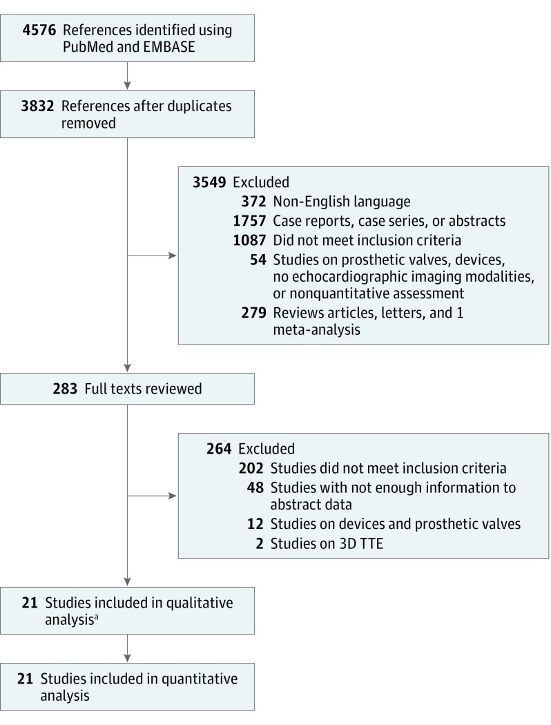
We performed a computerized literature search of all publications in the PubMed and EMBASE databases from inception to May 1, 2017. We then manually searched the reference lists of included articles. This search was last assessed as being up to date on May 1, 2017 (Figure 1). Our aim was to include all randomized and nonrandomized studies conducted on patients hospitalized for infective endocarditis with a comparison between those with and without a vegetation size greater than 10 mm with regard to the outcome of embolic events. Inclusion of vegetation size equal to 10 mm varied, with some studies including it in the group with vegetation sizes greater than 10 mm and others including it in the group with vegetation sizes less than 10 mm (Table). Search terms included varying combinations of the following keywords: infective endocarditis, emboli, vegetation size, pulmonary infarct, stroke, splenic emboli, renal emboli, retinal emboli, and mesenteric emboli.
Figure 1. PRISMA Diagram of Search Strategy for Meta-analysis.
The search was last assessed as being up to date on May 1, 2017. TTE indicates 2-dimensional transthoracic echocardiography.
aTwo studies were added after manual review.
Table. Description of Studies Included in the Meta-analysis.
| Source | Years of Study | Study Type | Patients With IE, No. | Patients With Measured Vegetation Sizes, No. | Valves Involved | Type of EE | Inclusion Cutoff Above or Below 10 mm | Criteria for IE | Patients With Staphylococcus aureus Bacteremia, No. (%) |
|---|---|---|---|---|---|---|---|---|---|
| Wong et al, 1983 | 1978-1981 | Prospective | 34 | 27 | Aortic, mitral, tricuspid | Splenic, bone, cerebral, pulmonary, renal | >10 | 3 of the Following present: fever, regurgitant murmur, EE, or bacteremia | 16/34 Patients (47.1) |
| Robbins et al, 1986 | 1979-1982 | Prospective | 21 | 17 | Tricuspid, pulmonary, prosthetic | Pulmonary | >10 | Major: echocardiographic evidence of vegetations, fever; minor: bacteremia, EE, murmur (need 2 major or 1 major +3 minor) | 11/23 Patients (47.8) |
| Lutas et al, 1986 | 1979-1982 | Retrospective | 77 | 42 | Aortic, mitral, tricuspid, prosthetic | NA | <10 | Not clear, mentions bacteremia and vegetations | 22/76 Patients with blood cultures (28.9) |
| Buda et al, 1986 | NA | Prospective | 50 | 18 | Aortic, mitral, tricuspid | Cerebral, peripheral | NA | Presence of murmur with ≥2 positive blood culture results obtained at separate times yielding the same organism and ≥1 of the following: a new or changed murmur, peripheral stigmata of IE, or laboratory evidence of IE | 21/50 Patients (42.0) |
| Erbel et al, 1988 | NA | Prospective | 96 | 51 | Aortic, mitral, tricuspid, mural left ventricle/right ventricle | NA | <10 | ≥1 of the Following: fever, chills, night sweats, arthralgia, murmur, EE, bacteremia, anemia, or elevated ESR | 16/39 Patients with blood cultures (41.0) (all staphylococcus) |
| Mügge et al, 1989 | 1984-1987 | Prospective | 105 | 96 | Aortic, mitral, tricuspid, prosthetic | Cerebral, lung, spleen, kidney | >10 | Fever, regurgitant murmur, or anemia associated with bacteremia | 24/97 Patients with blood cultures (24.7) |
| Hwang et al, 1993 | 1989-1992 | Prospective | 41 | 41 | Aortic, mitral, tricuspid, pulmonic | Cerebral, extremity, spleen, kidney, retinal artery | >10 | Histologic findings of IE or clinical diagnosis of probable or possible IE by the Von Reyn criteria | 7/50 Patients with blood cultures (14.0) |
| Jung et al, 1994 | 1983-1993 | Retrospective | 80 | 37 | Aortic, mitral, tricuspid, pulmonic, prosthetic | Cerebral, extremity, kidney, lung, spleen, coronary vessel | >10 | ≥1 of the Following: direct evidence of IE at time of surgery or culture of embolus; ≥2 positive blood culture results with ≥3 of fever, underlying heart disease, new murmur, vegetations, or EE; positive blood culture result with underlying heart disease or new regurgitant murmur; negative blood culture result with fever, heart disease, vegetations, or EE | 12/51 Patients with blood cultures (23.5) |
| De Castro et al, 1997 | 1993-1995 | Retrospective | 57 | 54 | Aortic, mitral, tricuspid, pulmonic | Peripheral artery, cerebral, lung, coronary artery | NA | Duke criteria | 16/57 Patients with blood cultures (28.1) (all staphylococcus) |
| Di Salvo et al, 2001 | 1993-2000 | Prospective | 178 | 133 | Aortic, mitral, tricuspid, pulmonic, prosthetic | Cerebral, lung, spleen, kidney, peripheral arteries, eye coronaries | >10 | Duke criteria | 43/178 Patients (24.2) |
| Vilacosta et al, 2002 | 1996-2000 | Prospective | 211 | 169 | Aortic, mitral | Cerebral, upper extremity, lower extremity, kidney, spleen | >10 | Duke criteria | 38/211 Patients (18.0) |
| Deprèle et al, 2004 | 1995-2001 | Retrospective | 80 | 80 | Aortic, mitral, tricuspid | Cerebral, skin, spleen, kidney, lung, coronaries | NA | Duke criteria | 6/80 Patients (7.5) |
| Thuny et al, 2005 | 1993-2003 | Prospective | 384 | 384 | Aortic, mitral, tricuspid, pulmonic | Cerebral, spleen, kidney, peripheral arteries, eye coronaries, lung | <10 | Duke criteria | NA for cohort of new EE; 82/384 patients (21.4) for cohort of all EE |
| Gotsman et al, 2007 | 1991-2000 | Retrospective | 100 | 50 | Aortic, mitral, tricuspid, prosthetic | Arterial emboli, intracranial hemorrhage, pulmonary infarcts, mycotic aneurysms, splinter hemorrhage, Janeway lesions, conjunctival hemorrhage | >10 | Duke criteria | 16/96 Patients (16.7) |
| Pepin et al, 2009 | 1991-2006 | Retrospective | 241 | 101 | Aortic, mitral, prosthetic | Cerebral, coronary | >10 | Modified Duke criteria | 118/241 Patients (49.0) |
| Leitman et al, 2012 | 1998-2010 | Retrospective | 146 | 102 | Aortic, mitral, tricuspid, prosthetic, pacemaker, right atrial wall | Brain, kidney, extremities, lung, subarachnoid hemorrhage | >10 | Duke criteria | 39% (All staphylococcus)a |
| Hajihossainlou et al, 2013 | 1995-2010 | Retrospective | 286 | 286 | Aortic, mitral, tricuspid, prosthetic | NA | >10 | Duke criteria | 87/286 Patients (30.4) |
| García-Cabrera et al, 2013 | 1984-2009 | Prospective | 1345 | 1116 | Aortic, mitral, prosthetic | Ischemic stroke | >10 | Modified Duke criteria | 263/1345 Patients (19.6) |
| Misfeld et al, 2014 | 1995-2012 | Retrospective | 1571 | 1516 | Aortic, mitral, tricuspid, pulmonic, prosthetic | Cerebral embolism | >10 | Modified Duke criteria | 143/375 Patients with embolism (38.1) |
| Rizzi et al, 2014 | 2004-2011 | Retrospective | 1456 | 710 | Aortic, mitral, prosthetic, right sided | Cerebral, pulmonary, splenic, limbs | >10 | Modified Duke criteria | 283/1456 Patients (19.4) |
| Aherrera et al, 2016 | 2013-2016 | Prospective | 87 | 86 | Aortic, mitral, tricuspid, pulmonic, prosthetic | Arterial emboli, intracranial hemorrhage, pulmonary infarcts, mycotic aneurysms | >10 | Modified Duke criteria | 13/87 Patients (14.9) |
Abbreviations: CXR, chest x-ray; EE, embolic events; ESR, erythrocyte sedimentation rate; IE, infective endocarditis; NA, not available.
Numbers of patients or cases were not available in this study.
Study Selection
We applied the Preferred Reporting Items for Systematic Reviews and Meta-Analyses statement (PRISMA) to the methods for this study. We used the following inclusion criteria: (1) studies of adult patients with native valve infective endocarditis due to any organism, (2) studies that provided the information on embolic events in patients with a vegetation size less than 10 mm and a vegetation size greater than 10 mm, and (3) studies in which vegetation size was estimated by 2-dimensional transthoracic echocardiography (TTE) and/or transesophageal echocardiography (TEE). We used the following exclusion criteria: (1) studies on prosthetic valve infective endocarditis and device-associated infective endocarditis unless data for native valve infective endocarditis were also present, (2) studies in which vegetation size was not quantified but rather qualitatively assessed (such as small vs large), (3) studies in which data for no vegetation were included with vegetation size less than 10 mm and separate estimates of embolic events for vegetation size less than 10 mm could not be extracted from available information, (4) studies in which a hazard ratio or an adjusted odds ratio (OR) was available but enough data were not present to extract or calculate an unadjusted OR, (5) studies in which multiple prognostic markers were tested and data for a complete cohort of patients with a vegetation size less than 10 mm or greater than 10 mm were unavailable, (6) studies in which vegetation size was measured with modalities other than 2-dimensional TTE or TEE, (7) conference abstracts, (8) case reports and case series, and (9) non–English language literature.
Study End Points
The primary aim of this meta-analysis was to study the association of a vegetation size greater than 10 mm with embolic events in patients with infective endocarditis. As a secondary analysis, we also compared the odds of all-cause mortality in patients with vegetation sizes less than and greater than 10 mm.
Data Extraction and Study Quality Appraisal
Two of us (D.M. and A.M.) abstracted data from all included studies on a standardized worksheet. The following data were collected: name of the author(s), study title, year of publication, years of study, type of study (retrospective vs prospective), type of echocardiography used, percentage of male patients, percentage of type of valve involved (aortic, mitral, tricuspid, or prosthetic), percentage of patients undergoing surgery, percentage of patients with Staphylococcus aureus bacteremia, and mean age. Data used to calculate the OR for systemic embolization among different vegetation sizes were also obtained. For the study by Thuny et al, data on embolic events were available before and after antibiotic administration. We used the postadministration data to calculate embolic events. For the study by Leitman et al, data on short- and long-term mortality were available. Herein we used the data on short-term mortality to maintain uniformity with other studies and to avoid incorporation of effect from other confounding factors that may influence long-term mortality in these patients. Wherever separate values for TTE and TEE were provided, the values with the maximum number of patients were incorporated into our analysis.
Two of us (D.M. and A.M.) independently assessed the risk of bias among the included studies using the standardized Newcastle-Ottawa Scale. This validated instrument for appraising observational studies measures risk of bias in 8 categories: representativeness of the exposed cohort, selection of the nonexposed cohort, ascertainment of exposure, demonstration that the outcome of interest was not present at the start of the study, comparability, assessment of outcome, follow-up long enough for outcomes to occur, and adequacy of follow-up of cohorts (eTable 1 in the Supplement). All discrepancies in data abstraction or quality appraisal were resolved by discussion or adjudication by another of us (M.Y.D.). eTable 2 in the Supplement provides the PRISMA checklist for the meta-analysis.
Data Synthesis and Analysis
We summarized categorical dichotomous data across treatment arms using the Mantel-Haenszel OR with 95% CI. We evaluated heterogeneity of effects using the Higgins I2 statistic. We also used Mantel-Haenszel risk difference to calculate summary effects for the primary and secondary outcomes. Random effects were used for all our analyses. We also performed meta-regression analyses for the primary outcome to assess whether the association of vegegation size with embolic risk is modulated by prespecified study-level factors such as age, male sex, type of valve involved, and prosthetic valve involvement. This analysis was not possible for the secondary outcome owing to the smaller number of included studies for that analysis. We also performed a sensitivity analysis to evaluate how removal of each study affected the overall outcome and a prespecified subgroup analysis stratifying the primary outcome by type of study (prospective vs retrospective), years of publication (1980-1999 vs 2000-2016), and use of Duke (or modified Duke) criteria. To address publication bias, we used visual inspection of funnel plots and the Egger test. Comprehensive Meta-analysis software (version 3.3.070; https://www.meta-analysis.com) was used for meta-analysis and meta-regression. A 2-tailed P = .05 was considered to be significant for all our analyses.
Results
Our search yielded 21 unique studies published from 1983 to 2016, including a total of 6646 unique patients with infective endocarditis and a total of 5116 vegetation specimens with available dimensions. Characteristics of the studies are listed in the Table.
Association of Vegetation Size Greater Than 10 mm With Embolic Events
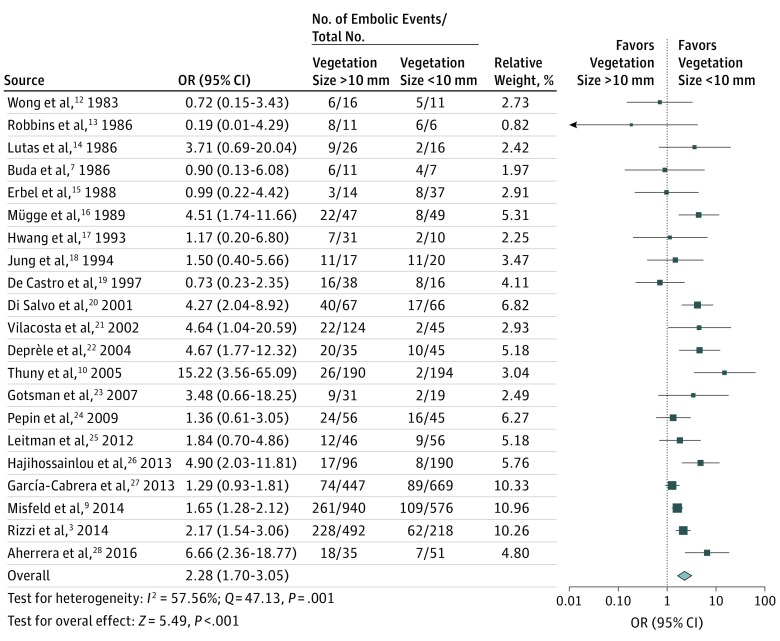
We observed that patients with a vegetation size greater than 10 mm had increased odds of embolic events compared with patients with a vegetation size less than 10 mm (OR, 2.28; 95% CI, 1.71-3.05; P < .001) (Figure 2). The risk difference was 0.13 (95% CI, 0.09-0.18; P < .001). We further explored the heterogeneity among the included studies by performing subgroup analysis by period and study method. We observed that the odds of embolic events were comparable between patients with and without a vegetation size greater than 10 mm when studies in the subgroup of studies published from 1983 to 1999 (OR, 1.41; 95 CI, 0.79-2.53; P = .24) were considered. However, we found a markedly increased likelihood of embolic events with a vegetation size greater than 10 mm in the studies published from 2000 to 2016 (OR, 2.70; 95% CI, 1.91-3.81; P < .001), but the difference between the 2 subgroups was not significant (Q = 3.49; P = .06) (eFigure 1 in the Supplement). A meta-analysis of prospective (OR, 2.44; 95% CI, 1.31-4.53) and retrospective (OR, 2.05; 95% CI, 1.53-2.75) studies showed increased odds of embolic events with a vegetation size greater than 10 mm, with no differences between the subgroups (Q = 0.24; P = .62) (eFigure 2 in the Supplement). We also observed no difference in the subgroup of studies that used Duke or modified Duke criteria (OR, 2.52; 95% CI, 1.81-3.52; P < .001) compared with studies that did not (OR, 1.61; 95% CI, 0.86-3.01; P = .13) (Q = 1.53; P = .21). When the primary outcome was stratified by left- and right-sided vegetation specimens, in the 5 studies that allowed for calculation of primary outcome for left-sided vegetation specimens, the association was not significant (OR, 1.37; 95% CI, 1.02-1.83; P = .06). Only 2 studies had isolated data for right-sided vegetation specimens available. The association did not reach significance for these studies, and the CIs were large (OR, 1.42; 95% CI, 0.17-11.49) (eFigure 3 in the Supplement).
Figure 2. Forest Plot for Comparative Odds of Embolic Events.
Data are stratified by vegetation size less than and greater than 10 mm. Squares represent odds ratios (ORs), with their size proportional to the weight of the study using the Mantel Haenszel test; horizontal lines, 95% CIs; diamond overall OR and 95% CI; Q, Cochrane Q statistic.
Use of meta-regression revealed that odds of embolic events with a vegetation size greater than 10 mm were not significantly associated with the mean age of the study population, percentage of prosthetic valve involvement, percentage of aortic valve involvement, percentage of mitral valve involvement, or percentage of male patients. Although meta-regression by percentage of tricuspid valve involvement seemed to show nonsignificant lower odds with greater tricuspid valve involvement, visual inspection of the scatterplot revealed that this result was owing to a single study by Robbins et al that included patients with only right-sided endocarditis. Removal of this study resulted in loss of this finding. Details of meta-regression analyses are shown in eTable 3 in the Supplement, and meta-regression scatterplots are presented in eFigure 4 in the Supplement.
Cumulative meta-analysis showed that with serial addition of studies by publication year, overall effect was statistically significant only after the 2001 publication (Di Salvo et al). Sensitivity analysis using the 1-study-removal method failed to show that removal of any 1 study significantly influenced the overall effect (eFigures 5 and 6 in the Supplement). Considering only high-quality (Newcastle-Ottawa Scale score ≥7) studies did not change the effect significantly (OR, 2.54; 95% CI, 1.79-3.59).
To evaluate for association of increasing vegetation sizes, we sought to compare embolic events with vegetation size cutoffs of 5 mm and 15 mm. We found that with a cutoff of 5 mm, odds were similar to those with a cutoff of 10 mm (OR, 2.52; 95% CI, 1.78-3.55) but were greater with a cutoff of 15 mm (OR, 4.25; 95% CI, 1.65-10.93) (eFigure 7 in the Supplement).
Association of Vegetation Size Greater Than 10 mm With All-Cause Mortality
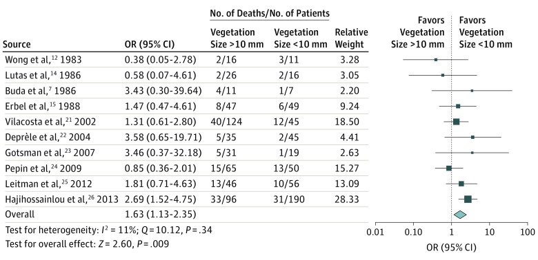
We found that a vegetation size greater than 10 mm was associated with increased odds of all-cause mortality (OR, 1.63; 95% CI, 1.13-2.35; P = .009) (Figure 3). The risk difference was 0.08 (95% CI, 0.02-0.13; P = .006). Cumulative meta-analysis showed that with serial addition of studies by publication year, overall effect (as measured by OR) was statistically significant only after the publication year 2013 (Hajihossainlou et al). On sensitivity analysis by the 1-study-removal method, we found that removal of the study by Hajihossainlou et al made the overall effect not significant (P = .17) (eFigures 8 and 9 in the Supplement).
Figure 3. Forest Plot for Comparative Odds of All-Cause Mortality.
Data are stratified by vegetation size less than and greater than 10 mm. Squares represent odds ratios (ORs), with their size proportional to the weight of the study using the Mantel Haenszel test; horizontal lines, 95% CIs; diamond, overall OR and 95% CI; and Q, Cochrane Q statistic.
Publication Bias
Visual inspection of funnel plots and quantitative assessment using the Egger test revealed no publication bias in the primary or secondary outcome. Results of assessment for publication bias are found in eFigure 10 in the Supplement.
Discussion
In our large meta-analysis of 21 studies with more than 6500 cases of infective endocarditis and more than 5000 measured vegetations, we revealed that patients with a vegetation size greater than 10 mm had significantly increased odds of embolic events and mortality. This increased association was not found to depend on age, sex, or type of valve involvement. We also reported that the strength of association of a vegetation size greater than 10 mm with embolic outcomes increased over time.
Fragmentation of vegetation specimens or cardiac tissue in patients with infective endocarditis leads to systemic embolic events. This devastating complication can occur in as many as 80% of cases of infective endocarditis. The brain and spleen are the most frequent sites of embolism in left-sided infective endocarditis, whereas pulmonary embolism is frequent in right-sided infective endocarditis of a native valve. Large vegetation sizes have been linked in multiple studies with an increased risk of embolic events. These vegetations are often dichotomized at a seemingly arbitrary cutoff greater than 10 mm. To prevent occurrence of embolic events, the American Heart Association guidelines suggest consideration of surgical options when the vegetation size is greater than 10 mm, particularly when involving the anterior leaflet of the mitral valve and when associated with other relative indications for surgery (class IIb, level of evidence C). Echoing this sentiment, the European Society of Cardiology guidelines suggest consideration of surgical options in aortic or mitral vegetations greater than 10 mm with 1 or more embolic event despite antibiotic therapy (class I, level of evidence B). However, the evidence behind these recommendations comes from relatively small observational studies with varying degrees of bias. Our study therefore adds to the existing literature by systematically analyzing individual studies and their risk of bias and by providing pooled odds of embolic events. Clinicians often need to balance the risk of embolic events with the risk of surgery, and our analysis will benefit those discussions by providing quality evidence behind the odds of embolic events in patients with vegetation size greater than 10 mm.
A previous meta-analysis by Tischler and Vaitkus revealed that patients with a vegetation size greater than 10 mm had significantly increased odds of embolic events and all-cause mortality that did not reach statistical significance. Their analysis of 10 studies had several limitations, including variable definitions of large vegetation and lack of assessment of publication bias, study quality, or degree of heterogeneity using the I2 statistic. We aimed in our analysis to systematically address these shortcomings and other additional limitations of a meta-analysis based purely on observational data. First, we included only studies in which using a cutoff of 10 mm to calculate odds of embolic events was possible. We assessed study quality using the standardized Newcastle-Ottawa Scale and found that a sensitivity analysis using only high-quality studies did not change the overall effect for the primary outcome significantly. In addition, our evaluation using funnel plots and the Egger test did not reveal any evidence of publication bias.
We used the Higgins I2 statistic to assess the degree of heterogeneity and found that heterogeneity for the primary outcome was moderately large (I2 = 58%). We further explored the cause of this heterogeneity by performing subgroup analyses by period of publication and study methods.
Our analysis by publication year revealed that studies before 2000 were more homogenous (I2 = 27%), and the pooled odds of embolic events in this subgroup, although increased for those with a vegetation size greater than 10 mm, did not attain statistical significance. In contrast, when studies after the year 2000 were considered, a vegetation size greater than 10 mm was associated with significantly increased odds of embolic events. Our cumulative meta-analysis (with serial addition of studies by publication year) showed concordant findings and revealed that the pooled effect only attained statistical significance after 2001. This finding appears to be in contrast to those of Tischler and Vaitkus, who owing to their publication date in 1997, only included studies before 2001 and found a vegetation size greater than 10 mm to be significantly associated with embolic events. This finding can likely be explained by their inclusion of patients with no vegetation to the cohort with a vegetation size less than 10 mm. The presence of vegetation specimens has been shown to have negative prognostic implications, and therefore inclusion of no vegetation with a vegetation size less than 10 mm likely overestimates the odds of embolic events with a vegetation size greater than 10 mm. Several possible reasons explain this increased association of vegetation size greater than 10 mm with embolic events over time. Among the studies in our analysis, Hajihossainlou et al noted that the prevalence of S aureus infection and intravenous drug use increased over time from 1995 to 2010. In addition, Vilacosta et al noted that the increased risk of embolic events in patients with a vegetation size greater than 10 mm was only significant for those with staphylococcal infections. Therefore, a change in microbiology may be responsible for the increased effect of a vegetation size greater than 10 mm seen in our meta-analysis after 2001. However, given the overlapping years of study, it is difficult to analyze this increase in our meta-analysis. Our systematic review of the percentage of S aureus infections among included studies (Table) did not show any clear increasing or decreasing temporal trend. Another possible explanation is that the use of Duke and modified Duke criteria helped to better categorize patients with infective endocarditis after 1994 (publication of Duke criteria). In our review of inclusion criteria in the Table, we found that all studies after De Castro et al (published in 1997) used Duke or modified Duke criteria. In subgroup analysis by inclusion criteria, we noted that the summary effect (odds of embolic events with a vegetation size >10 mm) was significant only in studies that used the Duke or modified Duke criteria. This finding may partially explain the temporal trend of increasing effect with publication year. Another possibility is that with early echocardiography techniques, smaller vegetations were missed, thereby overestimating the risk of embolism in vegetation size less than 10 mm.
In addition, technological advancement (and increasing use of TEE) has led not only to detection of smaller vegetations but also to more accurate determination of vegetation size. In a cohort that included more than 300 episodes of infective endocarditis, Luaces et al reported that their mean vegetation size of 14 mm was markedly higher than that found in classic series, and they attributed this greater size to technological advancements in the field of echocardiography. Although the European Society of Cardiology and the American Heart Association guidelines suggest that aortic or mitral valve involvement may enhance the risk of embolic events in patients with a vegetation size greater than 10 mm, our meta-regression analysis failed to show any significant effect of type of valve involvement on association of a vegetation size greater than 10 mm and embolic events. Although we could perform this analysis with only study-level data and the analysis does not reflect individual risk, it provides evidence that further prospective research is needed to validate the effect of localization of vegetation on strength of association of large vegetation size with embolic events.
Limitations
Our study has several limitations. First, this meta-analysis was performed on study-level data encompassing varying degrees of selection bias that is difficult to ascertain. Second, the effect of antibiotic use and microbiology could not be incorporated into the analysis owing to lack of sufficient data. Although 3 studies provided detailed information on the trend of embolic events after initiation of targeted antimicrobial therapy and unanimously noted a markedly decreased risk of embolic events in the second week after initiation of antimicrobial therapy, only Thuny et al provided information on the comparative risk of embolic events (with a vegetation size >10 mm) before and after initiation of antibiotics. However, even in that study, no statistical testing was performed to determine whether the risk of embolic events with a vegetation size greater than 10 mm changes after initiation of antibiotic therapy.
Another limitation is that the association of vegetation size of exactly 10 mm with embolic events remains ambiguous. This ambiguity occurs because some studies included these patients among the group with a vegetation size less than 10 mm, whereas others included them in the group with a vegetation size greater than 10 mm (Table). In addition, although most studies included patients with varied sites of systemic embolism, different sites of embolization may have different prognostic implications. Last, although we aimed to study only native valves and extracted data only on native valves wherever feasible, 14 of the 21 included studies had prosthetic valve involvement ranging from 2% to 30%. However, our meta-regression analysis showed that the presence of these cases did not affect the overall results in a significant manner. Our search was designed to capture studies evaluating embolic events in infective endocarditis, and it was not designed to evaluate the secondary outcome of mortality. Despite these limitations, we believe that the large size of our dataset, standardized protocol-based analysis, use of multiple subgroup and sensitivity analysis, and use of cumulative meta-analysis make our results robust and provide strength to our analysis.
Conclusions
In our meta-analysis of 21 studies, we showed that patients with a vegetation size greater than 10 mm had significantly increased odds of embolic events and mortality. We also showed that the strength of association of a vegetation size greater than 10 mm with embolic events was greater in subgroups of publications from 2000 to 2016 (compared with those from 1980-1999) and was unaffected by age, sex, and type of valve involved.
eTable 1. Assessment of Quality of Studies Using the Newcastle-Ottawa Scale
eTable 2. PRISMA Checklist
eTable 3. Results of meta-regression
eFigure 1. Subgroup Analysis of Primary Outcome by Time Period
eFigure 2. Subgroup Analysis of Primary Outcome by Study Type (Prospective vs Retrospective)
eFigure 3. Primary Outcome in Left-sided Endocarditis Only and Right-sided Endocarditis Only
eFigure 4. Metaregression Scatter Plots Depicting Association of Log Odds Ratio (of Primary Outcome) With Baseline Characteristics
eFigure 5. Cumulative Meta-analysis for Primary Outcome Obtained by Sequential Analysis of Increasing Evidence by Publication Year
eFigure 6. Sensitivity Analysis by 1-Study-Removal Method for Primary Outcome
eFigure 7. Primary Outcome Stratified by Vegetation Size Cut Off of 5 mm Above and 15 mm Below
eFigure 8. Sensitivity Analysis by 1-Study-Removal Method for Secondary Outcome
eFigure 9. Cumulative Meta-analysis for Secondary Outcome
eFigure 10. Funnel Plots for Primary and Secondary Outcomes
References
- 1.Hoen B, Alla F, Selton-Suty C, et al. ; Association pour l’Etude et la Prévention de l’Endocardite Infectieuse (AEPEI) Study Group . Changing profile of infective endocarditis: results of a 1-year survey in France. JAMA. 2002;288(1):75-81. [DOI] [PubMed] [Google Scholar]
- 2.Cabell CH, Jollis JG, Peterson GE, et al. Changing patient characteristics and the effect on mortality in endocarditis. Arch Intern Med. 2002;162(1):90-94. [DOI] [PubMed] [Google Scholar]
- 3.Rizzi M, Ravasio V, Carobbio A, et al. ; Investigators of the Italian Study on Endocarditis . Predicting the occurrence of embolic events: an analysis of 1456 episodes of infective endocarditis from the Italian Study on Endocarditis (SEI). BMC Infect Dis. 2014;14:230. [DOI] [PMC free article] [PubMed] [Google Scholar]
- 4.Sonneville R, Mourvillier B, Bouadma L, Wolff M. Management of neurological complications of infective endocarditis in ICU patients. Ann Intensive Care. 2011;1(1):10. [DOI] [PMC free article] [PubMed] [Google Scholar]
- 5.Kang DH, Kim YJ, Kim SH, et al. Early surgery versus conventional treatment for infective endocarditis. N Engl J Med. 2012;366(26):2466-2473. [DOI] [PubMed] [Google Scholar]
- 6.Baddour LM, Wilson WR, Bayer AS, et al. ; American Heart Association Committee on Rheumatic Fever, Endocarditis, and Kawasaki Disease of the Council on Cardiovascular Disease in the Young; Council on Clinical Cardiology; Council on Cardiovascular Surgery and Anesthesia; Stroke Council . Infective endocarditis in adults: diagnosis, antimicrobial therapy, and management of complications: a scientific statement for healthcare professionals from the American Heart Association. Circulation. 2015;132(15):1435-1486. [DOI] [PubMed] [Google Scholar]
- 7.Buda AJ, Zotz RJ, LeMire MS, Bach DS. Prognostic significance of vegetations detected by two-dimensional echocardiography in infective endocarditis. Am Heart J. 1986;112(6):1291-1296. [DOI] [PubMed] [Google Scholar]
- 8.Hwang JJ, Shyu KG, Chen JJ, Tseng YZ, Kuan P, Lien WP. Usefulness of transesophageal echocardiography in the treatment of critically ill patients. Chest. 1993;104(3):861-866. [DOI] [PubMed] [Google Scholar]
- 9.Misfeld M, Girrbach F, Etz CD, et al. Surgery for infective endocarditis complicated by cerebral embolism: a consecutive series of 375 patients. J Thorac Cardiovasc Surg. 2014;147(6):1837-1844. [DOI] [PubMed] [Google Scholar]
- 10.Thuny F, Di Salvo G, Belliard O, et al. Risk of embolism and death in infective endocarditis: prognostic value of echocardiography: a prospective multicenter study. Circulation. 2005;112(1):69-75. [DOI] [PubMed] [Google Scholar]
- 11.Tischler MD, Vaitkus PT. The ability of vegetation size on echocardiography to predict clinical complications: a meta-analysis. J Am Soc Echocardiogr. 1997;10(5):562-568. [DOI] [PubMed] [Google Scholar]
- 12.Wong D, Chandraratna AN, Wishnow RM, Dusitnanond V, Nimalasuriya A. Clinical implications of large vegetations in infectious endocarditis. Arch Intern Med. 1983;143(10):1874-1877. [PubMed] [Google Scholar]
- 13.Robbins MJ, Frater RW, Soeiro R, Frishman WH, Strom JA. Influence of vegetation size on clinical outcome of right-sided infective endocarditis. Am J Med. 1986;80(2):165-171. [DOI] [PubMed] [Google Scholar]
- 14.Lutas EM, Roberts RB, Devereux RB, Prieto LM. Relation between the presence of echocardiographic vegetations and the complication rate in infective endocarditis. Am Heart J. 1986;112(1):107-113. [DOI] [PubMed] [Google Scholar]
- 15.Erbel R, Rohmann S, Drexler M, et al. Improved diagnostic value of echocardiography in patients with infective endocarditis by transoesophageal approach: a prospective study. Eur Heart J. 1988;9(1):43-53. [PubMed] [Google Scholar]
- 16.Mügge A, Daniel WG, Frank G, Lichtlen PR. Echocardiography in infective endocarditis: reassessment of prognostic implications of vegetation size determined by the transthoracic and the transesophageal approach. J Am Coll Cardiol. 1989;14(3):631-638. [DOI] [PubMed] [Google Scholar]
- 17.Hwang JJ, Shyu KG, Chen JJ, et al. Infective endocarditis in the transesophageal echocardiographic era. Cardiology. 1993;83(4):250-257. [DOI] [PubMed] [Google Scholar]
- 18.Jung HO, Seung KB, Kang DH, et al. A clinical consideration of systemic embolism complicated to infective endocarditis in Korea. Korean J Intern Med. 1994;9(2):80-87. [DOI] [PMC free article] [PubMed] [Google Scholar]
- 19.De Castro S, Magni G, Beni S, et al. Role of transthoracic and transesophageal echocardiography in predicting embolic events in patients with active infective endocarditis involving native cardiac valves. Am J Cardiol. 1997;80(8):1030-1034. [DOI] [PubMed] [Google Scholar]
- 20.Di Salvo G, Habib G, Pergola V, et al. Echocardiography predicts embolic events in infective endocarditis. J Am Coll Cardiol. 2001;37(4):1069-1076. [DOI] [PubMed] [Google Scholar]
- 21.Vilacosta I, Graupner C, San Román JA, et al. Risk of embolization after institution of antibiotic therapy for infective endocarditis. J Am Coll Cardiol. 2002;39(9):1489-1495. [DOI] [PubMed] [Google Scholar]
- 22.Deprèle C, Berthelot P, Lemetayer F, et al. Risk factors for systemic emboli in infective endocarditis. Clin Microbiol Infect. 2004;10(1):46-53. [DOI] [PubMed] [Google Scholar]
- 23.Gotsman I, Meirovitz A, Meizlish N, Gotsman M, Lotan C, Gilon D. Clinical and echocardiographic predictors of morbidity and mortality in infective endocarditis: the significance of vegetation size. Isr Med Assoc J. 2007;9(5):365-369. [PubMed] [Google Scholar]
- 24.Pepin J, Tremblay V, Bechard D, et al. Chronic antiplatelet therapy and mortality among patients with infective endocarditis. Clin Microbiol Infect. 2009;15(2):193-199. [DOI] [PubMed] [Google Scholar]
- 25.Leitman M, Dreznik Y, Tyomkin V, Fuchs T, Krakover R, Vered Z. Vegetation size in patients with infective endocarditis. Eur Heart J Cardiovasc Imaging. 2012;13(4):330-338. [DOI] [PubMed] [Google Scholar]
- 26.Hajihossainlou B, Heidarnia MA, Sharif Kashani B. Changing pattern of infective endocarditis in Iran: a 16 years survey. Pak J Med Sci. 2013;29(1):85-90. [DOI] [PMC free article] [PubMed] [Google Scholar]
- 27.García-Cabrera E, Fernández-Hidalgo N, Almirante B, et al. ; Group for the Study of Cardiovascular Infections of the Andalusian Society of Infectious Diseases; Spanish Network for Research in Infectious Diseases . Neurological complications of infective endocarditis: risk factors, outcome, and impact of cardiac surgery: a multicenter observational study. Circulation. 2013;127(23):2272-2284. [DOI] [PubMed] [Google Scholar]
- 28.Aherrera JA, Abola MT, Balabagno MM, et al. Prediction of symptomatic embolism in Filipinos with infective endocarditis using the embolic risk French calculator. Cardiol Res. 2016;7(4):130-139. [DOI] [PMC free article] [PubMed] [Google Scholar]
- 29.Moher D, Liberati A, Tetzlaff J, Altman DG; PRISMA Group . Preferred Reporting Items for Systematic Reviews and Meta-analyses: the PRISMA statement. PLoS Med. 2009;6(7):e1000097. [DOI] [PMC free article] [PubMed] [Google Scholar]
- 30.Li JS, Sexton DJ, Mick N, et al. Proposed modifications to the Duke criteria for the diagnosis of infective endocarditis. Clin Infect Dis. 2000;30(4):633-638. [DOI] [PubMed] [Google Scholar]
- 31.Durack DT, Lukes AS, Bright DK; Duke Endocarditis Service . New criteria for diagnosis of infective endocarditis: utilization of specific echocardiographic findings. Am J Med. 1994;96(3):200-209. [DOI] [PubMed] [Google Scholar]
- 32.Habib G, Lancellotti P, Antunes MJ, et al. ; Task Force per il Trattamento dell’Endocardite Infettiva della Società Europea di Cardiologia (ESC) . 2015 ESC guidelines for the management of infective endocarditis [in Italian]. G Ital Cardiol (Rome). 2016;17(4):277-319. [DOI] [PubMed] [Google Scholar]
- 33.Wann LS, Hallam CC, Dillon JC, Weyman AE, Feigenbaum H. Comparison of M-mode and cross-sectional echocardiography in infective endocarditis. Circulation. 1979;60(4):728-733. [DOI] [PubMed] [Google Scholar]
- 34.Luaces M, Vilacosta I, Fernández C, et al. Vegetation size at diagnosis in infective endocarditis: influencing factors and prognostic implications. Int J Cardiol. 2009;137(1):76-78. [DOI] [PubMed] [Google Scholar]
Associated Data
This section collects any data citations, data availability statements, or supplementary materials included in this article.
Supplementary Materials
eTable 1. Assessment of Quality of Studies Using the Newcastle-Ottawa Scale
eTable 2. PRISMA Checklist
eTable 3. Results of meta-regression
eFigure 1. Subgroup Analysis of Primary Outcome by Time Period
eFigure 2. Subgroup Analysis of Primary Outcome by Study Type (Prospective vs Retrospective)
eFigure 3. Primary Outcome in Left-sided Endocarditis Only and Right-sided Endocarditis Only
eFigure 4. Metaregression Scatter Plots Depicting Association of Log Odds Ratio (of Primary Outcome) With Baseline Characteristics
eFigure 5. Cumulative Meta-analysis for Primary Outcome Obtained by Sequential Analysis of Increasing Evidence by Publication Year
eFigure 6. Sensitivity Analysis by 1-Study-Removal Method for Primary Outcome
eFigure 7. Primary Outcome Stratified by Vegetation Size Cut Off of 5 mm Above and 15 mm Below
eFigure 8. Sensitivity Analysis by 1-Study-Removal Method for Secondary Outcome
eFigure 9. Cumulative Meta-analysis for Secondary Outcome
eFigure 10. Funnel Plots for Primary and Secondary Outcomes


