Abstract
This case report assesses 2 patients with asteroid hyalosis with novel multicolor imaging technology; results highlight the value of this approach relative to color fundus photography and spectral-domain optical coherence tomography.
Asteroid hyalosis is a degenerative change of the vitreous that consists of yellow-white opacities that reflect light and impede the quality of color fundus photography (CFP). Eyes that have retinal disease along with dense asteroid hyalosis usually need spectral-domain optical coherence tomography (SD-OCT) or fluorescein angiography (FA) for adequate assessment of the retina. Although SD-OCT and FA do provide information about anatomy of retinal layers and retinal blood vessels, they do not provide information about the spatial distribution of retinal lesions.
Multicolor (MC) imaging is a novel imaging technique available with Spectralis SD-OCT (Heidelberg Engineering) that uses 3 different laser lights (blue [488 nm], green [515 nm], and infrared [820 nm]) to create an image of the retina. The blue laser is used to view the vitreoretinal interface and superficial retinal layers. The green laser is used for creating images of the middle retinal layers, including retinal blood vessels, hemorrhages, and exudates. The infrared laser produces images of the outer retinal layers and the choroid. A composite image of these 3 colored lasers results in a multicolor image, which is different from conventional CFP. We herein report the potential usefulness of MC imaging in eyes with dense asteroid hyalosis.
Methods
We report the cases of 2 patients with asteroid hyalosis who visited our clinic for evaluation for diabetic retinopathy. Clinicians noted each patient’s best-corrected visual acuity using a Snellen distance visual acuity chart. Multicolor imaging, FA, and SD-OCT were performed using the Spectralis high-resolution angiography and SD-OCT system (Heidelberg Engineering).
This study was approved by the institutional review board of Sankara Nethralaya Kolkata. Permission for the inclusion of clinical data was obtained from each patient via a written consent form.
Results
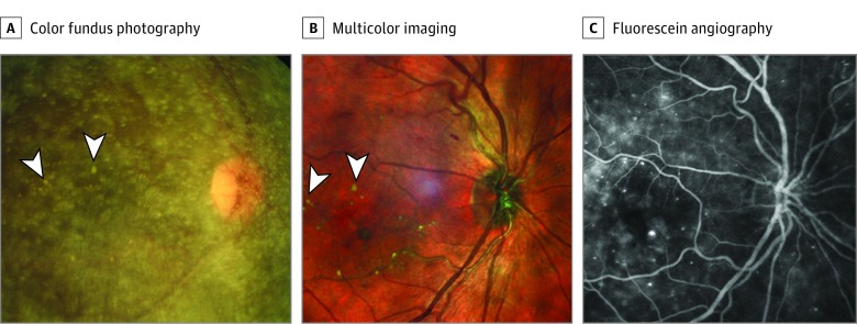
Patient 1 was a man in his late 40s whose visual acuity was 20/40 vision OU. His right eye showed asteroid hyalosis. The view of the fundus on CFP was poor; the optic disc and major retinal vessels appeared hazy. A few yellowish spots were noted at a level deeper than the asteroid hyalosis crystals, which appeared to be hard exudates at the macula (Figure 1A). The additional characteristics of the macular status were not visible. However, an MC image of the right eye revealed a clear view of the macula. The hard, yellowish exudates that were barely visible on CFP were clearly seen on the MC image. In addition, an FA showed multiple leaking microaneurysms at the macula (Figure 1B and C).
Figure 1. Images by Color Fundus Photography, Multicolor Imaging, and Fluorescein Angiography of Patient 1.
A, A color fundus photograph of the right eye shows asteroid hyalosis; interspersed yellowish spots appear as hard exudate (marked by white arrowheads). B, A multicolor image shows a clear view of the macula, including hard exudates (marked with white arrowheads). C, Fluorescein angiography using the Spectralis system (Heidelberg Engineering) shows multiple leaking microaneurysms.
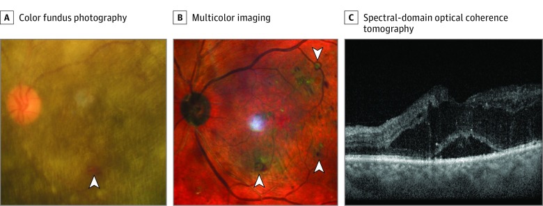
Patient 2 was a man in his early 50s whose visual acuity was 20/200 OU. The view of the fundus of the left eye of the patient was limited because of his asteroid hyalosis. Apart from the optic disc and major retinal arcade vessels, a retinal hemorrhage could be seen at the inferior macula on CFP (Figure 2A). However, the macula was clearly visible on the MC image, and retinal hemorrhages could be seen in the inferior, temporal, and superior macula (Figure 2B). Additionally, an SD-OCT image showed cystoid macular edema (Figure 2C).
Figure 2. Images by Color Fundus Photography, Multicolor Imaging, and Spectral-Domain Optical Coherence Tomography of Patient 2.
A, A color fundus photograph of the left eye shows asteroid hyalosis; retinal hemorrhage is marked with a white arrowhead. B, A multicolor image shows a clear view of the macula with multiple retinal hemorrhages (marked by white arrowheads); the visible white patch from nasal to fovea is an artifact. C, Spectral-domain optical coherence tomography shows macular edema.
Discussion
Multicolor imaging has been used to view various retinal diseases (eg, epiretinal membrane, age-related macular degeneration, and dominant cystoid macular dystrophy). In these 2 cases of asteroid hyalosis, MC imaging was used to create an image of the retina when CFP could not reveal adequate retinal details. The characteristics of the MC image technology make it suitable for imaging the retina in cases of asteroid hyalosis because it uses confocal scanning technology. In contrast, conventional CFP uses white light, which is reflected by asteroid crystals and reduces the view of the retina. The additional information that can be obtained via MC imaging may be important for the documentation of qualities of the fundus at baseline and at follow-up visits, permitting clinicians to monitor the progression of disease.
This study has a limitation in that additional equipment was not used for comparisons of image quality. Imaging with an ultrawide-field camera system can provide good images in eyes with media opacities; its high-resolution central framing might provide similar quality to multicolor imaging.
This article shows the potential usefulness of MC imaging in eyes with asteroid hyalosis. Using MC imaging in the eyes of patients with this condition can obviate the difficulties with CFP and provide clinicians a simpler way to assess the fundus.
References
- 1.Fawzi AA, Vo B, Kriwanek R, et al. . Asteroid hyalosis in an autopsy population: the University of California at Los Angeles (UCLA) experience. Arch Ophthalmol. 2005;123(4):486-490. [DOI] [PubMed] [Google Scholar]
- 2.Alasil T, Adhi M, Liu JJ, Fujimoto JG, Duker JS, Baumal CR. Spectral-domain and swept-source OCT imaging of asteroid hyalosis: a case report. Ophthalmic Surg Lasers Imaging Retina. 2014;45(5):459-461. [DOI] [PMC free article] [PubMed] [Google Scholar]
- 3.Hwang JC, Barile GR, Schiff WM, et al. . Optical coherence tomography in asteroid hyalosis. Retina. 2006;26(6):661-665. [DOI] [PMC free article] [PubMed] [Google Scholar]
- 4.Kilic Muftuoglu I, Bartsch DU, Barteselli G, Gaber R, Nezgoda J, Freeman WR. Visualization of macular pucker by multicolor scanning laser imaging. Retina. 2018;38(2):352-358. [DOI] [PMC free article] [PubMed] [Google Scholar]
- 5.Graham KW, Chakravarthy U, Hogg RE, Muldrew KA, Young IS, Kee F. Identifying features of early and late age-related macular degeneration: a comparison of multicolor vs traditional color fundus photography [published online August 22, 2017]. Retina. [DOI] [PubMed] [Google Scholar]
- 6.Roy R, Saurabh K, Bhattacharyya S, Thomas NR, Datta K. Multimodal imaging in dominant cystoid macular dystrophy. Indian J Ophthalmol. 2017;65(9):865-866. [DOI] [PMC free article] [PubMed] [Google Scholar]


