Description
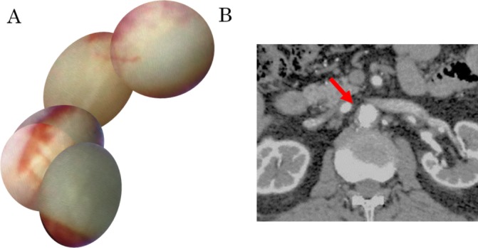
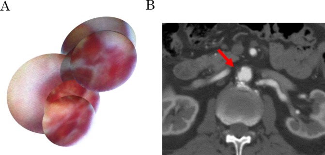
An 81-year-old man was referred to our hospital for intermittent claudication for 2 months. He had a history of hypertension, dyslipidaemia, renal insufficiency and percutaneous coronary intervention of the left circumflex coronary artery 3 years ago. Non-obstructive angioscopy (NOA) of the aorta had been performed to evaluate aortic atherosclerosis 2 years earlier.1 At the suprarenal abdominal artery, a partially peeled section of the intima was detected via NOA using the caterpillar method2 (figure 1A). At this site, blood flow filled the space between the intima and the peeled surface with pulsation (video 1). This was thought to be a subintimal intraplaque haemorrhage. CT angiography did not demonstrate any sign of aortic dissection at the corresponding area except for intimal thickening (figure 1B). Since the last catheterisation, the patient has not experienced chest or back pain. He has experienced intermittent claudication from the progression of right common iliac artery stenosis, apart from the lesion. Angioscopically, the lesion cavity has become larger, the structure has become complicated, and areas covered with red thrombi have been exposed (figure 2A, video 2). CT angiography demonstrated a slight progression of intimal thickening, but still no sign of aortic dissection (figure 2B). We presumed that NOA demonstrated the beginning of plaque rupture with a subintimal haemorrhage and cavity and showed disrupted plaque 2 years later.
Figure 1.

The subintimal haemorrhage at the suprarenal abdominal artery. (A) Angioscopic image. The intima was partially peeled. At this site, blood flow filled the space between the intima and the peeled surface with pulsation. (B) Axial CT image of the corresponding area. The aorta did not show dilatation. Hazy intimal thickening was identified.
Video 1.
The subintimal haemorrhage from the peeled intima at the suprarenal abdominal artery as seen on non-obstructive angioscopy
Figure 2.

A large cavity 2 years later at the site of the subintimal haemorrhage. (A) Angioscopic image. A larger cavity with a complicated structure and frames covered with red thrombi were seen. (B) Axial CT image of the corresponding area. Slightly progressed hazy intimal thickening was found without any sign of aortic dissection or obvious penetrating atherosclerotic ulcer.
Video 2.
A large cavity 2 years later at the site of the subintimal haemorrhage as seen on non-obstructive angioscopy
Learning points.
A subintimal intraplaque haemorrhage may be the beginning of aortic plaque rupture.
Angioscopy may detect the fragility of an asymptomatic aorta, such as a subintimal intraplaque haemorrhage or an aortic plaque rupture.
Long-term follow-up and more evidence are needed to clarify the significance of a subintimal intraplaque haemorrhage and aortic plaque rupture.
Footnotes
Contributors: SK wrote the first version of the manuscript. MT and ST performed the supervision of the manuscript, gave expert opinion, looked for the images and corrected the final version. KK supervised all the process, gave expert opinion and gave final approval for the version of the manuscript.
Funding: This research received no specific grant from any funding agency in the public, commercial or not-for-profit sectors.
Competing interests: None declared.
Patient consent: Obtained.
Provenance and peer review: Not commissioned; externally peer reviewed.
References
- 1. Komatsu S, Ohara T, Takahashi S, et al. . Early detection of vulnerable atherosclerotic plaque for risk reduction of acute aortic rupture and thromboemboli and atheroemboli using non-obstructive angioscopy. Circ J 2015;79:742–50. doi:10.1253/circj.CJ-15-0126 [DOI] [PubMed] [Google Scholar]
- 2. Komatsu S, Takahashi S, Ohara T, et al. . Aortic valve stenosis and atheromatous ascending aorta. Intern Med 2017;56:2685–6. doi:10.2169/internalmedicine.8486-16 [DOI] [PMC free article] [PubMed] [Google Scholar]


