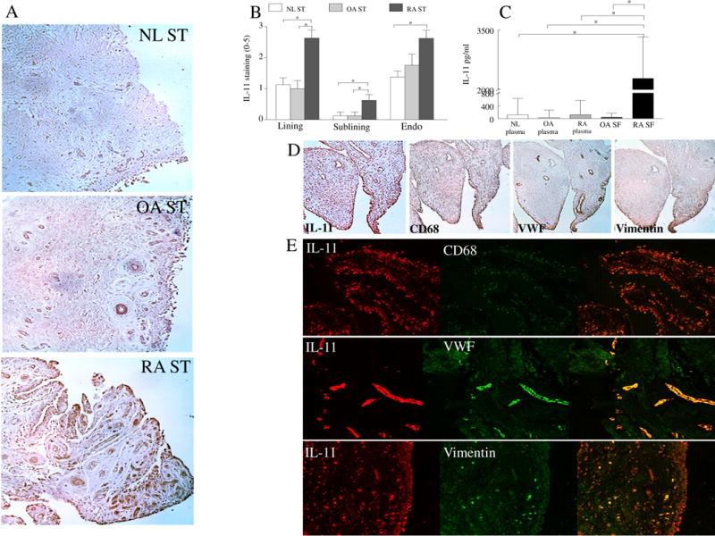
Fig. 1. IL-11 is overexpressed in RA ST lining fibroblasts and macrophages as well as sublining endothelial cells and macrophages and its level in RA SF is significantly higher than OA SF and plasma from RA, OA and NL individuals.

A. STs from NL, OA and RA were stained with anti-IL-11 Ab (original magnification x 200) and (B) staining was scored on a scale of 0–5 in the lining, sublining and endothelial cells, n=8−9. C. IL-11 protein concentration was determined in RA SF (n=40) and OA SF (n=40) as well as in plasma from RA (n=40), OA (n=35) and NL (n=35) individuals. D. RA ST serial sections were stained with Abs to anti-IL-11, CD68, VWF or Vimentin to establish which cell types produce IL-11, n=5. E. To confirm serial section studies, individual (red or green) as well as the overlapping (yellow) immunofluorescence staining was shown of RA STs that were stained with Abs to anti-IL-11 (green) or cell markers per slide including CD68 (red), VWF (red) or Vimentin (red) (original magnification x 400), n=5. Values are the mean ± SE. * represents p <0.05.
