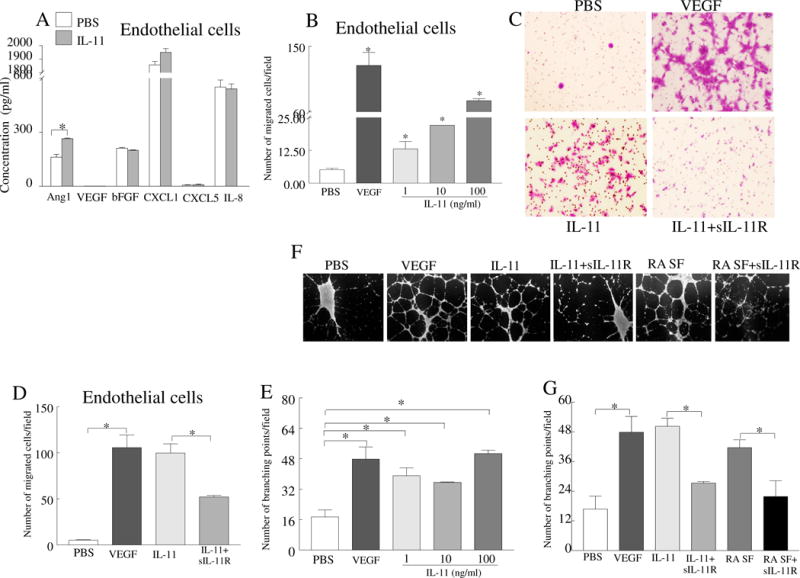
Fig. 5. IL-11 promotes endothelial cell migration and tube formation that is mediated through IL-11Rα ligation.

A. Protein levels of Ang-1, VEGF, bFGF, CXCL1, CXCL5 and IL-8 were quantified in supernatants obtained from HUVECs stimulated with PBS or IL-11 (100ng/ml) for 48h, n=5. B. Effect of IL-11 (1–100ng/ml) was determined on HUVEC migration using inserts of 8μm following 24h incubation. C. Representative images from HUVEC migration in response to IL-11 (100ng/ml) alone or IL-11 plus IL-11Rα-Fc chimera (10μg/ml). D. Number of endothelial cells that migrated in response to different treatment conditions shown in C. E. IL-11 (1–100ng/ml) effect was examined on HUVECs (25,000 cells/well) tube formation following 18h. The number of branching points were counted in each treatment and compared to negative control (PBS, 1% FBS in medium). F. Representative images of tube formation when HUVECs were treated with IL-11 (100ng/ml) or RA SF (10%) in absence or presence of IL-11Rα-Fc chimera (10μg/ml). G. The number of branching points formed in different treatment groups shown in F. In all experiments, VEGF (100ng/ml) was regarded as positive control. Experiments were performed in triplicates and repeated six times. Values are the mean ± SD. * represents p <0.05.
