Abstract
Günter Blobel was a scientific colossus who dedicated his career to understanding the mechanisms for protein sorting to membrane organelles. His monumental contributions established research paradigms for major arenas of molecular cell biology. For this work, he received many accolades, including the Nobel Prize in Medicine or Physiology in 1999. He was a scientist of extreme passion and a nurturing mentor for generations of researchers, imbuing them with his deep love of cell biology and galvanizing them to continue his scientific legacy. Günter passed away on February 18, 2018, at the age of 81.

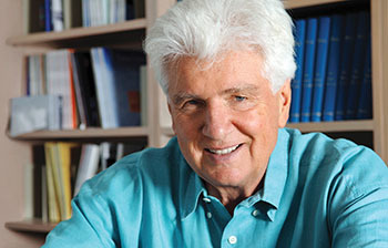
Günter Blobel.
Günter was a Lebenskünstler—a master of the art of living. He had a tremendous presence; when he walked into a room, he couldn’t help but command attention. A towering man, with a flock of white hair and a jovial nature, he loved to tell stories: of experiences in the laboratory, of life in New York, and of his time growing up in Germany. Günter was born in 1936, as the son of a veterinarian. He considered his early childhood to be idyllic, raised with his seven siblings in a rural and remote part of Silesia, then in the eastern part of Germany. There, largely isolated from the horrific developments overtaking Europe, Günter recalled long afternoons in beautiful manor houses adorned with hunting trophies, winter days of sleighing, and summer days in horse-drawn carriages. However, this life was shattered during the final months of the Second World War, when he and his family were forced to flee the advancing Soviet army. Passing through Dresden at this chaotic time, Günter experienced two life-changing events: seeing for the first time the magnificent baroque splendor of this “great jewel of a city,” and days later, witnessing its leveling by Allied firebombing. The family picked up the threads of their former lives in Freiberg, a medieval town located to the west of Dresden. Again, Günter has fond recollections of his time there. He immersed himself in the town’s rich cultural legacy, centered around baroque and classical music performances in its Gothic cathedral. Unfortunately, the new communist regime of East Germany was inhospitable to the Blobel family, who were seen as part of the bourgeois class and were therefore denied access to higher education. Forced to leave Freiberg by these circumstances, Günter moved to Tübingen in West Germany to study medicine. Though he completed an MD degree, he was not inspired to become a practicing physician. Instead, he decided to try a career in research science.
Accordingly, Günter joined the laboratory of Van Potter at the University of Wisconsin to obtain a PhD. As a graduate student, he developed an interest in cell structure and function, and became engaged with a problem that subsequently became an obsession: how the cell sorts secretory proteins to the rough ER. To pursue these interests, he moved to the laboratory of George Palade at the Rockefeller University for postdoctoral studies. Günter’s time with Palade was a critical and formative experience, as the Palade laboratory was an intellectual epicenter for study of the ER. Palade and coworkers had described the secretory pathway during the 1960s in an exquisite series of studies that combined cell fractionation, biochemistry, and electron microscopy. Indeed, this and related work on the functions of membrane organelles earned Palade, Christian De Duve, and Albert Claude the Nobel Prize in Medicine or Physiology in 1974. Günter revered Palade, and used his mentor’s work and conceptualization of cell function as a touchstone throughout his career.
As a newly promoted faculty member at the Rockefeller University, Günter set out to address the question of how mRNAs that encode secretory proteins are selected for synthesis on the rough ER. Despite a lack of experimental evidence, in 1971, Günter and David Sabatini proposed an amazingly prescient hypothesis (1). They suggested that ER targeting of secretory proteins is specified by the presence of a signal peptide on the N terminus of proteins destined to be translocated into the ER. This concept was subsequently termed the “signal hypothesis” (2). ‘‘At first it was just a wonderful idea,” Günter remembered; “It was quite a bold thing to say because nothing hinted at a signal sequence. But it was by far the best thing we could come up with.”
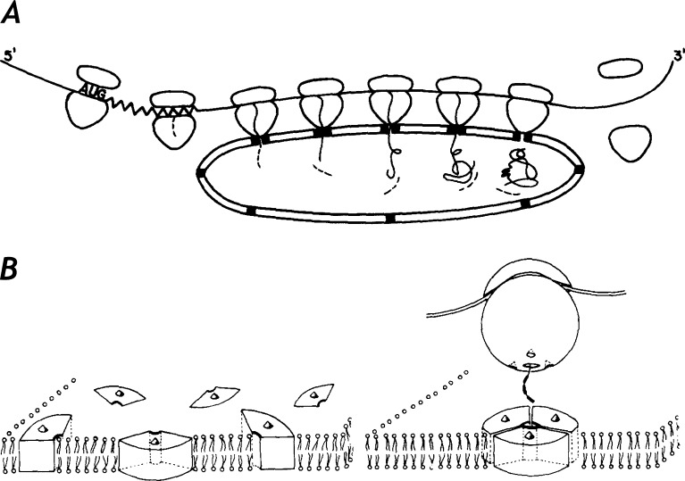
Inspired by his work with Palade, Günter knew that only a complete in vitro reconstitution of the ER translocation process with purified components—a Herculean challenge at the time—could validate this hypothesis. Encouraged by studies from several laboratories suggesting that the IgG light chain might be synthesized as a precursor polypeptide larger than the secreted form (1972–1975), he worked intensively to develop methods to biochemically dissect the process of ER secretory protein translocation. He provided the first strong experimental support for the signal hypothesis in two landmark papers in 1975 (2, 3). As with many of his most remarkable discoveries, these were published in the Journal of Cell Biology. By analyzing IgG light chain synthesis using in vitro assays with isolated ER membranes, he obtained striking evidence for the presence of an N-terminal signal sequence on the nascent chains that was involved in cotranslational translocation across the ER membrane, and that was cotranslationally removed (Fig. 1). He predicted that the signal sequence communicated with specific receptor proteins to mediate ER attachment of ribosomes and induce transient assembly of a proteinaceous channel to conduct the nascent polypeptide across the ER membrane. He also proposed that the mechanisms and components involved in secretory protein translocation could be used for the insertion of membrane proteins into the ER. These ideas were controversial, particularly the notion that a proteinaceous channel was involved in signal sequence–mediated membrane translocation, and became the focus of heated scientific debates for the subsequent two decades. But, as was typical of Günter’s outlook on science, he was unfazed: “I thought my ideas were reasonable. So why not propose them?”
Figure 1.
The original signal hypothesis (adapted from reference 2). (A) Illustration of the essential features of the signal hypothesis for the transfer of proteins across membranes. Signal codons after the initiation codon AUG are indicated by a zigzag region in the mRNA. The signal sequence region of the nascent chain is indicated by a dashed line. Endoproteolytic removal of the signal sequence is indicated by the presence of signal peptides (indicated by short dashed lines). (B) Model for the formation of a transient proteinaceous tunnel in the membrane through which the nascent chain is transferred.
Indeed, the controversies surrounding the signal hypothesis were laid to rest, one by one, by a series of elegant papers from the Blobel laboratory and from others over the next two decades. This work identified a cytosolic ribonucleoprotein particle (termed the SRP) that interacts with the signal sequence, a specific SRP receptor in the ER membrane involved in ribosome targeting, a signal peptidase that cotranslationally removes the signal peptide, and, finally, the proteinaceous channel (Sec61 complex) that mediates movement of the nascent chain across the membrane (4). The evolutionary significance of signal-mediated protein translocation across the ER was resoundingly underscored by the identification of conserved systems in diverse organisms including yeast and bacteria. The experimental approach used by Günter and associates for these studies, involving the use of an in vitro assay with a quantitative functional output for mechanistic analysis by fractionation and reconstitution, helped usher in the era of modern molecular cell biology.
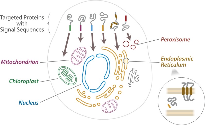
With the first experimental evidence for the signal hypothesis in hand, Günter prophetically speculated that systems analogous to those for ER translocation also might be deployed for targeting proteins to other membrane organelles (2). These speculations were decisively borne out in subsequent studies on protein transport into the mitochondrion, the chloroplast, the peroxisome, and the nucleus (Fig. 2), providing the ultimate vindication for these bold conjectures. This was a major element of Günter’s principle of “protein topogenesis”: that information for sorting of proteins to different membrane compartments, as well as for integration into membranes, is encoded in discrete classes of “topogenic” sequences recognized by various receptor systems, membrane-spanning protein conduits, and other effectors (5).
Figure 2.
The principles of protein targeting directed by signal sequences. A schematic of a cell is shown, with different membrane-bound organelles illustrated and labeled. Newly synthesized proteins carry signal sequences, often but not always at one end of the protein, which can direct that protein to the correct organelle within the cell and allow them to cross the organellar membranes. The lower right inset depicts how additional classes of topogenic sequences can specify the membrane integration of proteins (brown shading) instead of simple membrane translocation with signal sequence cleavage (orange shading).
Propelled by his polymathic character, Günter brought his enthusiasm and brilliance to bear on several other areas, particularly nucleocytoplasmic transport and the nuclear lamina. In the mid-1970s, Günter and coworkers made the remarkable discovery that nuclear pore complexes (NPCs) remain as intact structures after detergent extraction of nuclear envelopes, attached to a proteinaceous “nuclear lamina” derived from the inner surface of the nuclear envelope (Fig. 3, A and B; 6, 7). These results suggested that NPCs could be isolated and biochemically analyzed, a goal that was realized many years later by trainees from his laboratory. Günter enthusiastically promoted the view that the nuclear lamina is a widespread nuclear structural component of fundamental importance, albeit not evident in most cells by electron microscopy. These results initiated a stream of biochemical and functional studies that gradually became an experimental torrent involving numerous laboratories. This effort has firmly established the importance of the lamina in myriad nuclear functions in higher eukaryotes, including nuclear mechanics, signaling, and the dynamic 3D functional organization of chromatin. The relatively recent discoveries linking mutations in nuclear lamina proteins to at least 15 human genetic disorders speak to Günter’s visionary intuition on the importance of the nuclear lamina.
Figure 3.
The nuclear envelope, and associated structures. (A and B) Transmission electron micrographs of a thin section through an NPC–lamina fraction isolated from rodent liver, showing NPCs in lateral (single arrow) and frontal (double arrow) views, and the associated nuclear lamina (lA). Bars, 100 nm (adapted from reference 7). (C) An example of one of the most recent structures from the Blobel laboratory (PDB: 5SUP): the messenger ribonucleoprotein particles remodeling complex of Sub2 associated with an ATP analogue, RNA, and a C-terminal fragment of Yra1, required to process and package messenger ribonucleoprotein particles before export through the NPC.
In the arena of nucleocytoplasmic transport. Günter’s group was a frontrunner in the identification of nuclear transport factor proteins and NPC components, his competitive fervor helping to propel the field forward at an astounding rate during the 1990s. More recently, in keeping with his desire to understand the mechanistic principles of cells and fascinated by the molecular architecture of these players, he retailored his laboratory to solve the atomic structures for some of the proteins comprising these assemblies (Fig. 3 C). He drew great joy from solving molecular structures, seeing in them an elegance akin to that found in great architectural masterpieces, and he was adept in interpreting them in the context of cellular functions. Often he would retreat to his office for days, poring over stacks of literature and drawing on his experiences, to interpret their mechanisms and dynamics, ultimately explaining them with flamboyant imagery that reflected his love of cell biology.
Günter felt that doing science is a privilege and that it unifies humanity. He espoused da Vinci’s belief that “the noblest pleasure is the joy of understanding.” His enthusiasm was infectious and his laboratory was a hotbed of exciting ideas and around-the-clock activity. Sometimes the exciting ideas did not survive rigorous experimental scrutiny. But Günter was not afraid to miss the mark. Those who knew him well also knew how passionate he could become when a concept seemed particularly appealing. However, as he himself acknowledged, it sometimes became clear that (paraphrasing Thomas Huxley) “there are beautiful hypotheses killed by ugly facts.” He fully embraced this facet of scientific research, further stating that “one must not be wed to one’s fantasies.”
As trainees, it was a pleasure and privilege for us to be part of his life and to have been influenced by this extraordinary man. We started our careers on the fertile ground tilled by our time in Günter’s laboratory. He inspired us to think big and to ask the questions in biology that really mattered. Günter viewed his laboratory as the greatest of master artists viewed their studios, just as these studios apprenticed new artists while the paintings were produced, Günter sought both to produce new scientific discoveries and to train new scientific researchers. Only last year, five of Günter’s postdoctoral researchers and senior fellows gained assistant professor tenure track positions. His office door was always open to his trainees, past and present. They knew they could go in, even if defeated and distraught after long strings of failures, thrash through the issues, work with him on new ways of tackling them, and emerge reinvigorated.
Günter’s passions extended beyond the scientific. The simplest of things could ignite an exuberant outpouring. A walk with his dogs through Central Park, the flowers in the garden at the Rockefeller University, or an evening at his favorite haunt, Barbetta Restaurant. This sentiment and joie de vivre belied an intensely competitive spirit, but at heart Günter was a kind gentleman who was enormously generous. Spurred by his childhood experiences, he became the founder and president of the nonprofit organization Friends of Dresden. Indeed, he donated his Nobel Prize money to Dresden, devoted to the rebuilding of the Frauenkirche—a Lutheran church and baroque architectural masterpiece—and the New Synagogue, to replace the synagogue destroyed by the Nazis.
With deep sadness, we accept that eventually Günter gracefully succumbed to the self-described “noble injuries of time,” maintaining his enthusiasm until the end. There is so much more we could say about this incredibly inspiring man; we feel we got to know him well (Fig. 4). But, as Günter himself often liked to say, “less is more.” For his memorial, one can view the architectural splendors in Dresden reborn through his efforts, his towering masterpieces of scientific insight that underlie countless medical therapies and treatments being pioneered today, and the generations of researchers inspired by him who are continuing his work and who are passing on his baton to the next generations.
Figure 4.
Blobel laboratory trainees were polled for up to five one-word descriptors they have used to describe Günter to their friends, family, and colleagues. Responses are shown in word cloud format produced using software from WordArt.com. The size of each word reflects its frequency of usage.
References
- 1.Blobel G., and Sabatini D.D.. 1971. Ribosome-membrane interaction in eukaryotic cells. In Biomembranes. Manson L.A., editor. Springer, Boston, MA: 193–195. 10.1007/978-1-4684-3330-2_16 [DOI] [Google Scholar]
- 2.Blobel G., and Dobberstein B.. 1975. Transfer of proteins across membranes. I. Presence of proteolytically processed and unprocessed nascent immunoglobulin light chains on membrane-bound ribosomes of murine myeloma. J. Cell Biol. 67:835–851. 10.1083/jcb.67.3.835 [DOI] [PMC free article] [PubMed] [Google Scholar]
- 3.Blobel G., and Dobberstein B.. 1975. Transfer of proteins across membranes. II. Reconstitution of functional rough microsomes from heterologous components. J. Cell Biol. 67:852–862. 10.1083/jcb.67.3.852 [DOI] [PMC free article] [PubMed] [Google Scholar]
- 4.Blobel G. 2000. Protein targeting (Nobel lecture). ChemBioChem. 1:86–102. [DOI] [PubMed] [Google Scholar]
- 5.Blobel G. 1980. Intracellular protein topogenesis. Proc. Natl. Acad. Sci. USA. 77:1496–1500. 10.1073/pnas.77.3.1496 [DOI] [PMC free article] [PubMed] [Google Scholar]
- 6.Aaronson R.P., and Blobel G.. 1975. Isolation of nuclear pore complexes in association with a lamina. Proc. Natl. Acad. Sci. USA. 72:1007–1011. 10.1073/pnas.72.3.1007 [DOI] [PMC free article] [PubMed] [Google Scholar]
- 7.Dwyer N., and Blobel G.. 1976. A modified procedure for the isolation of a pore complex–lamina fraction from rat liver nuclei. J. Cell Biol. 70:581–591. 10.1083/jcb.70.3.581 [DOI] [PMC free article] [PubMed] [Google Scholar]


