PICTURE OF THE MONTH
Fetal Intelligent Navigation Echocardiography (FINE) is a method that automatically generates and displays nine standard fetal echocardiography views from volume datasets obtained with spatiotemporal image correlation1,2. Color Doppler FINE or 5D Heart Color (UGEO WS80A; Samsung Healthcare, Seoul, Korea) allows the same cardiac views to be displayed with color Doppler information3. The Picture of the Month illustrates how the diagnostic planes shown by color Doppler FINE allow a precise diagnosis of hypoplastic left heart with coarctation of the aorta.
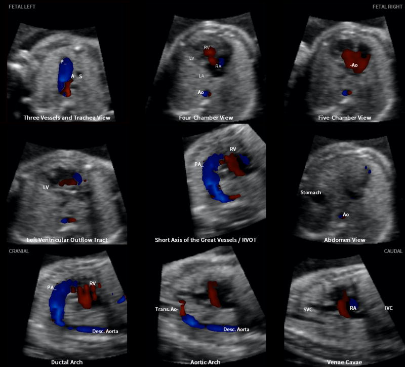
The characteristic features of this congenital heart defect were visualized in the following seven echocardiography views (Figure 1):
The three vessels and trachea view showed a hypoplastic transverse aortic arch with retrograde flow (red color), along with a dilated pulmonary artery demonstrating antegrade flow (blue color).
In the four-chamber view, the left side of the heart was severely hypoplastic. There was antegrade flow through the tricuspid valve during diastole, but absent flow through the atretic mitral valve.
The five-chamber view also showed a severely hypoplastic left side, antegrade flow through the tricuspid valve, and absence of a color Doppler signal in the atretic aortic root.
The left ventricular outflow tract view confirmed absence of color Doppler flow through the mitral valve, as well as an atretic aortic valve with absent flow. However, antegrade flow was seen through the tricuspid valve.
In the short-axis view of the great vessels/right ventricular outflow tract, the cross-section of the aorta was small when compared with the pulmonary artery. There was systolic perfusion across the pulmonary valve and trunk.
The ductal arch view demonstrated similar findings.
The aortic arch view showed a very narrow transverse aortic arch (coarctation), with reversed color Doppler flow in this area, as well as in the isthmus.
Figure 1.
Color Doppler Fetal Intelligent Navigation Echocardiography method in 26-week fetus with hypoplastic left heart and coarctation of aorta (automatic labeling shown). A, transverse aortic arch; Ao, aorta; Desc., descending; IVC, inferior vena cava; LA, left atrium; LV, left ventricle; P, pulmonary artery; PA, pulmonary artery; RA, right atrium; RV, right ventricle; RVOT, right ventricular outflow tract; S, superior vena cava; SVC, superior vena cava; Trans., transverse.
The images obtained using color Doppler FINE allowed a precise diagnosis of fetal hypoplastic left heart and coarctation of the aorta at 26 weeks of gestation. More details about the case and color Doppler FINE can be found in an accompanying article in this issue of UOG.3
References
- 1.Yeo L, Romero R. Fetal Intelligent Navigation Echocardiography (FINE): a novel method for rapid, simple, and automatic examination of the fetal heart. Ultrasound Obstet Gynecol. 2013;42:268–284. doi: 10.1002/uog.12563. [DOI] [PMC free article] [PubMed] [Google Scholar]
- 2.Garcia M, Yeo L, Romero R, Haggerty D, Giardina I, Hassan SS, Chaiworapongsa T, Hernandez-Andrade E. Prospective evaluation of the fetal heart using Fetal Intelligent Navigation Echocardiography (FINE) Ultrasound Obstet Gynecol. 2016;47:450–459. doi: 10.1002/uog.15676. [DOI] [PMC free article] [PubMed] [Google Scholar]
- 3.Yeo L, Romero R. Color and power Doppler combined with Fetal Intelligent Navigation Echocardiography (FINE) to evaluate the fetal heart. Ultrasound Obstet Gynecol. 2017;50:476–491. doi: 10.1002/uog.17522. [DOI] [PMC free article] [PubMed] [Google Scholar]


