Abstract
Competing endogenous RNA (ceRNA) has emerged as an important post-transcriptional mechanism that simultaneously alters expressions of thousands genes in cancers. However, only a few ceRNA genes have been studied for their functions to date. To understand the major biological functions of thousands ceRNA genes as a whole, we designed Model-based Analysis of Competing-endogenous Pathways (MACPath) to infer pathways co-regulated through ceRNA mechanism (cePathways). Our analysis on breast tumors suggested that NGF (nerve growth factor)-induced tumor cell proliferation might be associated with tumor-related growth factor pathways through ceRNA. MACPath also identified indirect cePathways, whose ceRNA relationship is mediated by mediating ceRNAs. Finally, MACPath identified mediating ceRNAs that connect the indirect cePathways based on efficient integer linear programming technique. Mediating ceRNAs are unexpectedly enriched in tumor suppressor genes, whose down-regulation is suspected to disrupt indirect cePathways, such as between DNA replication and WNT signaling pathways. Altogether, MACPath is the first computational method to comprehensively understand functions of thousands ceRNA genes, both direct and indirect, at the pathway level.
Author summary
Competing endogenous RNAs (ceRNA) are RNAs known to co-regulate each other’s expression levels by competing for the binding of shared microRNAs. In human cancer, thousands genes are known to be in the ceRNA relationship. To understand the major biological functions of the ceRNA genes as a whole, we offer Model-based Analysis of Competing-endogenous Pathways (MACPath). Based on a network model representing ceRNA relationship changes in tumor, MACPath identifies biological pathways co-regulated through the ceRNA relationship (cePathways), since a biological pathway represents a group of genes coordinating to carry out the same biological function. In breast tumor data, MACPath shows that extensive ceRNA relationship changes in tumor are associated with previously under-characterized tumorigenic crosstalk e.g. between nerve growth factor and other tumor-related growth factor pathways. MACPath also identified indirect cePathways, whose ceRNA relationship is mediated by mediating ceRNAs. When usually thousands ceRNAs mediate indirect cePathways, MACPath employed efficient integer linear programming technique to characterize the mediation. In the breast tumor data, the characterization provides insights into how indirect cePathways and the mediating ceRNAs promote the tumorigenic process. To the best of our knowledge, MACPath is the first computational framework that can comprehensively identify cePathway relationship, both direct and indirect.
This is a PLOS Computational Biology Methods paper.
Introduction
Competing endogenous RNAs (ceRNAs) are transcripts that share microRNA (miRNA) binding sites. Through competition for the binding of shared miRNAs, ceRNAs co-regulate each others’ expression levels[1] (ceRNA relationship). Although its scope is not fully understood[2], ceRNA has been shown to play a prominent role in regulating gene expression in diverse physiological[3, 4] and pathological conditions, especially for human cancers (reviewed in [5]). Tumor-associated ceRNA function has been identified for a number of individual genes. For example, as CNOT6L and VAPA have ceRNA relationship with PTEN, they regulate PTEN itself and phenocopy its tumor suppressive properties[6].
Recently, several studies reported widespread ceRNA relationship (ceRNA network) in tumors involving thousands ceRNA genes[7, 8]. To identify the tumor-associated function of thousands ceRNA genes as a whole rather than arbitrarily chosen individual genes, we developed Model-based Analysis of Competing-endogenous Pathways (MACPath). MACPath groups ceRNA relationships by competing-endogenous pathways (cePathway), i.e. pairs of biological pathways enriched with ceRNA relationships in-between. Since a biological pathway represents a group of genes coordinating to carry out the same biological function, a cePathway relationship may represent a co-regulation between biological functions (crosstalk) through enriched ceRNA relationships. Furthermore, MACPath also identifies dysregulated cePathway relationships between cancer and normal cells. For example, cePathway relationships gained in tumors might be oncogenic, while those lost in tumors might be tumor-suppressive. Finally MACPath can identify dysregulation in indirect cePathway relationship, i.e. co-regulation between pathways mediated by (direct) ceRNAs[9, 10]. When applied to TCGA breast cancer data, MACPath discovered thousands of lost and gained (direct and indirect) cePathway relationships, which collectively provide novel biological insights into ceRNA regulations during tumorigenesis.
Results
Direct cePathways dysregulated in tumors
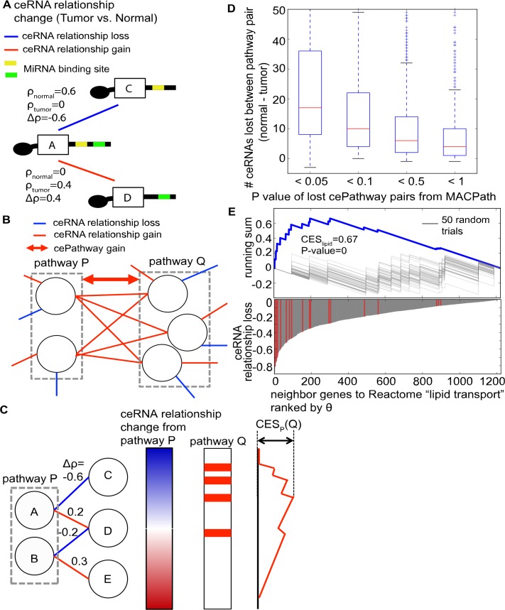
To identify direct cePathway relationships dysregulated in tumors, MACPath first builds a network model representing changes in ceRNA relationship between cancer and normal tissues (ceRNA relationship change network, Def. 1). Since co-expression between genes that share the same miRNA binding sites indicates the level of their ceRNA relationship[7, 10], their co-expression change (tumor vs. normal) can represent their ceRNA relationship change (Fig 1A). In the network model, MACPath identifies dysregulated cePathway relationship between pathway P and Q (Fig 1B, for example) in three steps. First, MACPath calculates the ceRNA relationship changes between all genes within pathway P and their ceRNA genes. Second, MACPath estimates if pathway Q is over-represented in the ceRNA relationship changes (ceRNA relationship change enrichment score (CESP(Q)), Fig 1C). Third, MACPath estimates the statistical significance (empirical p-value) of the CES score by running a permutation test (see Materials and Methods). Pathways P and Q can gain cePathway relationship if they are significantly over-represented in the ceRNA relationship gains in tumors (Fig 1B, for example), or can lose cePathway relationship if over-represented in the ceRNA relationship losses.
Fig 1. Identifying direct cePathway relationship dysregulations in tumors.
(a). An illustration of ceRNA relationship change network. RNA A shares a significant number of miRNA binding sites with RNA C and D, where ρnormal and ρtumor represent their co-expression in normal and tumor samples, respectively. Between normal and tumor samples, co-expression decrease in tumors (Δρ = ρtumor-ρnormal<0) between A and C could represent their ceRNA relationship loss and co-expression increase (Δρ>0) between A and D represent their ceRNA relationship gain. (b). An example of a cePathway relationship gain between pathway P and Q in the ceRNA relationship change network. CePathway relationship loss would be identified in the same fashion.(c). An illustration of MACPath to identify cePathway relationship dysregulation between pathway P and Q. Genes showing ceRNA relationship changes from pathway P are sorted by the magnitude of the changes (θP from Def. 2, blue for loss and red for gain in the left bar). On the genes ranked by θP, a running sum is calculated for genes in pathway Q (in the right bar). The maximum variation of the running sum from 0 is the ceRNA relationship change enrichment score (CESP(Q)). (d). The number difference of ceRNAs between pathway pairs (normal vs. tumor) divided by P value MACPath estimated for the pairs. (e). Upper panel shows behavior of running sum between Reactome "lipid transport" pathway and PID "HIF1 TF" pathway. Gray lines represent running sum of 50 permutation trials. Lower panel shows θ of neighbor genes to Reactome "lipid transport" pathways ranked by their θ. Red lines indicate where genes in PID “HIF1 TF” are placed.
In 97 breast tumor and their matched normal samples in TCGA, MACPath identified 1,019 lost and 1,698 gained cePathway relationships (FDR-corrected P value < 0.01 and CES > 0.6, see Materials and Methods) from all combinations (1330*1329/2 = 883,785) of 1,330 mSigDB canonical pathways[11] (S1 Table). To validate the use of MACPath, we identified 377,446 and 36,320 ceRNAs in the normal and tumor data based on co-expression (>0.6 in Pearson's correlation coefficient) and miRNA binding site share (< 0.05 in B-H corrected P-value)[7]. Since ceRNAs between pathways were suggested to indicate crosstalk between pathways[7], checking the ceRNA number difference from normal to tumor would be another way to represent cePathway dysregulation. In particular, pathway pairs losing ceRNAs in-between may represent lost cePathways, and those gaining ceRNAs represent gained cePathways. However, while MACPath generally identified pathway pairs losing many ceRNAs as lost cePathways, some lost cePathways do not lose many ceRNAs (43.3% of the lost cePathways lost less than 10 ceRNAs, S1 Table, Fig 1D). For example, Reactome "lipid transport" lost cePathway relationship with PID "HIF1 TF" pathway with a strong over-representation in the ceRNA relationship loss in tumor (P-value = 0, Fig 1E). However, they lost only 2 ceRNAs in tumor. Further, the ceRNA number difference cannot effectively identify gained cePathways, since 10-fold decrease of ceRNAs in tumor (377,446 vs. 36,320) makes it very difficult for pathway pairs to gain ceRNAs (only 8 of the 1,698 gained cePathways (0.4%) gain ceRNAs). Additionally, the ceRNA number difference cannot control for false positives, since it does not estimate significance. Altogether, MACPath identifies statistically significant and biologically reasonable cePathway dysregulation, which is impossible with a naive, yet the only other available method.
Dysregulated cePathways may underlie NGF-induced breast tumor cell proliferation
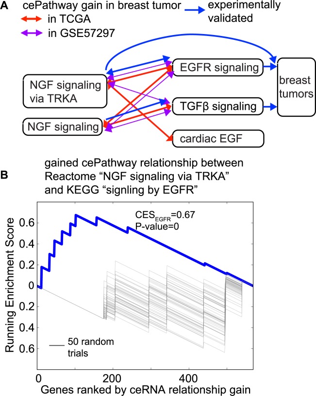
Many of the gained cePathway relationships involve nerve growth factor (NGF) signaling and tumor-related growth factor pathways, such as TGFβ[12] (transforming growth factor β) and EGFR[13] (epidermal growth factor receptor) (Fig 2A). An example is the gained cePathway relationship between REACTOME “NGF signaling via TRKA” and REACTOME “signaling by EGFR” (Fig 2B). NGF stimulates cell proliferation in tumor, but not in normal cells[14]. The mechanism may involve miRNAs, since NGF interacts with miRNAs for cell survival[15], which usually co-occurs with cell proliferation in cancer cells[16]. Therefore, we speculate that the mechanism underlying the NGF-induced cell proliferation might be due to the gained cePathway relationships between NGF and growth factor pathways. This is consistent with previous literature, in which crosstalk between NGF/TrkA and EGFR has been reported for cell activation[17], and NGF has been reported to up-regulate TGFβ in a cell differentiation experiment[18]. Furthermore, most of the cePathway gains in TCGA data are also found in an independent breast tumor data (GSE57297)[19] using MACPath with the same cutoff (Fig 2A, see Materials and Methods), suggesting the cePathway crosstalk between NGF and the growth factors is a common mechanism in breast cancers.
Fig 2. Dysregulated cePathways suggest NGF-induced cell proliferation.
(a). Growth factor pathways that gained cePathway relationship in tumors with NGF signaling pathways in the TCGA data (red) and in GSE57297[19] (purple). (b). Behavior of running sum between Reactome “signaling by NGF” pathway and KEGG “TGFβ signaling” pathway. Gray lines represent running sum of 50 random trials (Actual p-values are estimated from 2,000 such random trials).
Indirect cePathways dysregulated in tumors
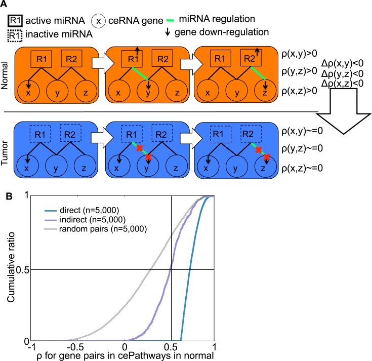
The ceRNA relationship change network that models direct ceRNA relationship (Def. 1) can further be used to model indirect ceRNA relationship. For genes x, y, and z, direct ceRNA relationship (x, y) can propagate to direct ceRNA relationship (y, z), making an indirect ceRNA relationship (x, z)[9, 10] (e.g. x, y, and z in normal condition in Fig 3A). Inspection on the same number (n = 5,000) of the ceRNA relationships showed that indirect ceRNA relationships are substantially co-expressed (ρ>0.5 for 53.8% of the relationships, Fig 3B), although less co-expressed than direct ceRNA relationships that are identified based on high co-expression. Additionally, indirect ceRNAs are more highly expressed than direct ceRNAs in the normal samples (P-value = 8.04e-44, S2B Fig). MACPath identifies indirect cePathway relationship based on indirect ceRNAs (x, z) that show the same direction of co-expression change as in (x, y) and (y, z) in the ceRNA network (see Materials and Methods). For TCGA breast cancer, MACPath identifies 509 lost and 84 gained indirect cePathway relationships (S2 Table, FDR-corrected P value <0.01 in ForwardStop[20]).
Fig 3.
Identifying gained/lost indirect cePathway relationships (a). In normal when regulating miRNAs (R1 and R2) are active (in abundance relative to other competing molecules[21]), down-regulation of x would direct more copies of R1 to repress y. In turn, repressed y would direct R2 to repress z. Overall, ceRNA relationship on (x, y) and (y, z) would yield indirect ceRNA relationship on (x, z). In tumor when (x, y) lost ceRNA relationship possibly in association to inactivation of the regulating miRNAs, down-regulation of x would not repress y and z, representing the loss of not only direct ceRNA relationship in (x, y) and (y, z) but also indirect ceRNA relationship in (x, z). From normal and tumor condition, (x, z) would lose co-expression in tumors (Δρ<0) altogether with (y, z) and (x, z). (b). Accumulative distribution of co-expression (ρ) in direct and indirect ceRNA relationships (found in direct and indirect cePathway relationships) in normal samples, compared to the same number of random pairs (n = 5,000).
Mediating ceRNAs drive indirect cePathway relationships
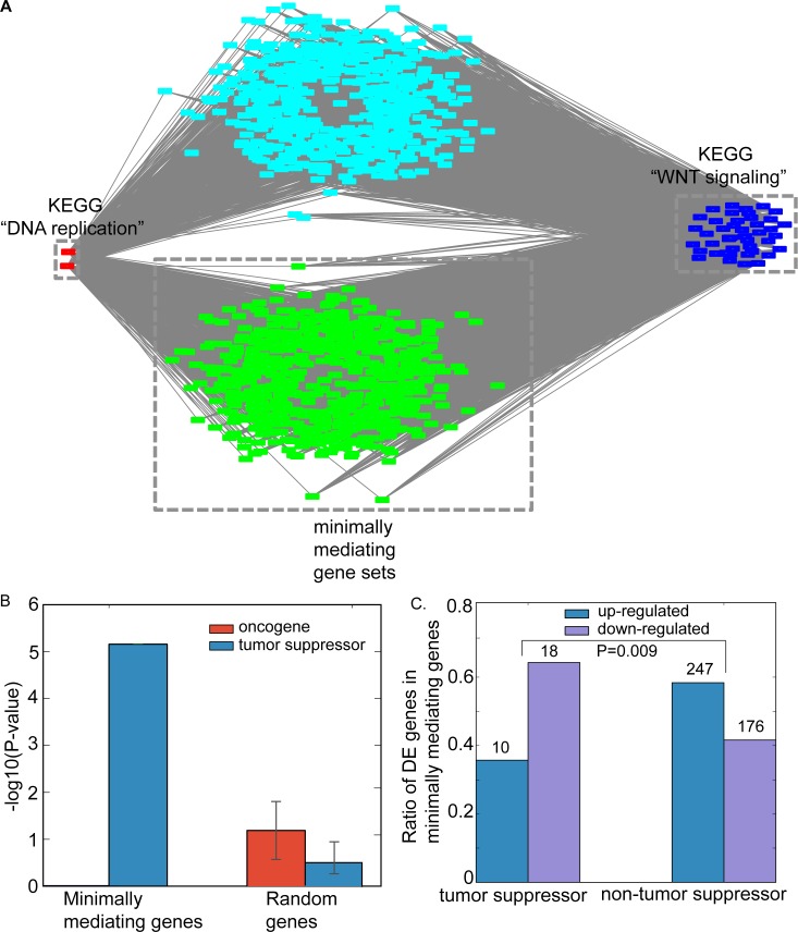
Extensive ceRNA relationship in tumors[7, 8] implies that many genes would mediate indirect ceRNA relationships. Consistently, our network model identifies on average 840 mediating ceRNAs for each indirect cePathway relationship (S3 Table). For example, 1,132 genes are inferred to mediate the significant loss of indirect cePathway relationship between KEGG “DNA replication” and KEGG “WNT signaling” (P = 0.0005, S2 Table, Fig 4A). In normal cells, both WNT signaling and DNA replication were reported to crosstalk to the same genes, such as those in DNA damage pathways[22, 23], which would yield indirect crosstalk between DNA replication and WNT signaling. In addition, these two pathways were shown to suppress tumor in a miRNA dependent manner[24, 25], possibly through their indirect cePathway relationship.
Fig 4. The lost indirect cePathway relationship between KEGG “DNA replication” and KEGG “WNT signaling”.
(a). Red nodes are ceRNA genes of KEGG “DNA replication” and blue nodes are those of KEGG “WNT signaling”. 1,132 genes (green and light blue nodes) mediate the ceRNA loss between the pathways, where green nodes are 615 minimally mediating gene set. Visualization is taken from Cytoscape[27]. (b). Enrichment of the minimally mediating genes to oncogenes (red) and tumor suppressors (blue) compared to the same number of random genes. Error bar represents standard variation values from 100 random trials. (c). Ratio of differentially expressed tumor suppressors in the set of the mediating genes compared to other genes in the set. EdgeR estimates differential expression with FDR-corrected P value < 0.05.
One important question is to identify which of the 1,132 mediating ceRNAs would exert strong mediation effect for the loss of indirect cePathway. We address this problem by finding the minimum number of mediating genes covering all genes in the indirect cePathways. Finding minimally mediating genes is an NP-hard problem (see Materials and Methods), which requires trying all combinations of mediating genes. We formulated the problem such that it is amenable to efficient integer linear programming (ILP) solvers (see Materials and Methods) and found minimally mediating genes within a second for most indirect cePathway relationship dysregulation. In particular, MACPath employed GNU Linear Programming Kit (GLPK) solver, which returns a different set of minimally mediating genes for a different permutation of the same input values. Using the solver on multiple (10,000) permutations on the input values for the lost indirect cePathway relationship KEGG "DNA replication" and KEGG "WNT signaling", MACPath yielded 615 minimally mediating genes, which are surprisingly enriched for tumor suppressors, but not oncogenes (Fig 4B). Tumor suppressors that are also minimally mediating genes are more likely to be down-regulated than other non-tumor-suppressor minimally mediating genes (P-value = 0.009, Fig 4C). While tumor suppressor down-regulation is a hallmark of cancer[26], one of its tumorigenic effects may be to disrupt indirect cePathway relationships, for example between KEGG "DNA replication" and KEGG "WNT signaling".
YAP, TAZ, and vitamin D receptor promote or inhibit lung adenocarcinoma possibly through ceRNA
To ascertain its general applicability, we ran MACPath on 58 TCGA lung adenocarcinoma (LUAD) and their matched normal samples, identifying direct cePathway dysregulation (13,247 gains and 7,121 losses in tumor) (S3 Fig, S4 Table). Further analyses on the dysregulation provide new insights into some tumor-associated crosstalks in lung adenocarcinoma (AC). Reactome “YAP1 and TAZ stimulated gene expression” that gained the most number (339) of cePathway relationships is an example. Overexpression of YAP and TAZ promotes growth signals in cancer types including lung AC [28]. Their expressions were found to be regulated by miRNAs in diverse cancer types [29], but their post-transcriptional tumorigenic mechanisms remain elusive. Since Reactome “YAP1 and TAZ stimulated gene expression” gains cePathway relationships with several tumor-associated growth factor pathways (e.g. EGFR, TGFβ and IGF), YAP and TAZ would promote the growth factor pathways possibly through ceRNA relationships. Further, cePathway dysregulations suggest their potential roles as prognostic biomarkers. As an example, vitamin D receptor pathways gained many cePathway relationships (5th and 11th highest in the number of gains). Vitamin D receptor is known to regulate cell cycle [30], whose high expression correlating with longer survival through antiproliferative effects and cell cycle arrest [31]. Since its function as cell cycle regulator involve miRNAs in diverse types of cancers [30], vitamin D receptor pathways gaining cePathway relationships to 23 cell cycle checkpoint pathways may enhance the antiproliferative and cell cycle arrest mechanisms at the ceRNA level.
Discussion
CeRNA plays an important role in the pathogenesis of cancers by forming extensive ceRNA network (ceRNET)[32] involving thousands ceRNA genes[21, 33]. For the ceRNETs to produce appropriate tumor-associated responses, the ceRNA genes are expected to coordinate to function in the level of biological pathways. Therefore, identifying ceRNA relationship between biological pathways (cePathway) would be a useful tool to understand tumor-associated ceRNA functions as a whole. For this purpose, we developed a computational method MACPath that identifies pathway pairs enriched in a differential network that encodes the ceRNA relationship changes (gain/loss) in tumor (cePathway relationship gain/loss).
A few computational methods have been proposed to identify pathway pairs enriched in a differential network[34, 35], which can be adapted to identify direct cePathway relationship dysregulation. However, they are not directly applicable for indirect cePathway relationships, since they do not consider indirect relationships. To the best of our knowledge, MACPath is the first computational method that can comprehensively identify both direct and indirect cePathway relationship dysregulation in a unified computational platform.
Materials and methods
MiRNA binding site database
Predicted miRNA binding sites were obtained from TargetScanHuman version 6.2[36]. For accurate analyses, those with preferentially conserved targeting score (Pct) more than 0 were used. Experimentally validated miRNA binding sites were obtained from TarBase version 5.0[37], miRecords version 4[38], and miRTarBase version 4.5[39]. The binding sites found in indirect studies such as microarray experiments and high-throughput proteomics measurements were filtered out[40]. Another source is microRNA target atlas composed of public AGO-CLIP data[41]. As suggested, we employed only significant binding sites (q-value < 0.05). The predicted and validated binding site information was combined for use[40]. Note that all miRNAs are converted to miRNA family ID[36], and representative miRNA names for the families (appeared first in the ID) are presented.
TCGA breast tumor RNA-seq data
Quantified gene expression files (RNASeqV1) for 97 primary breast tumors (TCGA sample code 01) and their matching normal samples (TCGA sample code 11) were downloaded from TCGA Data Portal[42]. Tumor samples are composed of various subtypes. 10,868 expressed RefSeq genes (FPKM ≥ 1 in > 80% of all samples) were selected for downstream analyses. To better quantify gene expression in the presence of 3′ UTR-shortening, we only counted reads in the coding regions, approximated by taking maximum CDS region for each exon across different CDS annotations of multiple transcripts. Exon and CDS annotation for TCGA data were downloaded from Sage Bionetworks’ Synapse database system.
CeRNA relationship change network
Given gene expression levels in normal and tumor samples, MACPath first builds ceRNA relationship change network in the following.
Definition 1. CeRNA relationship change network G is an undirected graph (V, E), where V is a set of genes and E ⊂ V × V represents gene pairs with significant miRNA binding site overlap (S1A Fig).
Since, for a gene pair with significant miRNA binding site overlap (x,y) ∈ E, co-expression increase/decrease in tumors would represent ceRNA relationship gain (Δρtumor–normal(x,y) > 0) or loss (Δρtumor–normal(x,y) < 0), G would encode ceRNA relationship change by holding the co-expression change value for (x,y) ∈ E. In building G for the breast tumor data (S1B and S1C Fig), we used only expressed mRNAs (FPKM>1) and miRNAs with moderate expression (FPKM between 0.01 and 100), since these are known to facilitate active ceRNA relationship[10]. After removing genes with less than 6 such miRNA binding sites, gene pairs with significant miRNA binding site overlap (< 0.05 in B-H corrected p-value) were selected, where significance is estimated from hypergeometric test.
Identifying direct cePathway relationships gained/lost in tumors
Given the ceRNA relationship change network (Def. 1), MACPath assesses if pathway P and Q would gain or lose the cePathway relationship in tumors in the three steps.
Step 1. Estimating ceRNA changes
ceRNA relationship change between P and its neighbor gene y is measured.
Definition 2. θP(y) = ∑x∈P Δρ(x,y), if (x,y) ∈ E, where Δρ(x,y) = ρtumor(x,y) − ρnormal(x,y).
θP(y) represents gain or loss of co-expression (and thus ceRNA relationship) in tumors between y and P; negative values represent loss and positive values represent gain. θP(y) is calculated for all genes connected to P in G and they are ranked by θP (Fig 1C).
Step 2. Calculating ceRNA relationship enrichment score
If pathway Q is not enriched in the neighbor genes, MACPath calls that they are not in a cePathway relationship. If pathway Q is enriched, we walk down the ranked list produced in Step 1, as a running sum is increased when we encounter a gene in Q and decreased when otherwise (Fig 1C). From the running sum, ceRNA relationship enrichment score (CESP(Q)) is the maximum deviation from zero. To avoid yielding high scores for Q that is clustered near the middle of the ranked list, the amount of increase is set proportional to θP. However, the use of weighted increase (θP) can cause an asymmetric distribution of CES scores, complicating the interpretation of the scores[11]. Therefore, we followed Subramanian et al.[11] and considered genes with the negative and positive θP values separately for lost and gained cePathway relationships, respectively.
Step 3. Estimating significance of CES
From 2,000 permutations on the gene labels in the ranked list, p-value is the fraction of the permutations where the calculated CESp values are greater than or equal to the observed CESP.
For the TCGA breast tumor data, we selected significantly lost or gained cePathway relationships based on the CES values corresponding to top 5% of the empirical distribution (>0.6) and the multiple-testing corrected significance (B-H P value < 0.01).
GEO breast tumor microarray data
To validate our findings from the TCGA breast tumor data using an independent data set, we downloaded gene expression profiles on 32 human breast tissues of multiple cancer subtypes (including 19 Luminal A, 3 Luminal B, 3 Triple Negative) and 7 (non-matched) normal controls in microarray data (GSE57297) [19]. Although TCGA RNA-Seq data contain matched normal samples, this data do not. We used the normalized signal intensity of genes provided in the series matrix file.
Identifying indirect cePathway relationships gained/lost in tumors
To identify indirect cePathway relationship dysregulation in tumors, we define κP(z), indirect ceRNA relationship change between pathway P and its indirect ceRNA partner z through mediating genes in the ceRNA relationship change network (Def. 1).
Definition 3. κP(z) = ∑x∈PΔρ(x,z), if
-
i)
(x,y) ∈ E and (y,z) ∈ E for some y,
-
ii)
Δρ(x,y), Δρ(y,z), and Δρ(x,z) are in the same direction.
Since indirect ceRNA change between x and z would be propagated from ceRNA (x, y) and (y,z), the second condition filters out invalid indirect ceRNA changes. On the estimated indirect ceRNA changes using κP(z), MACPath identifies indirect cePathway dysregulations by following Step2 and 3 defined for direct cePathway relationship dysregulation.
Controlling false discovery rate in an adaptive manner
Between direct and indirect cePathways, it would be more difficult to identify true signals for indirect cePathways, because it involves another dimension of data, mediating RNAs. To identify true signals for indirect cePathways in a more sophisticated way, we tested for FDR control in an adaptive way[43]. Among adaptive methods showing higher statistical power, we selected ForwardStop[20] with α = 0.01 to be stringent.
Pathway annotation in the analyses
We annotate pathways based on keyword search in the following way: A(_B) pathways are identified with keyword “A” (and “B”) in the corresponding table. For example, cancer pathways are with keyword “CANCER”. After collecting pathways with the keywords, manual inspection was conducted to filter out irrelevant pathways. Manual inspection indicated that the keyword-based identification mostly identified only relevant pathways.
Minimum number of mediating genes for indirect cePathway relationship dysregulation
Assuming that all mediating genes cover genes both in P and Q, this problem is identical to Minimum Set Cover Problem (MSCP)[44]. For multiple sets covering items in a universe, MSCP finds the minimum number of sets that covers the whole universe. With each mediating gene taken as a set covering genes in P and Q, seeking the minimum number of sets covering P and Q is identical to MSCP. Hence, this problem is also NP-hard. We formulate the problem into integer linear programming, in which the objective function and the constraints are given as follows.
We define a binary variable as follows:
Bpm, ∀p ∈ (P ∪ Q), ∀m ∈ V − (P ∪ Q). Bpm will take value 1 if (p,m) ∈ E. Bpm = 1 represents the case where gene p ∈ (P ∪ Q) is connected to m.
Then, the ILP program is:
Minimize |M|
Subject to Bpm = 1, ∀p ∈ (P ∪ Q), m ∈ M.
To solve based on the ILP program, we used GNU Linear Programming Kit (GLPK) solver (version 5.1.3). Although the GLPK solver does not return a comprehensive set of solutions, permutation on the input values returns multiple solutions of the same size. Based on the multiple solutions, we conducted functional analysis (Fig 3B and 3C).
Tumor suppressors and oncogenes
The tumor suppressors and oncogenes used in this study were defined by the TUSON algorithm from genome sequencing of >8,200 tumor-normal pairs[45], namely residue-specific activating mutations for oncogenes and discrete inactivating mutations for tumor suppressors. TUSON is a computational method that analyzes patterns of mutation in tumors and predicts the likelihood that any individual gene functions as a tumor suppressor or oncogene. We ranked genes by their TUSON prediction P values from the most to the least significant and used the top 500 genes (P-value < 0.01) as the reference tumor suppressors or oncogenes. After removing 30 genes in common, 470 tumor suppressors and oncogenes were used for the enrichment analysis. Please note that there were very few breast tumor-specific tumor suppressors and oncogenes (36 and 3 with breast_q-value ≤ 0.5, respectively) and 90% of them were found in the top 500 pan-cancer predictions.
Supporting information
(a). Illustration of the pipeline to build the ceRNA relationship change network (Def. 1) for the TCGA breast tumor data. (b). Connectivity (number of neighbor genes) of genes in the network against the number of miRNA binding sites residing in their 3′UTR. (c). Distribution of Δρtumor–normal values in the network. 63.8% of edges lose co-expression (Δρ < 0) in tumors, marked by gray vertical line. Red line indicates Δρ value for the case of r = 0.01.
(TIF)
(a). An illustration of invalid indirect ceRNA change. In tumor samples (column), gene x, y, z are expressed (white for lowly expressed and black for highly expressed). If they were not correlated at all in the matched normal samples, co-expression change (tumor vs. normal) of (x,y) and of (y,z) would be positive with their correlation gain in the half of the tumor samples. However, co-expression of (x,z) would not be gained in tumor (thus, not functional), because (x,y) and (y,z) were correlated in the other half of the tumor samples (red rectangles). (b). Average expression levels of genes across normal samples (y-axis) belonging to direct ceRNAs and equally correlated (ρ > 0.6) indirect ceRNAs (t-statistic -14.1 and P-value 8.04e-44).
(TIF)
(TIF)
(XLSX)
(XLSX)
(XLSX)
(XLSX)
Acknowledgments
We thank Benjamin Rodriguez in our laboratory for his valuable suggestions.
Data Availability
The open source MACPath program is freely available at https://github.com/thejustpark/MACPath.
Funding Statement
This work was supported by the US National Institutes of Health (NIH) grants R01HG007538 and R01CA193466, Cancer Prevention Research Institute of Texas (CPRIT) grant RP150292 to WL. The funders had no role in study design, data collection and analysis, decision to publish, or preparation of the manuscript.
References
- 1.Fabian MR, Sonenberg N. The mechanics of miRNA-mediated gene silencing: a look under the hood of miRISC. Nature Structural & Molecular Biology. 2012;19:586–93. doi: 10.1038/nsmb.2296 . [DOI] [PubMed] [Google Scholar]
- 2.Broderick Ja, Zamore PD. Competitive endogenous RNAs cannot alter microRNA function in vivo. Molecular cell. 2014;54:711–3. doi: 10.1016/j.molcel.2014.05.023 . [DOI] [PubMed] [Google Scholar]
- 3.Cesana M, Cacchiarelli D, Legnini I, Santini T, Sthandier O, Chinappi M, et al. A long noncoding RNA controls muscle differentiation by functioning as a competing endogenous RNA. Cell. 2011;147:358–69. doi: 10.1016/j.cell.2011.09.028 . [DOI] [PMC free article] [PubMed] [Google Scholar]
- 4.Wang Y, Xu Z, Jiang J, Xu C, Kang J, Xiao L, et al. Endogenous miRNA Sponge lincRNA-RoR Regulates Oct4, Nanog, and Sox2 in Human Embryonic Stem Cell Self-Renewal. Developmental Cell. 2013;25:69–80. doi: 10.1016/j.devcel.2013.03.002 . [DOI] [PubMed] [Google Scholar]
- 5.Wang Y, Hou J, He D, Sun M, Zhang P, Yu Y, et al. The Emerging Function and Mechanism of ceRNAs in Cancer. Trends in Genetics. 2016;32:211–24. doi: 10.1016/j.tig.2016.02.001 . [DOI] [PMC free article] [PubMed] [Google Scholar]
- 6.Tay Y, Kats L, Salmena L, Weiss D, Tan SM, Ala U, et al. Coding-independent regulation of the tumor suppressor PTEN by competing endogenous mRNAs. Cell. 2011;147:344–57. doi: 10.1016/j.cell.2011.09.029 . [DOI] [PMC free article] [PubMed] [Google Scholar]
- 7.Sumazin P, Yang X, Chiu H-S, Chung W-J, Iyer A, Llobet-Navas D, et al. An extensive microRNA-mediated network of RNA-RNA interactions regulates established oncogenic pathways in glioblastoma. Cell. 2011;147:370–81. doi: 10.1016/j.cell.2011.09.041 . [DOI] [PMC free article] [PubMed] [Google Scholar]
- 8.Xu J, Li Y, Lu J, Pan T, Ding N, Wang Z, et al. The mRNA related ceRNA-ceRNA landscape and significance across 20 major cancer types. Nucleic Acids Research. 2015;43:8169–82. doi: 10.1093/nar/gkv853 . [DOI] [PMC free article] [PubMed] [Google Scholar]
- 9.Nitzan M, Steiman-Shimony A, Altuvia Y, Biham O, Margalit H. Interactions between distant ceRNAs in regulatory networks. Biophysical Journal. 2014;106:2254–66. doi: 10.1016/j.bpj.2014.03.040 . [DOI] [PMC free article] [PubMed] [Google Scholar]
- 10.Ala U, Karreth Fa, Bosia C, Pagnani A, Taulli R, Léopold V, et al. Integrated transcriptional and competitive endogenous RNA networks are cross-regulated in permissive molecular environments. Proceedings of the National Academy of Sciences of the United States of America. 2013;110:7154–9. doi: 10.1073/pnas.1222509110 . [DOI] [PMC free article] [PubMed] [Google Scholar]
- 11.Subramanian A, Tamayo P, Mootha VK, Mukherjee S, Ebert BL. Gene set enrichment analysis: A knowledge-based approach for interpreting genome-wide expression profiles. 2005. doi: 10.1073/pnas.0506580102 [DOI] [PMC free article] [PubMed] [Google Scholar]
- 12.Gorsch SM, Memoli VA, Stukel TA, Gold LI, Arrick BA. Immunohistochemical staining for transforming growth factor beta 1 associates with disease progression in human breast cancer. Cancer Res. 1992;52:6949–52. . [PubMed] [Google Scholar]
- 13.Masuda H, Zhang D, Bartholomeusz C, Doihara H, Hortobagyi GN, Ueno NT. Role of epidermal growth factor receptor in breast cancer. Breast Cancer Research and Treatment. 2012;136:331–45. doi: 10.1007/s10549-012-2289-9 . [DOI] [PMC free article] [PubMed] [Google Scholar]
- 14.Descamps S, Lebourhis X, Delehedde M, Boilly B, Hondermarck H. Nerve growth factor is mitogenic for cancerous but not normal human breast epithelial cells. The Journal of biological chemistry. 1998;273:16659–62. doi: 10.1074/jbc.273.27.16659 . [DOI] [PubMed] [Google Scholar]
- 15.Terasawa K, Ichimura A, Sato F, Shimizu K, Tsujimoto G. Sustained activation of ERK1/2 by NGF induces microRNA-221 and 222 in PC12 cells. FEBS Journal. 2009;276:3269–76. doi: 10.1111/j.1742-4658.2009.07041.x . [DOI] [PubMed] [Google Scholar]
- 16.Aktipis CA, Boddy AM, Gatenby RA, Brown JS, Maley CC. Life history trade-offs in cancer evolution. Nature reviews Cancer. 2013;13:883–92. doi: 10.1038/nrc3606 . [DOI] [PMC free article] [PubMed] [Google Scholar]
- 17.El Zein N, D'Hondt S, Sariban E. Crosstalks between the receptors tyrosine kinase EGFR and TrkA and the GPCR, FPR, in human monocytes are essential for receptors-mediated cell activation. Cellular Signalling. 2010;22:1437–47. doi: 10.1016/j.cellsig.2010.05.012 . [DOI] [PubMed] [Google Scholar]
- 18.Chung J, Kubota H, Ozaki YI, Uda S, Kuroda S. Timing-dependent actions of NGF required for cell differentiation. PLoS ONE. 2010;5 doi: 10.1371/journal.pone.0009011 . [DOI] [PMC free article] [PubMed] [Google Scholar]
- 19.Fu J, Allen W, Xia A, Ma Z, Qi X. Identification of biomarkers in breast cancer by gene expression profiling using human tissues. Genomics Data. 2014;2:299–301. doi: 10.1016/j.gdata.2014.09.004 . [DOI] [PMC free article] [PubMed] [Google Scholar]
- 20.G'Sell MG, Wager S, Chouldechova A, Tibshirani R. Sequential Selection Procedures and False Discovery Rate Control. arXiv preprint. 2013. doi: 10.1111/rssb.12122 [Google Scholar]
- 21.Thomson DW, Dinger ME. Endogenous microRNA sponges: evidence and controversy. Nature Reviews Genetics. 2016;17:272–83. doi: 10.1038/nrg.2016.20 [DOI] [PubMed] [Google Scholar]
- 22.Karimaian A, Majidinia M, Bannazadeh Baghi H, Yousefi B. The crosstalk between Wnt/β-catenin signaling pathway with DNA damage response and oxidative stress: Implications in cancer therapy. DNA Repair. 2017;51:14–9. doi: 10.1016/j.dnarep.2017.01.003 [DOI] [PubMed] [Google Scholar]
- 23.Willis N, Rhind N. Regulation of DNA replication by the S-phase DNA damage checkpoint. Cell division. 2009;4:13 doi: 10.1186/1747-1028-4-13 . [DOI] [PMC free article] [PubMed] [Google Scholar]
- 24.Song JL, Nigam P, Tektas SS, Selva E. MicroRNA regulation of Wnt signaling pathways in development and disease. Cellular Signalling. 2015;27:1380–91. doi: 10.1016/j.cellsig.2015.03.018 . [DOI] [PMC free article] [PubMed] [Google Scholar]
- 25.Zhang X, Xiao D, Wang Z, Zou Y, Huang L, Lin W, et al. MicroRNA-26a/b Regulate DNA Replication Licensing, Tumorigenesis, and Prognosis by Targeting CDC6 in Lung Cancer. Molecular cancer research: MCR. 2014:1535–46. doi: 10.1158/1541-7786.MCR-13-0641 . [DOI] [PubMed] [Google Scholar]
- 26.Hanahan D, Weinberg Ra. Hallmarks of cancer: the next generation. Cell. 2011;144:646–74. doi: 10.1016/j.cell.2011.02.013 . [DOI] [PubMed] [Google Scholar]
- 27.Shannon P, Markiel A, Ozier O, Baliga NS, Wang JT, Ramage D, et al. Cytoscape: a software environment for integrated models of biomolecular interaction networks. Genome research. 2003;13:2498–504. doi: 10.1101/gr.1239303 . [DOI] [PMC free article] [PubMed] [Google Scholar]
- 28.Zanconato F, Cordenonsi M, Piccolo S. YAP/TAZ at the Roots of Cancer. Cancer Cell. 2016;29:783–803. doi: 10.1016/j.ccell.2016.05.005 . [DOI] [PMC free article] [PubMed] [Google Scholar]
- 29.Zhou X, Lei QY. Regulation of TAZ in cancer. Protein and Cell. 2016;7:548–61. doi: 10.1007/s13238-016-0288-z . [DOI] [PMC free article] [PubMed] [Google Scholar]
- 30.Fleet JC, Desmet M, Johnson R, Li Y. Vitamin D and Cancer: A review of molecular mechanisms James. Biochem Journal. 2012;441:61–76. doi: 10.1042/BJ20110744.Vitamin [DOI] [PMC free article] [PubMed] [Google Scholar]
- 31.Kim SH, Chen G, King AN, Jeon CK, Christensen PJ, Zhao L, et al. Characterization of vitamin D receptor (VDR) in lung adenocarcinoma. Lung Cancer. 2012;77:265–71. doi: 10.1016/j.lungcan.2012.04.010 . [DOI] [PMC free article] [PubMed] [Google Scholar]
- 32.Cheng D-l, Xiang Y-y, Ji L-j, Lu X-j. Competing endogenous RNA interplay in cancer: mechanism, methodology, and perspectives. 2015. doi: 10.1007/s13277-015-3093-z [DOI] [PubMed] [Google Scholar]
- 33.Jens M, Rajewsky N. Competition between target sites of regulators shapes post-transcriptional gene regulation. Nature Reviews Genetics. 2014;16:113–26. doi: 10.1038/nrg3853 [DOI] [PubMed] [Google Scholar]
- 34.Hung J-H, Whitfield TW, Yang T-H, Hu Z, Weng Z, DeLisi C. Identification of functional modules that correlate with phenotypic difference: the influence of network topology. Genome biology. 2010;11:R23 doi: 10.1186/gb-2010-11-2-r23 . [DOI] [PMC free article] [PubMed] [Google Scholar]
- 35.Han J, Shi X, Zhang Y, Xu Y, Jiang Y, Zhang C, et al. ESEA: Discovering the Dysregulated Pathways based on Edge Set Enrichment Analysis. Scientific Reports. 2015;5:13044 doi: 10.1038/srep13044 . [DOI] [PMC free article] [PubMed] [Google Scholar]
- 36.Lewis BP, Burge CB, Bartel DP. Conserved seed pairing, often flanked by adenosines, indicates that thousands of human genes are microRNA targets. Cell. 2005;120:15–20. doi: 10.1016/j.cell.2004.12.035 . [DOI] [PubMed] [Google Scholar]
- 37.Papadopoulos GL, Reczko M, Simossis Va, Sethupathy P, Hatzigeorgiou AG. The database of experimentally supported targets: a functional update of TarBase. Nucleic acids research. 2009;37:D155–8. doi: 10.1093/nar/gkn809 . [DOI] [PMC free article] [PubMed] [Google Scholar]
- 38.Xiao F, Zuo Z, Cai G, Kang S, Gao X, Li T. miRecords: an integrated resource for microRNA-target interactions. Nucleic acids research. 2009;37:D105–10. doi: 10.1093/nar/gkn851 . [DOI] [PMC free article] [PubMed] [Google Scholar]
- 39.Hsu S-D, Tseng Y-T, Shrestha S, Lin Y-L, Khaleel A, Chou C-H, et al. miRTarBase update 2014: an information resource for experimentally validated miRNA-target interactions. Nucleic acids research. 2014;42:D78–85. doi: 10.1093/nar/gkt1266 . [DOI] [PMC free article] [PubMed] [Google Scholar]
- 40.Dvinge H, Git A, Gräf S, Salmon-Divon M, Curtis C, Sottoriva A, et al. The shaping and functional consequences of the microRNA landscape in breast cancer. Nature. 2013;497:378–82. doi: 10.1038/nature12108 . [DOI] [PubMed] [Google Scholar]
- 41.Hamilton MP, Rajapakshe K, Hartig SM, Reva B, McLellan MD, Kandoth C, et al. Identification of a pan-cancer oncogenic microRNA superfamily anchored by a central core seed motif. Nature communications. 2013;4:2730 doi: 10.1038/ncomms3730 . [DOI] [PMC free article] [PubMed] [Google Scholar]
- 42.Goldman M, Craft B, Swatloski T, Ellrott K, Cline M, Diekhans M, et al. The UCSC Cancer Genomics Browser: update 2013. Nucleic acids research. 2013;41:D949–54. doi: 10.1093/nar/gks1008 . [DOI] [PMC free article] [PubMed] [Google Scholar]
- 43.Li A, Barber RF. Accumulation tests for FDR control in ordered hypothesis testing. arXiv:statME/150507352. 2015. PubMed PMID: arXiv:stat.ME/1505.07352. [Google Scholar]
- 44.Gens G, Levner E. Complexity of approximation algorithms for combinatorial problems: a survey. ACM SIGACT News. 1980;12:52–65. [Google Scholar]
- 45.Davoli T, Xu AW, Mengwasser KE, Sack LM, Yoon JC, Park PJ, et al. Cumulative Haploinsufficiency and Triplosensitivity Drive Aneuploidy Patterns and Shape the Cancer Genome. Cell. 2013;155:948–62. doi: 10.1016/j.cell.2013.10.011 . [DOI] [PMC free article] [PubMed] [Google Scholar]
Associated Data
This section collects any data citations, data availability statements, or supplementary materials included in this article.
Supplementary Materials
(a). Illustration of the pipeline to build the ceRNA relationship change network (Def. 1) for the TCGA breast tumor data. (b). Connectivity (number of neighbor genes) of genes in the network against the number of miRNA binding sites residing in their 3′UTR. (c). Distribution of Δρtumor–normal values in the network. 63.8% of edges lose co-expression (Δρ < 0) in tumors, marked by gray vertical line. Red line indicates Δρ value for the case of r = 0.01.
(TIF)
(a). An illustration of invalid indirect ceRNA change. In tumor samples (column), gene x, y, z are expressed (white for lowly expressed and black for highly expressed). If they were not correlated at all in the matched normal samples, co-expression change (tumor vs. normal) of (x,y) and of (y,z) would be positive with their correlation gain in the half of the tumor samples. However, co-expression of (x,z) would not be gained in tumor (thus, not functional), because (x,y) and (y,z) were correlated in the other half of the tumor samples (red rectangles). (b). Average expression levels of genes across normal samples (y-axis) belonging to direct ceRNAs and equally correlated (ρ > 0.6) indirect ceRNAs (t-statistic -14.1 and P-value 8.04e-44).
(TIF)
(TIF)
(XLSX)
(XLSX)
(XLSX)
(XLSX)
Data Availability Statement
The open source MACPath program is freely available at https://github.com/thejustpark/MACPath.


