eLife logoLink to eLife
. 2018 Mar 19;7:e30388. doi: 10.7554/eLife.30388

Synapse maintenance and restoration in the retina by NGL2

Florentina Soto 1,, Lei Zhao 1, Daniel Kerschensteiner 1,2,3,4,
Editor: Carol A Mason5
PMCID: PMC5882244  PMID: 29553369

Abstract

Synaptic cell adhesion molecules (CAMs) promote synapse formation in the developing nervous system. To what extent they maintain and can restore connections in the mature nervous system is unknown. Furthermore, how synaptic CAMs affect the growth of synapse-bearing neurites is unclear. Here, we use adeno-associated viruses (AAVs) to delete, re-, and overexpress the synaptic CAM NGL2 in individual retinal horizontal cells. When we removed NGL2 from horizontal cells, their axons overgrew and formed fewer synapses, irrespective of whether Ngl2 was deleted during development or in mature circuits. When we re-expressed NGL2 in knockout mice, horizontal cell axon territories and synapse numbers were restored, even if AAVs were injected after phenotypes had developed. Finally, overexpression of NGL2 in wild-type horizontal cells elevated synapse numbers above normal levels. Thus, NGL2 promotes the formation, maintenance, and restoration of synapses in the developing and mature retina, and restricts axon growth throughout life.

Research organism: Mouse

Introduction

Nervous system functions rely on the precise connectivity of neural circuits. Several synaptic CAMs have been shown to regulate circuit development (de Wit and Ghosh, 2016; Siddiqui and Craig, 2011). Discrepancies with results obtained in vitro highlight the importance of analyzing the function of synaptic CAMs in vivo (Südhof, 2017). Most in vivo studies of synaptic CAMs have compared connectivity of wild-type and germline knockout mice, which remove synaptic CAMs from all cells throughout life. Whether synaptic CAMs act cell-autonomously or not; and whether they maintain synapses and can restore them in mature circuits, therefore, remains unknown. Synapse loss can long precede cell death in neurodegenerative diseases, and the ability to establish connections with mature circuits limits benefits from transplantation of stem-cell-derived neuronal replacements (Buckingham et al., 2008; Gamm et al., 2015; Hong et al., 2016; Scheff et al., 2006). Identifying molecular mechanisms that maintain synapses and can promote their restoration is, therefore, an important goal of therapeutic neuroscience.

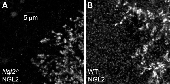
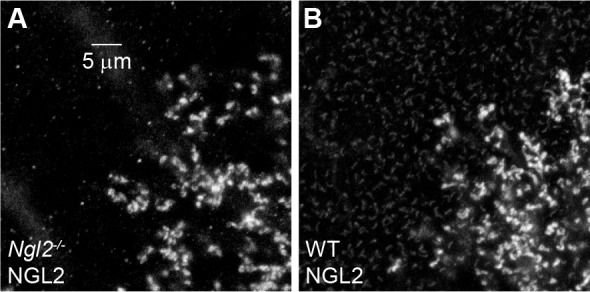
Netrin-G ligand 2 (NGL2), a synaptic CAM with an extracellular leucine-rich repeat (LRR) domain, regulates synapse development in the hippocampus and the retina (DeNardo et al., 2012; Nishimura-Akiyoshi et al., 2007; Soto et al., 2013). In the hippocampus, NGL2 localizes to the proximal segments of CA1 pyramidal neuron dendrites and promotes the formation of synapses with Schaffer collateral axons (DeNardo et al., 2012; Nishimura-Akiyoshi et al., 2007). In the retina, NGL2 localizes to the axon tips of horizontal cells and promotes the formation of synapses with rod photoreceptors (Figure 1—figure supplement 1) (Soto et al., 2013). Whether NGL2 acts cell-autonomously or not, and whether it maintains synapses and can promote their restoration in mature circuits is unknown.

Here we use AAVs to delete (CRISPR/Cas9), re-, and overexpress NGL2 in individual horizontal cells in the developing and mature retina to address these questions.

Results

A strategy for temporally controlled removal of NGL2

We devised the following AAV-mediated CRISPR/Cas9 strategy to delete Ngl2 with temporal control in individual horizontal cells in vivo. We identified two short guide RNAs (sgRNAs) that reliably introduced frame-shifting insertions and deletions (indels) near the start of the open reading frame of Ngl2 (see Materials and methods). We generated AAVs (serotype: 1/2) expressing these sgRNAs from a Pol III U6 promoter and tdTomato (tdT) from a Pol II CAG promoter (AAV-sgNGL2-tdT). We injected AAV-sgNGL2-tdT into the vitreous chamber of mice ubiquitously expressing the Cas9 endonuclease (Platt et al., 2014) (Cas9 mice, Figure 1A). To assess the efficiency of NGL2 removal, we injected AAV-sgNGL2-tdT in newborn (postnatal day 0, P0) Cas9 mice and stained flat-mounted retinas at P30 for NGL2. The NGL2 intensity at axon tips of tdT-positive horizontal cells in Cas9 mice was lower than at neighboring axon tips in 19 of 20 cells (i.e., 95% of cells, Figure 1B and C), whereas NGL2 intensity at axon tips of AAV-YFP-infected cells was indistinguishable from neighboring axon tips (Figure 1C). At many axon tips of AAV-sgNGL2-tdT-infected cells in Cas9 mice, NGL2 staining was reduced rather than absent. This could be, either because some NGL2 protein remained in horizontal cells expressing sgRNAs, or because multiple horizontal cells contributed to the NGL2 staining at each tip. Given that we injected AAV-sgNGL2-tdT at P0, nearly two weeks before NGL2 is first expressed (Soto et al., 2013), residual protein seemed an unlikely explanation. Co-injection of AAVs expressing spectrally separable fluorophores (cyan fluorescent protein [CFP] and tdT) revealed that overlapping horizontal cell axons co-innervate more than 40% of the rods in their shared territory (Figure 1D and E). As a population, horizontal cell axons cover the retina approximately ninefold (Soto et al., 2013; Keeley et al., 2014). Thus, multiple horizontal cells innervate most rods, which likely explains the remaining NGL2 staining at axon tips labeled by infection of single horizontal cells with AAV-sgNGL2-tdT. We conclude that our AAV-mediated CRISPR/Cas9 strategy removed NGL2 from horizontal cells with high efficiency (i.e., in 95% of infected cells).

Figure 1. AAV-mediated knockout of Ngl2 in horizontal cells.

(A) Schematic illustrating AAV-mediated CRISPR/Cas9 strategy for Ngl2 knockout in horizontal cells. In AAV-sgNGL2-tdT, small guide RNAs targeting NGL2 (sgNGL2) were expressed from a Pol III U6 promoter, and the red fluorescent protein tdT was expressed from a Pol II CAG promoter. AAV-sgNGL2-tdT was injected intravitreally into Cas9 mice (Platt et al., 2014). (B) Representative images of an axon of a horizontal cell infected with AAV-sgNGL2-tdT (injection at P0, analysis at P30) in a Cas9 retina. Left, overview of the axon labeled by tdT; right, magnified excerpts showing NGL2 staining at tips of this axon and overlapping axons of uninfected horizontal cells. (C) Relative NGL2 intensity in axon tips of infected vs. uninfected horizontal cell, for AAV-sgNGL2-tdT (sgNGL2) and AAV-YFP (YFP). Dots show data from single cells compared to its neighbors, the circle (errorbar) indicates the mean (±SEM) of the population. In 19 of 20 horizontal cells (3 mice) infected with AAV-sgNGL2-tdT, the NGL2 intensity was significantly reduced (p<0.01 for each, Wilcoxon rank sum test), whereas NGL2 intensity was unchanged in five of five horizontal cells (2 mice) infected with AAV-YFP. (D) Representative images of two overlapping horizontal cell axons labeled with CFP and tdT, respectively. Left, overview image; right, magnified excerpts from rods contacted by tips of either (top and middle) or both (bottom) axons. (E) Summary data of shared rod contacts (i.e., overlapping axon tips) within the overlapping territory of two horizontal cell axons. Dots show data from individual horizontal cell pairs, the circle (errorbar) indicates the mean (±SEM) of the population.

Figure 1.

Figure 1—figure supplement 1. NGL2 localizes to tips of horizontal cell axons, not rod bipolar cell dendrites.

Figure 1—figure supplement 1.

(A and B) Representative images of axon tips of horizontal cells infected with AAV-YFP (A, injection at P0, imaging at P30) or dendrites of rod bipolar cells infected with AAV-Grm6-YFP (B) in wild-type retinas stained for NGL2.

NGL2 regulates horizontal cell axon development cell autonomously

We first used this strategy, to analyze the effects of early postnatal NGL2 removal from individual horizontal cells on their development. In wild-type mice, P0 injection of AAV-sgNGL2-tdT affected neither the size of horizontal cell axons nor the density of their tips at P30 (Figure 2A, B, F and G). In Cas9 mice, axons of horizontal cells infected with AAV-sgNGL2-tdT were larger and had fewer tips than axons of horizontal cells infected with AAV-YFP (Figure 2C–2G). Both in wild-type mice and in Cas9 mice injected with AAV-sgNGL2-tdT, nearly all horizontal cell axon tips apposed ribbon release sites of rod photoreceptors (Figure 2—figure supplement 1). We, therefore, use axon tips throughout this study as an indicator of synapses between horizontal cells and rods. Changes in horizontal cell axon size and tip density in Cas9 mice injected with AAV-sgNGL2-tdT matched those observed in germline Ngl2 knockout mice (Ngl2-/- mice) (Soto et al., 2013). They were stable over time (Figure 2—figure supplement 2) and indistinguishable between both sgRNAs (Figure 2C–2G). Consistent with our findings in Ngl2-/- mice (Soto et al., 2013), removal of NGL2 from individual horizontal cells did not affect the size of horizontal cell dendrites or the number of their contacts with cones (Figure 2H–2N). Thus, during development, NGL2 appears to selectively and cell-autonomously regulate the growth of horizontal cell axons and the formation of synapses between horizontal cell axons and rods.

Figure 2. NGL2 regulates horizontal cell axon size and tip density cell autonomously.

(A–E) Representative images of horizontal cell axons labeled by AAV-YFP (YFP) or AAV-sgNGL2-tdT (sgNGL21 and sgNGL22) in wild-type (WT) and Cas9 mice. Overview images are maximum intensity projections of the complete axons; insets show maximum intensity projections limited to axon tips at higher magnification. (F and G) Summary data of axon territories (F) and the density of axon tips in these territories (G). Dots show data from single cells, circles (errorbars) indicate means (±SEM) of the respective populations. AAV-sgNGL2-tdT infection in wild-type mice did not affect horizontal cell axon size (WT YFP n = 29, 8 mice, WT sgNGL21n = 37, 12 mice, p=1) or tip density (WT YFP n = 10, 3 mice, WT sgNGL21n = 23, 8 mice, p=0.25). Horizontal cell axons in Cas9 mice were similar in size (WT YFP n = 29, 8 mice, Cas9 YFP n = 20, 8 mice, p=0.11) and tip densities (WT YFP n = 10, 3 mice, Cas9 YFP n = 8, 4 mice, p=1) to horizontal cell axons in wild-type mice. Both sgRNAs tested drastically increased horizontal cell axon size in Cas9 mice (Cas9 sgNGL21n = 35, 6 mice, p<10−7 for comparison to Cas9 YFP, Cas9 sgNGL22n = 32, 7 mice, p=10−9 for comparison to Cas9 YFP) and reduced tip densities (Cas9 sgNGL21n = 29, 5 mice, p < 10-12 for comparison to Cas9 YFP, Cas9 sgNGL22n = 16, 3 mice, p<0.001 for comparison to Cas9 YFP). (H – L) Analogous to (A – E) for horizontal cell dendrites and their contacts with cones. (M and N) Analogous to (F and G) for horizontal cell dendrites territories (WT YFP n = 15, 8 mice, WT sgNGL21n = 15 , 9 mice, Cas9 YFP n = 12, 8 mice, Cas9 sgNGL21n = 47, 6 mice, Cas9 sgNGL22n = 15, 7 mice) and terminal clusters within these territories (WT YFP n = 12, 6 mice, WT sgNGL21n = 15, 9 mice, Cas9 YFP n = 12, 8 mice, Cas9 sgNGL21n = 14, 3 mice, Cas9 sgNGL22n = 14, 6 mice). No significant differences between genotypes and AAVs were observed for horizontal cell dendrites (p>0.9, for all comparisons). P-values reported in this figure legend are from ANOVA tests with Bonferroni correction for multiple comparisons.

Figure 2.

Figure 2—figure supplement 1. Nearly all horizontal cell axon tips are synaptic.

Figure 2—figure supplement 1.

(A and B) Representative images of axon tips of horizontal cells infected with AAV-YFP in a wild-type retina (A) and with AAV-sgNGL2-tdT in a Cas9 retina (B) stained for the presynaptic ribbon marker Bassoon. (C) Summary data of axon tips overlapping with Bassoon staining in horizontal cells infected with AAV-YFP wild-type retinas and horizontal cells infected with AAV-sgNGL2-tdT in Cas9 retinas (YFP 96.7 ± 1.3%, n = 7, 3 mice, sgNGL2 97.7 ± 1.4%, n = 4, 2 mice, p=0.94, Wilcoxon rank sum test). Dots show data from single cells, circles (errorbars) indicate means (±SEM) of the respective populations.
Figure 2—figure supplement 2. Stable changes in horizontal cell axon size and axon tip density after NGL2 removal.

Figure 2—figure supplement 2.

(A) Schematic illustrating experimental timeline. AAVs were intravitreally injected into newborn (P0) mice and retinas collected on P30, P60, and P90. (B and C) Representative images of horizontal cell axons labeled by AAV-YFP (YFP) or AAV-sgNGL2-tdT (sgNGL2) in Cas9 mice, imaged at P30. Overview images are maximum intensity projections of the complete axons; insets show maximum intensity projections limited to axon tips at higher magnification. (D and E) Summary data of axon territories (D) and the density of axon tips in these territories (E) at P30. Dots show data from single cells, circles (errorbars) indicate means (±SEM) of the respective populations. At P30, axons of horizontal cells infected with AAV-sgNGL2-tdT occupied larger territories (Cas9 YFP n = 14, 8 mice, Cas9 sgNGL2 n = 35, 17 mice, p<10−5), and had lower axon tip densities (Cas9 YFP n = 8, 4 mice, Cas9 sgNGL2 n = 29, 8 mice, p=10−4) compared to horizontal cells infected with AAV-YFP. (F and G) Representative images analogous to (B and C) for tissue collected at P60. (H and I) Summary data analogous to (D and E) for tissue collected at P60. At P60, axons of horizontal cells infected with AAV-sgNGL2-tdT were larger (Cas9 YFP n = 35, 15 mice, Cas9 sgNGL2 n = 34, 11 mice, p<0.001), and the density of axon tips was reduced (Cas9 YFP n = 10, 3 mice, Cas9 sgNGL2 n = 20, 6 mice, p<10−4) compared to horizontal cells infected with AAV-YFP. (J and K) Representative images analogous to (B and C) for tissue collected at P90. (L and M) Summary data analogous to (D and E) for tissue collected at P90. At P90, axons of horizontal cells infected with AAV-sgNGL2-tdT were larger (Cas9 YFP n = 22, 12 mice, Cas9 sgNGL2 n = 17, 8 mice, p<0.001), and the density of axon tips was reduced (Cas9 YFP n = 9, 3 mice, Cas9 sgNGL2 n = 11, 5 mice, p<0.001) compared to horizontal cells infected with AAV-YFP. P-values in this figure legend are from Wilcoxon rank sum tests.
Figure 2—figure supplement 3. Effects of NGL2 removal on horizontal cell axon targeting.

Figure 2—figure supplement 3.

(A–F) Representative images of horizontal cell axons – rotated maximum intensity projections of confocal image stacks from retinal whole mounts – labeled by AAV-YFP (YFP, (A, C, F) or AAV-sgNGL2-tdT (sgNGL21, B, D; sgNGL22 E) in wild-type (WT, (A, B), Cas9 (C - E), and Ngl2-/- (F) mice. (G) Summary data indicating the fraction of all horizontal cells examined in the different combinations of AAVs and genotypes (WT YFP n = 12, 3 mice, WT sgNGL21n = 35, 12 mice, Cas9 YFP n = 19, 6 mice, Cas9 sgNGL21, n = 28, 6 mice, Cas9 sgNGL22, n = 31, 7 mice, Ngl2-/- YFP n = 13, 4 mice) with no (green), one to five (orange), and more than five (red) mistargeted tips in the outer nuclear layer (ONL). Horizontal cells infected with AAV-sgNGL2-tdT (sgNGL22, not sgNGL21) in Cas9 mice had more severe mistargeting phenotypes than horizontal cells in wild-type mice infected with AAV-YFP (p<0.02) or AAV-sgNGL2-tdT (p<0.004), or horizontal cells in Cas9 mice infected with AAV-YFP (p<0.004). Horizontal cells in Cas9 mice infected with AAV-sgNGL2-tdT (sgNGL22 and sgNGL21) in Cas9 mice had less severe mistargeting phenotypes than horizontal cells in Ngl2-/- mice infected with AAV-YFP (p<0.002). P-values in this figure legend are from Χ2 tests.

In Ngl2-/- mice, horizontal cell axons frequently stray into the outer nuclear layer (Soto et al., 2013). Stray processes were less abundant in horizontal cells targeted by our AAV-mediated CRISPR/Cas9 strategy (Figure 2—figure supplement 3), indicating either that mistargeting involves non-cell-autonomous actions of NGL2, or that delays in the AAV-mediated removal of NGL2 reduced the severity of laminar targeting deficits compared to germline knockouts.

NGL2 maintains horizontal cell axon tips and restrains axon growth in mature circuits

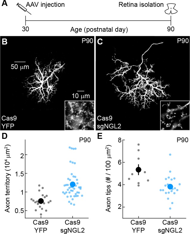
In mice, neuronal morphology and connectivity in the outer retina are mature 3–4 weeks after birth (Huckfeldt et al., 2009; Poché et al., 2007; Blanks et al., 1974). To test whether NGL2 contributes to the maintenance of horizontal cell axons and synapses, we injected AAV-sgNGL2-tdT in P30 (i.e., young adult) Cas9 mice. By P60, axon arbors of tdT-positive horizontal cells had expanded and lost many tips, compared to Cas9 mice injected with AAV-YFP (Figure 3A–3E). Axon expansion and tip loss were stable at P90 (Figure 3—figure supplement 1), indicating that the same phenotypes produced by developmental NGL2 removal emerge rapidly and persist after removal of NGL2 in mature circuits.

Figure 3. NGL2 restrains horizontal cell axon growth and maintains axon tips in young adult mice.

(A) Schematic of the experimental timeline. AAVs were injected into the vitreous chamber of P30 mice and retinas collected on P60. (B and C) Representative images of horizontal cell axons labeled by AAV-YFP (YFP) or AAV-sgNGL2-tdT (sgNGL2) in Cas9 mice, imaged at P60. Overview images are maximum intensity projections of the complete axons; insets show maximum intensity projections limited to axon tips at higher magnification. (D and E) Summary data of axon territories (D) and the density of axon tips in these territories (E) at P60. Dots show data from single cells, circles (errorbars) indicate means (±SEM) of the respective populations. At P60, axons of horizontal cells infected with AAV-sgNGL2-tdT occupied larger territories (Cas9 YFP n = 35, 15 mice, Cas9 sgNGL2 n = 46, 6 mice, p<10−9, Wilcoxon rank sum test), and had lower densities of axon tips (Cas9 YFP n = 10, 3 mice, Cas9 sgNGL2 n = 12, 4 mice, p<0.001, Wilcoxon rank sum test) compared to horizontal cells infected with AAV-YFP.

Figure 3.

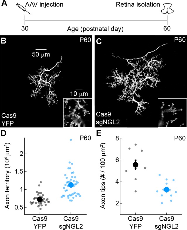
Figure 3—figure supplement 1. NGL2 restrains HC axon growth and maintains axon tips in adult mice.

Figure 3—figure supplement 1.

(A) Schematic of the experimental timeline. AAVs were injected into the vitreous chamber of P30 mice and retinas collected at P90. (B and C) Representative images of axons of horizontal cells infected with AAV-YFP (YFP) or AAV-sgNGL2-tdT (sgNGL2) in Cas9 mice, imaged at P90. Overview images are maximum intensity projections of the complete axons; insets show maximum intensity projections limited to axon tips at higher magnification. (D and E) Summary data of axon territories (D) and axon tip densities (E) at P90. Dots show data from single cells, circles (errorbars) indicate means (±SEM) of the respective populations. At P90, axons of horizontal cells infected with AAV-sgNGL2-tdT had expanded territories (Cas9 YFP n = 22, 8 mice, Cas9 sgNGL2 n = 48, 12 mice, p<10−6), and lost tips compared to horizontal cells infected with AAV-YFP (Cas9 YFP n = 9, 3 mice, Cas9 sgNGL2 n = 28, 7 mice p<0.003). P-values in this figure legend are from Wilcoxon rank sum tests.

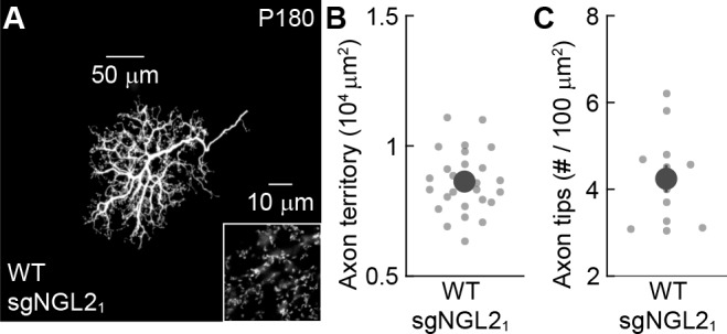
Many neurons exhibit heightened plasticity during critical periods that can extend several weeks beyond their initial neurite growth and synaptogenesis (Hong et al., 2014; Hensch, 2005). To assess whether injection of AAV-sgNGL2-tdT at P30 fell within such a critical period or whether NGL2 continues to be required throughout adulthood, we injected AAV-sgNGL2-tdT in mature adult (P150) Cas9 mice. Strikingly, by P180, horizontal cell axons had expanded and lost tips to the same degree observed for developmental and young adult manipulations (Figure 4A–4E). Injection of AAV-sgNGL2-tdT in P150 wild-type mice produced no phenotypes (Figure 4—figure supplement 1). Thus, NGL2 signaling appears to restrain horizontal cell axon growth and to maintain synapses between horizontal cells and rods throughout adulthood.

Figure 4. NGL2 restrains horizontal cell axon growth and maintains axon tips in mature adult mice.

(A) Schematic of the experimental timeline. AAVs were injected into the vitreous chamber of P150 mice and retinas collected on P180. (B and C) Representative images of horizontal cell axons labeled by AAV-YFP (YFP) or AAV-sgNGL2-tdT (sgNGL2) in Cas9 mice. Overview images are maximum intensity projections of the complete axons; insets show maximum intensity projections limited to axon tips at higher magnification. (D and E) Summary data of axon territories (D) and the density of axon tips in these territories (E). Dots show data from single cells, circles (errorbars) indicate means (±SEM) of the respective populations. Axons of horizontal cells infected with AAV-sgNGL2-tdT occupied larger territories (Cas9 YFP n = 34, 9 mice, Cas9 sgNGL2 n = 18, 11 mice, p<10−6, Wilcoxon rank sum test), and had lower densities of axon tips (Cas9 YFP n = 11, 3 mice, Cas9 sgNGL2 n = 10, 3 mice, p<0.001, Wilcoxon rank sum test) compared to horizontal cells infected with AAV-YFP.

Figure 4.

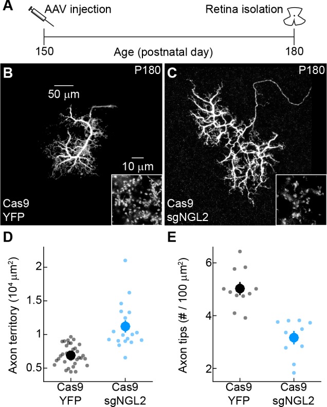
Figure 4—figure supplement 1. Small guide RNAs targeting Ngl2 do not affect horizontal cell axons in mature adult wild-type mice.

Figure 4—figure supplement 1.

(A) Representative image of the axon of a horizontal cell infected with AAV-sgNGL2-tdT (sgNGL2) in a wild-type retina. Overview images are maximum intensity projections of the complete axons; insets show maximum intensity projections limited to axon tips at higher magnification. Summary data of axon territories (B, n = 26, 5 mice) and axon tip densities (C, n = 16, 3 mice). Dots show data from single cells, circles (errorbars) indicate means (±SEM) of the respective populations.

AAV-mediated NGL2 expression in developing Ngl2-/- horizontal cells normalizes axon growth and enhances axon tip density

Because removal of NGL2 from individual horizontal cells disrupted axon and synapse development, we wanted to test whether expression of NGL2 in individual horizontal cells in Ngl2-/- mice could rescue axon and synapse development. We generated AAVs expressing full-length NGL2 (AAV-NGL2) in horizontal cells and injected AAV-NGL2 or AAV-YFP intravitreally at P0. At P30, we counted axon tips of infected horizontal cells by immunostaining for NGL2 (AAV-NGL2) or YFP (AAV-YFP), and measured axon territories as the area of the smallest convex polygon to encompass all tips of an arbor. AAV-mediated expression of NGL2 exceeded wild-type protein levels at P30 (Figure 5—figure supplement 1). In Ngl2-/- mice, AAV-NGL2 restored the size of horizontal cell axons to wild-type levels and increased the density of axon tips beyond wild-type levels (Figure 5A–5D, F and G). In wild-type mice, AAV-NGL2 did not significantly change horizontal cell axon size but elevated axon tip density (Figure 5D–5G). The most parsimonious explanation for these findings is that a threshold amount of NGL2 is required to restrict horizontal cell axon growth and that NGL2 protein levels control synapse density bidirectionally. These results also further support the notion that NGL2 regulates horizontal cell development cell autonomously.

Figure 5. AAV-mediated NGL2 expression normalizes axon growth and enhances axon tip formation of horizontal cells in developing Ngl2-/- mice.

(A) Schematic of the experimental timeline. AAVs were injected into the vitreous chamber of P0 mice and retinas collected at P30. (B–E) Representative images of axons of horizontal cells infected with AAV-YFP (YFP, (B and D) or AAV-NGL2 (NGL2, C, and E) in Ngl2-/- mice (B and C) or wild-type littermates (D and E). Overview images are maximum intensity projections of the complete axons; insets show maximum intensity projections limited to axon tips at higher magnification. (F and G) Summary data of axon territories (F) and the density of axon tips in these territories (G). Dots show data from single cells, circles (errorbars) indicate means (±SEM) of the respective populations. AAV-mediated expression of NGL2 reduced horizontal cell axon territories (Ngl2-/- YFP n = 12, 4 mice, Ngl2-/- NGL2 n = 21, 4 mice, p<10−7), restoring them to wild-type levels (WT YFP n = 31, 5 mice, p=1 for comparison to Ngl2-/- NGL2). In wild-type mice, axon territories of AAV-NGL2-infected horizontal cells were not significantly different from AAV-YFP-infected horizontal cells (WT NGL2, n = 15, 3 mice, p=0.48 for comparison to WT YFP). AAV-mediated NGL2 expression increased axon tip density in Ngl2-/- mice (Ngl2-/- YFP n = 10, 3 mice, Ngl2-/- NGL2 n = 12, 3 mice, p<10−10) beyond wild-type levels (WT YFP n = 12, 3 mice, p<10−6 for comparison to Ngl2-/- NGL2). AAV-mediated expression of NGL2 in wild-type mice similarly increased the axon tip density of horizontal cell axons (WT NGL2, n = 10, 3 mice, p<0.001 for comparison to WT YFP). P-values reported in this figure legend are from ANOVA tests with Bonferroni correction for multiple comparisons.

Figure 5.

Figure 5—figure supplement 1. AAV-mediated NGL2 expression in Ngl2-/- and wild-type P30 retinas.

Figure 5—figure supplement 1.

(A and B) Higher magnification excerpts of overview images in Figure 5 (C) and (E). We adjusted the brightness of the images in this figure supplement to reveal wild-type NGL2 expression in (B) and document its absence in (A) at the expense of saturating signals of AAV-mediated NGL2 expression.

AAV-mediated NGL2 expression in adult Ngl2-/- horizontal cells shrinks axons and restores axon tip density

We next explored whether restoring NGL2 to individual horizontal cells in adult Ngl2-/- circuits could reverse axonal and synaptic phenotypes. Indeed, injection of AAV-NGL2 at P30 reduced axon size and increased the density of axon tips of infected horizontal cells in Ngl2-/- mice to wild-type levels by P60 (Figure 6A–D, F and G). In wild-type mice, AAV-NGL2 injection at P30 did not significantly change the axon size of infected horizontal cells but elevated the density of axon tips (Figure 6D–6G). AAV-mediated expression of NGL2 after P30 injections exceeded wild-type protein levels by P60 (Figure 6—figure supplement 1). Thus, re- and overexpression of NGL2 in mature horizontal cells shrink axons back to their normal size in Ngl2-/- mice, and can restore and even enhance their connectivity in Ngl2-/- and wild-type mice.

Figure 6. AAV-mediated NGL2 expression shrinks axon arbors and restores axon tips of horizontal cells in adult Ngl2-/- mice.

(A) Schematic of the experimental timeline. AAVs were injected into the vitreous chamber of P30 mice and retinas collected at P60. (B–E) Representative images of axons of horizontal cells infected with AAV-YFP (YFP, (B and D) or AAV-NGL2 (NGL2, (C and E) in Ngl2-/- mice (B and C) or wild-type littermates (D and E). Overview images are maximum intensity projections of the complete axons; insets show maximum intensity projections limited to axon tips at higher magnification. (F and G) Summary data of axon territories (F) and the density of axon tips in these territories (G). Dots show data from single cells, circles (errorbars) indicate means (±SEM) of the respective populations. AAV-mediated expression of NGL2 reduced horizontal cell axon territories (Ngl2-/- YFP n = 12, four mice, Ngl2-/- NGL2 n = 12, two mice, p<10−4), restoring them to wild-type levels (WT YFP n = 11, four mice, p=1 for comparison to Ngl2-/- NGL2). In wild-type mice, axon territories of AAV-NGL2-infected horizontal cells were not significantly different from AAV-YFP-infected horizontal cells (WT NGL2, n = 15, four mice, p=1 for comparison to WT YFP). AAV-mediated NGL2 expression increased axon tip density in Ngl2-/- mice (Ngl2-/- YFP n = 11, 4mice, Ngl2-/- NGL2 n = 10, two mice, p<10−7) to wild-type levels (WT YFP n = 11, four mice, p=1 for comparison to Ngl2-/- NGL2). AAV-mediated expression of NGL2 in wild-type mice similarly increased the axon tip density of horizontal cells (WT NGL2, n = 13, four mice, p<0.001 for comparison to WT YFP). P-values reported in this figure legend are from ANOVA tests with Bonferroni correction for multiple comparisons.

Figure 6.

Figure 6—figure supplement 1. AAV-mediated NGL2 expression in Ngl2-/- and wild-type P60 retinas.

Figure 6—figure supplement 1.

(A and B) Higher magnification excerpts of overview images in Figure 6 (C) and (E). We adjusted the brightness of the images in this figure supplement to reveal wild-type NGL2 expression in (B) and document its absence in (A) at the expense of saturating signals of AAV-mediated NGL2 expression.

Discussion

Synaptic CAMs like NGL2 typically localize to nascent connections during development and remain at synapses for the lifetime of the animal (de Wit and Ghosh, 2014; Siddiqui and Craig, 2011; Sytnyk et al., 2017). Because most in vivo studies have used knockout mice that remove targeted genes in the germline, it is unclear whether synaptic CAMs contribute to the maintenance of synapses as well as their development. Here, we devised an AAV-mediated CRISPR/Cas9 strategy to remove NGL2 from horizontal cells with temporal control (Figure 1). Developmental removal of NGL2 with this strategy reduced the density of synapses formed by horizontal cell axons to the same extent as Ngl2-/- mice (Figure 2) (Soto et al., 2013). When we removed NGL2 in one- and five-months-old mice, horizontal cell axons rapidly lost connections with rod photoreceptors (Figures 3 and 4). In the medial prefrontal cortex of adult mice, removal of neuroligin-2 reduces the number of inhibitory synapses (Liang et al., 2015). In the cerebellum, parallel fiber inputs are lost after deletion of GluRδ2 in mature Purkinje cells (Takeuchi et al., 2005). Together, these findings indicate that synaptic CAMs stabilize synapses and that the cues that maintain circuits overlap with those that govern their formation.

Removal of NGL2 from individual horizontal cells (Figure 2) had the same effects on connectivity as removal from all cells in Ngl2-/- mice (Soto et al., 2013), and AAV-mediated expression of NGL2 in individual horizontal cells of Ngl2-/- mice was sufficient to rescue their axon and synapse development (Figure 5). Thus, NGL2 regulates horizontal cell connectivity cell autonomously. NGL2 appears to act similarly in the hippocampus, where knockdown in individual pyramidal cells lowers spine density (DeNardo et al., 2012). The observation that AAV-mediated overexpression of NGL2 in horizontal cells in wild-type and Ngl2-/- mice elevates axon tip density above wild-type levels (Figure 5) suggests that NGL2 levels control connectivity bidirectionally and limit synapse formation in wild-type retinas. Similarly, spine density in the hippocampus is reduced in Cadm1-/- mice and increased in transgenic mice overexpressing SynCAM1, the protein encoded by Cadm1 (Robbins et al., 2010). NGL2 interacts trans-synaptically with netrin-G2 (DeNardo et al., 2012; Kim et al., 2006; Nishimura-Akiyoshi et al., 2007; Zhang et al., 2008). Whether this interaction localizes NGL2 to horizontal cell axon tips, as it localizes NGL2 to proximal dendrites of CA1 pyramidal neurons (Nishimura-Akiyoshi et al., 2007), and how netrin-G2 – NGL2 complexes transmit signals between rods and horizontal cells remains to be explored. We know very little about the signaling mechanisms of synaptic CAMs, whether they act cell-autonomously or not, and whether they control neuronal processes (e.g., synapse formation) bidirectionally. Advances in viral targeting and genome editing (Buchholz et al., 2015; Platt et al., 2014; Senís et al., 2014; Trapani et al., 2014), which we exploited in our study, should accelerate progress in this area.

We found that NGL2 restricts horizontal cell axon growth throughout life (Figures 24). Similarly, the auxiliary Ca2+ channel subunit α2δ2, which mediates trans-synaptic interactions (Fell et al., 2016), restricts axon growth in developing and mature mouse dorsal root ganglion neurons (Tedeschi et al., 2016) and Ig-domain-containing CAMs contribute to axon maintenance in C. elegans (Aurelio et al., 2002; Cherra and Jin, 2016). Together, these results indicate that synaptic CAMs can control the size of neurite arbors during development and at maturity.

We found that manipulations of NGL2 co-regulate horizontal cell axon growth and synapse development in seemingly homeostatic fashion: deletion of Ngl2 reduced synapse density and increased axons size (Figures 24), whereas re-expression of NGL2 in Ngl2-/- mice increased synapse density and reduced axon size (Figures 5 and 6). Whether neurite growth and synapse formation in horizontal cells are coupled or independently controlled remains to be tested. A homeostatic relationship between these processes was reported for aCC neurons in Drosophila embryos (Tripodi et al., 2008). Bidirectional manipulations in the number of input synapses on aCC dendrites elicit opposite changes in arbor size (Tripodi et al., 2008). By contrast, neurite growth appears positively coupled to synapse formation of axons and dendrites in the optic tectum of Xenopus and Zebrafish, respectively (Niell et al., 2004; Ruthazer et al., 2006). In the mouse retina, the size and morphology of bipolar cell axons and ganglion cell dendrites are established and maintained independent of connections between them (Johnson and Kerschensteiner, 2014; Kerschensteiner et al., 2009; Morgan et al., 2011). What accounts for the differences in the relationship of synapses and neurite arbors in different neurons, circuits, and species, remains to be identified.

AAV-mediated expression of NGL2 in individual horizontal cells in adult Ngl2-/- and wild-type mice restored and enhanced their connectivity (Figure 6). In neurodegenerative diseases, synapse loss can long precede cell death (Buckingham et al., 2008; Gamm et al., 2015; Hong et al., 2016; Scheff et al., 2006). Molecular mechanisms that maintain synapses could, therefore, be utilized to rescue circuit function in neurodegenerative diseases. We speculate that AAV-mediated expression of NGL2 might help preserve outer retinal connectivity in retinal degeneration, the most common heritable cause of visual impairment (Sohocki et al., 2001; Jones et al., 2012). Transplantation of stem-cell-derived photoreceptors is being explored as a treatment for late stages of retinal degeneration (Gamm et al., 2015; Wahlin et al., 2017; Chirco et al., 2017). Our results suggest that AAV-mediated expression of NGL2 in second-order neurons (i.e., bipolar and horizontal cells) could promote the integration of stem-cell-derived photoreceptors into host circuits and help restore vision.

Materials and methods

Key resources table.

Reagent type (species)
or resource
Designation Source or reference Identifiers Additional
information
Mouse strain Wild-type (C57Bl6/J) Jackson Laboratory RRID:IMSR_JAX000664
Mouse strain Cas9 Jackson Laboratory RRID:IMSR_JAX:024858
Mouse strain Ngl2-/- (Soto et al., 2013)
Plasmid pX330-U6-Chimeric_BB-CBh-hSpCas9 Addgene Plasmid # 42230
Plasmid pmaxGFP Lonza Catalog # VDC-1040
Virus AAV-YFP This paper
Virus AAV-Grm6-YFP (Johnson et al., 2017)
Virus AAV-sgNGL21-tdT This paper
Virus AAV-sgNGL22-tdT This paper
Virus AAV-NGL2 This paper
Antibody anti-NGL2 (mouse) NeuroMab RRID:AB_2137614 1:100 dilution
Antibody anti-Bassoon (mouse) Enzo RRID:AB_2313990 1:500 dilution
Antibody anti-DsRed (rabbit) Clontech Laboratories RRID:AB_10013483 1:1000 dilution
Antibody anti-GFP (chicken) ThermoFisher RRID:AB_2534023 1:500 dilution
Antibody anti-GFP (rabbit) ThermoFisher RRID:AB_221569 1:1000 dilution
Antibody anti-chicken IgY Alexa 488 ThermoFisher RRID:AB_2534096 1:1000 dilution
Antibody anti-rabbit IgG Alexa 488 ThermoFisher RRID:AB_2536097 1:1000 dilution
Antibody anti-rabbit IgG Alexa 568 ThermoFisher RRID:AB_143011 1:1000 dilution
Antibody anti-mouse IgG Alexa 568 ThermoFisher RRID:AB_2534072 1:1000 dilution
Antibody anti-mouse IgG Alexa 633 ThermoFisher RRID:AB_2535718 1:1000 dilution

Mice

In this study, we used mice in which the RNA-guided endonuclease Cas9 is ubiquitously expressed from a CAG promoter in the Rosa26 locus (Cas9 mice, Jackson Labs, RRID:IMSR_JAX:024858) (Platt et al., 2014). Cas9 mice were on a C57Bl6/N background. To confirm that sgRNAs had no effect in the absence of Cas9, we used wild-type mice on a C57Bl6/J background (RRID:IMSR_JAX000664). In addition, we used Ngl2-/- mice on a C57Bl6/J background. Mice of both sexes were used in our experiments. All procedures were approved by the Animal Studies Committee of Washington University School (protocol # 20170033) of Medicine and performed in compliance with the National Institutes of Health Guide for the Care and Use of Laboratory Animals.

DNA constructs and sgRNA testing

Single guide RNAs targeting the 5’ region of the mouse Ngl2 gene were designed using an open access web tool provided by the Zhang lab at MIT (http://crispr.mit.edu:8079/) to search the first 250 bp of the Ngl2 gene coding region. A pair of complementary oligonucleotides encoding the 20-nt guide sequences sgNGL21 (5’-gaatccacacttgcgccgtg-3’) and sgNGL22 (5’-aaggtggtgtgcacccgccg-3’) were synthetized. Oligonucleotides used in the generation of sgRNA templates were: Guide1-F 5’-ACCgaatccacacttgcgccgtg-3’, Guide1-R 5’-AACcacggcgcaagtgtggattc-3’, Guide2-F 5’-ACCaaggtggtgtgcacccgccg-3’, and Guide2-R 5’-AACcggcgggtgcacaccacctt-3’.

Plasmid pX330-U6-Chimeric_BB-CBh-hSpCas9 containing the U6 promoter, the sgRNA targeting sequence cloning site followed by the sgRNA scaffold, and the human codon-optimized Cas9 was obtained from Addgene (# 42230) and was used to clone the corresponding sgRNAs for testing in vitro. The Ngl2-/- cell line was generated from the mouse Neuro-2a line at the Genome Engineering and iPSC Center (GEiC), Washington University in St. Louis. Approximately 4 * 105 Neuro-2a cells were suspended in P3 primary buffer and electroporated using a 4D-Nucleofector (Lonza) with 0.5 µg of pmaxGFP (control, Lonza) or pX330-U6-Chimeric_BB-CBh-hSpCas9 containing sgNGL21 or sgNGL22, in 20 µL wells. Sixty hours following nucleofection, cells were harvested, and genomic DNA was isolated and screened with PCRs using tagged primer sets (5'-CACTCTTTCCCTACACGACGCTCTTCCGATCTgctcctagctcacttaagccggggt-3', 5'-GTGACTGGAGTTCAGACGTGTGCTCTTCCGATCTaggtgcctgaaggtgtcggcctgaa-3') specific to the targeted region. The targeted Ngl2 locus region was sequence-confirmed using next-generation sequencing. Approximately 1000 independent sequences were analyzed per plasmid used, and the percentage of non-homologous recombinant events detected was normalized to the total number of reads. Recombination rates were 60% and 68% for sgNGL21 or sgNGL22, respectively.

For constructing sgRNA-expressing AAV viral vectors, the original pX330 plasmid was altered by adding a SapI site before the sgRNA scaffold. Then, the U6-sgRNA region of pX330 was isolated with enzymes Acc65i-Afl III blunted with Klenow and cloned blunt upstream of the CAG promoter into an existing pAAV-CAG-tdT plasmid available in the lab. The new plasmid, pAAV-U6-sgRNA-CAG-tdT, was then digested with SapI to introduce the designed corresponding sgRNA oligos targeting Ngl2. For constructing the AAV viral vector expressing NGL2, full-length Ngl2 cDNA cloned in pEF-BOS (DeNardo et al., 2012) was amplified using EcoRI and NotI linkers and cloned using the same sites into the pAAV-CAG-YFP vector in place of YFP. The PCR product was verified by sequencing.

Adeno-associated viruses

Viral particles were packaged and purified as previously described (Grimm et al., 2003; Klugmann et al., 2005). Briefly, AAV1/2 chimeric virions, which readily infect horizontal cells (Soto et al., 2013), were produced by co-transfecting HEK293 cells with pAAV-U6-sgNGL21-CAG-tdT (AAV-sgNGL21-tdT), pAAV-U6-sgNGL22-CAG-tdT (AAV-sgNGL22-tdT), pAAV-CAG-YFP (AAV-YFP), pAAV-CAG-NGL2 (AAV-NGL2) or pAAV-Grm6-YFP (AAV-Grm6-YFP) (Johnson et al., 2017) and helper plasmids encoding Rep2 and the Cap for serotype one and Rep2 and the Cap for serotype 2. Forty-eight hours after transfection, cells and supernatant were harvested and viral particles purified using heparin affinity columns (Sigma, Saint Louis, MO). Viruses (250 nL) were injected with a Nanoject II (Drummond) into the vitreous chamber of newborn mice anesthetized on ice

Tissue preparation

Mice were deeply anesthetized with CO2, killed by cervical dislocation, and enucleated. Retinas were isolated in oxygenated mouse artificial cerebrospinal fluid (mACSFHEPES) containing (in mM): 119 NaCl, 2.5 KCl, 1 NaH2PO4, 2.5 CaCl2, 1.3 MgCl2, 20 HEPES, and 11 glucose (pH adjusted to 7.37 using NaOH) and mounted flat on black membrane discs (HABGO1300, Millipore, Burlington, MA), or left in the eyecup for 30 min fixation with 4% paraformaldehyde in mACSFHEPES.

Immunohistochemistry

After blocking for 2 hr with 5% Normal Donkey Serum in PBS, vibratome slices (thickness: 60 µm) embedded in 4% agarose (Sigma) were incubated overnight at 4°C with primary antibodies. Slices were then washed in PBS (3 × 20 min) and incubated with secondary antibodies for 2 hr at room temperature (RT). Flat-mount preparations were frozen and thawed three times after cryoprotection (1 hr 10% sucrose in PBS at RT, 1 hr 20% sucrose in PBS at RT, and overnight 30% sucrose in PBS at 4°C), blocked with 5% Normal Donkey Serum in PBS for 2 hr, and then incubated with primary antibodies for 5 d at 4°C and washed in PBS (3 × 1 hr) at RT. The following primary antibodies were used in this study: mouse anti-NGL2 (1:100, NeuroMab, Davis, CA, RRID:AB_2137614), mouse anti-Bassoon (1:500, Enzo, Farmingdale, NY, RRID:AB_2313990), rabbit anti-DsRed (1:1000, Clontech Laboratories, RRID:AB_10013483), chicken anti-GFP (1:500, ThermoFisher, Waltham, MA, RRID:AB_2534023), and rabbit anti-GFP (1:1000, ThermoFisher, RRID:AB_221569). Subsequently, flat mounts were incubated 1 d at 4°C with Alexa 488 (1:1000, ThermoFisher, anti-chicken IgY, RRID:AB_2534096, anti-rabbit IgG RRID:AB_2536097), Alexa 568 (1:1000, ThermoFisher, anti-rabbit IgG, RRID:AB_143011, anti-mouse IgG, RRID:AB_2534072), and Alexa 633 (1:1000, ThermoFisher, anti-mouse IgG, RRID:AB_2535718) secondary antibodies for and washed in PBS (3 × 1 hr) at RT.

Imaging

Image stacks were acquired on an Fv1000 laser scanning confocal microscope (Olympus) with 20 × 0.85 NA and 60 × 1.35 NA oil immersion objectives at a voxel sizes of 0.206 µm – 0.3 µm (x/y – z axis) and 0.082 µm – 0.3 µm (x/y – z axis), respectively, or on a Zeiss 810 laser scanning confocal microscope with an Airyscan detector through a 63 × 1.4 NA objective at a voxel size of 0.043 µm – 0.1 µm (x/y – z axis).

Image analysis

Horizontal cell axon and dendrite territories were defined as the areas of the smallest convex polygons to encompass the respective arbors (Figures 14) or all synaptic tips (Figures 5 and 6) in z-projections of confocal image stacks acquired in retinal flat mounts, and were measured using Fiji (Schindelin et al., 2012). Horizontal cell dendrite clusters at cone terminals and horizontal cell axon tips, which penetrate rod spherules (Peichl and González-Soriano, 1994; Wässle, 2004), were identified by eye in confocal image stacks and their positions (x, y, and z) noted to count synapses and analyze their stratification (Soto et al., 2013). To evaluate the efficiency of NGL2 removal in horizontal cells infected with AAV-sgNGL2-tdT, we acquired confocal images of their axons in retinas stained for NGL2. The intensity of NGL2 labeling in rod spherules penetrated by infected horizontal cells was then compared to the NGL2 signal in neighboring rod spherules. For visual clarity, in some of the representative images, horizontal cell axons and dendrites were digitally isolated using Amira (ThermoFisher) to remove Mueller glia labeled within the field of view.

Statistics

A power analysis using G*Power (Faul et al., 2009) on our initial data sets of Cas9 retinas injected with AAV-YFP and AAV-sgNGL2-tdT suggested that given the observed effect sizes for horizontal cell axon territories and synapses, total sample sizes exceed 16 for all comparisons between groups to achieve power of 0.95 at p<0.05. In our final data set, total sample sizes for all comparisons ranged from 18 to 71. To assess the statistical significance of differences between two groups, we used Wilcoxon rank sum for continuous quantitative data and Χ2 tests for frequency observations of categorical data. For comparisons of quantitative data between multiple groups, we used ANOVA tests with Bonferroni correction for multiple comparisons.

Acknowledgements

We thank members of the Kerschensteiner lab for helpful comments and suggestions throughout this study. We thank Michael Casey of the Vision Core at Washington University School of Medicine for help with the design of sgRNAs and the construction of plasmids. We acknowledge the Genome Engineering and iPSC Center (GEiC) at Washington University School of Medicine for validating sgRNAs in vitro. We are grateful to Drs. A Ghosh and L DeNardo for providing us with the mouse Ngl2 cDNA. This work was supported by the National Institutes of Health (EY027411 to FS and DK, EY026978 and EY023341 to DK, and the Vision Core Grant EY0268), and by the Research to Prevent Blindness Foundation via an unrestricted grant to the Department of Ophthalmology and Visual Sciences at Washington University School of Medicine.

Funding Statement

The funders had no role in study design, data collection and interpretation, or the decision to submit the work for publication.

Contributor Information

Florentina Soto, Email: sotof@wustl.edu.

Daniel Kerschensteiner, Email: kerschensteinerd@wustl.edu.

Carol A Mason, Columbia University, United States.

Funding Information

This paper was supported by the following grants:

  • National Eye Institute R01EY027411 to Florentina Soto, Daniel Kerschensteiner.

  • National Eye Institute EY0268 to Daniel Kerschensteiner, Florentina Soto.

  • National Eye Institute R01EY026978 to Daniel Kerschensteiner.

  • National Eye Institute R01EY023341 to Daniel Kerschensteiner.

Additional information

Competing interests

No competing interests declared.

Author contributions

Conceptualization, Formal analysis, Supervision, Funding acquisition, Investigation, Visualization, Writing—original draft, Writing—review and editing.

Investigation, Writing—review and editing.

Conceptualization, Formal analysis, Supervision, Funding acquisition, Visualization, Writing—original draft, Writing—review and editing.

Ethics

Animal experimentation: All procedures were approved by the Animal Studies Committee of Washington University School (protocol # 20170033) of Medicine and performed in compliance with the National Institutes of Health Guide for the Care and Use of Laboratory Animals.

Additional files

Transparent reporting form
DOI: 10.7554/eLife.30388.016

References

  1. Aurelio O, Hall DH, Hobert O. Immunoglobulin-domain proteins required for maintenance of ventral nerve cord organization. Science. 2002;295:686–690. doi: 10.1126/science.1066642. [DOI] [PubMed] [Google Scholar]
  2. Blanks JC, Adinolfi AM, Lolley RN. Synaptogenesis in the photoreceptor terminal of the mouse retina. The Journal of Comparative Neurology. 1974;156:81–93. doi: 10.1002/cne.901560107. [DOI] [PubMed] [Google Scholar]
  3. Buchholz CJ, Friedel T, Büning H. Surface-engineered viral vectors for selective and cell type-specific gene delivery. Trends in Biotechnology. 2015;33:777–790. doi: 10.1016/j.tibtech.2015.09.008. [DOI] [PubMed] [Google Scholar]
  4. Buckingham BP, Inman DM, Lambert W, Oglesby E, Calkins DJ, Steele MR, Vetter ML, Marsh-Armstrong N, Horner PJ. Progressive ganglion cell degeneration precedes neuronal loss in a mouse model of glaucoma. Journal of Neuroscience. 2008;28:2735–2744. doi: 10.1523/JNEUROSCI.4443-07.2008. [DOI] [PMC free article] [PubMed] [Google Scholar]
  5. Cherra SJ, Jin Y. A two-immunoglobulin-domain transmembrane protein mediates an epidermal-neuronal interaction to maintain synapse density. Neuron. 2016;89:325–336. doi: 10.1016/j.neuron.2015.12.024. [DOI] [PMC free article] [PubMed] [Google Scholar]
  6. Chirco KR, Worthington KS, Flamme-Wiese MJ, Riker MJ, Andrade JD, Ueberheide BM, Stone EM, Tucker BA, Mullins RF. Preparation and evaluation of human choroid extracellular matrix scaffolds for the study of cell replacement strategies. Acta Biomaterialia. 2017;57:293–303. doi: 10.1016/j.actbio.2017.05.011. [DOI] [PMC free article] [PubMed] [Google Scholar]
  7. de Wit J, Ghosh A. Control of neural circuit formation by leucine-rich repeat proteins. Trends in Neurosciences. 2014;37:539–550. doi: 10.1016/j.tins.2014.07.004. [DOI] [PMC free article] [PubMed] [Google Scholar]
  8. de Wit J, Ghosh A. Specification of synaptic connectivity by cell surface interactions. Nature Reviews Neuroscience. 2016;17:4–35. doi: 10.1038/nrn.2015.3. [DOI] [PubMed] [Google Scholar]
  9. DeNardo LA, de Wit J, Otto-Hitt S, Ghosh A. NGL-2 regulates input-specific synapse development in CA1 pyramidal neurons. Neuron. 2012;76:762–775. doi: 10.1016/j.neuron.2012.10.013. [DOI] [PMC free article] [PubMed] [Google Scholar]
  10. Faul F, Erdfelder E, Buchner A, Lang AG. Statistical power analyses using G*Power 3.1: tests for correlation and regression analyses. Behavior Research Methods. 2009;41:1149–1160. doi: 10.3758/BRM.41.4.1149. [DOI] [PubMed] [Google Scholar]
  11. Fell B, Eckrich S, Blum K, Eckrich T, Hecker D, Obermair GJ, Münkner S, Flockerzi V, Schick B, Engel J. α2δ2 controls the function and trans-synaptic coupling of cav1.3 channels in mouse inner hair cells and is essential for normal hearing. Journal of Neuroscience. 2016;36:11024–11036. doi: 10.1523/JNEUROSCI.3468-14.2016. [DOI] [PMC free article] [PubMed] [Google Scholar]
  12. Gamm DM, Wong R, and the AGI Workshop Panelists Report on the national eye institute audacious goals initiative: photoreceptor regeneration and integration workshop. Translational Vision Science & Technology. 2015;4:2. doi: 10.1167/tvst.4.6.2. [DOI] [PMC free article] [PubMed] [Google Scholar]
  13. Grimm D, Kay MA, Kleinschmidt JA. Helper virus-free, optically controllable, and two-plasmid-based production of adeno-associated virus vectors of serotypes 1 to 6. Molecular Therapy. 2003;7:839–850. doi: 10.1016/S1525-0016(03)00095-9. [DOI] [PubMed] [Google Scholar]
  14. Hensch TK. Critical period plasticity in local cortical circuits. Nature Reviews Neuroscience. 2005;6:877–888. doi: 10.1038/nrn1787. [DOI] [PubMed] [Google Scholar]
  15. Hong S, Beja-Glasser VF, Nfonoyim BM, Frouin A, Li S, Ramakrishnan S, Merry KM, Shi Q, Rosenthal A, Barres BA, Lemere CA, Selkoe DJ, Stevens B. Complement and microglia mediate early synapse loss in Alzheimer mouse models. Science. 2016;352:712–716. doi: 10.1126/science.aad8373. [DOI] [PMC free article] [PubMed] [Google Scholar]
  16. Hong YK, Park S, Litvina EY, Morales J, Sanes JR, Chen C. Refinement of the retinogeniculate synapse by bouton clustering. Neuron. 2014;84:332–339. doi: 10.1016/j.neuron.2014.08.059. [DOI] [PMC free article] [PubMed] [Google Scholar]
  17. Huckfeldt RM, Schubert T, Morgan JL, Godinho L, Di Cristo G, Huang ZJ, Wong RO. Transient neurites of retinal horizontal cells exhibit columnar tiling via homotypic interactions. Nature Neuroscience. 2009;12:35–43. doi: 10.1038/nn.2236. [DOI] [PMC free article] [PubMed] [Google Scholar]
  18. Johnson RE, Kerschensteiner D. Retrograde plasticity and differential competition of bipolar cell dendrites and axons in the developing retina. Current Biology. 2014;24:2301–2306. doi: 10.1016/j.cub.2014.08.018. [DOI] [PMC free article] [PubMed] [Google Scholar]
  19. Johnson RE, Tien NW, Shen N, Pearson JT, Soto F, Kerschensteiner D. Homeostatic plasticity shapes the visual system's first synapse. Nature Communications. 2017;8:1220. doi: 10.1038/s41467-017-01332-7. [DOI] [PMC free article] [PubMed] [Google Scholar]
  20. Jones BW, Kondo M, Terasaki H, Lin Y, McCall M, Marc RE. Retinal remodeling. Japanese Journal of Ophthalmology. 2012;56:289–306. doi: 10.1007/s10384-012-0147-2. [DOI] [PMC free article] [PubMed] [Google Scholar]
  21. Keeley PW, Whitney IE, Madsen NR, St John AJ, Borhanian S, Leong SA, Williams RW, Reese BE. Independent genomic control of neuronal number across retinal cell types. Developmental Cell. 2014;30:103–109. doi: 10.1016/j.devcel.2014.05.003. [DOI] [PMC free article] [PubMed] [Google Scholar]
  22. Kerschensteiner D, Morgan JL, Parker ED, Lewis RM, Wong RO. Neurotransmission selectively regulates synapse formation in parallel circuits in vivo. Nature. 2009;460:1016–1020. doi: 10.1038/nature08236. [DOI] [PMC free article] [PubMed] [Google Scholar]
  23. Kim S, Burette A, Chung HS, Kwon SK, Woo J, Lee HW, Kim K, Kim H, Weinberg RJ, Kim E. NGL family PSD-95-interacting adhesion molecules regulate excitatory synapse formation. Nature Neuroscience. 2006;9:1294–1301. doi: 10.1038/nn1763. [DOI] [PubMed] [Google Scholar]
  24. Klugmann M, Symes CW, Leichtlein CB, Klaussner BK, Dunning J, Fong D, Young D, During MJ. AAV-mediated hippocampal expression of short and long Homer 1 proteins differentially affect cognition and seizure activity in adult rats. Molecular and Cellular Neuroscience. 2005;28:347–360. doi: 10.1016/j.mcn.2004.10.002. [DOI] [PubMed] [Google Scholar]
  25. Liang J, Xu W, Hsu YT, Yee AX, Chen L, Südhof TC. Conditional neuroligin-2 knockout in adult medial prefrontal cortex links chronic changes in synaptic inhibition to cognitive impairments. Molecular Psychiatry. 2015;20:850–859. doi: 10.1038/mp.2015.31. [DOI] [PubMed] [Google Scholar]
  26. Morgan JL, Soto F, Wong RO, Kerschensteiner D. Development of cell type-specific connectivity patterns of converging excitatory axons in the retina. Neuron. 2011;71:1014–1021. doi: 10.1016/j.neuron.2011.08.025. [DOI] [PMC free article] [PubMed] [Google Scholar]
  27. Niell CM, Meyer MP, Smith SJ. In vivo imaging of synapse formation on a growing dendritic arbor. Nature Neuroscience. 2004;7:254–260. doi: 10.1038/nn1191. [DOI] [PubMed] [Google Scholar]
  28. Nishimura-Akiyoshi S, Niimi K, Nakashiba T, Itohara S. Axonal netrin-Gs transneuronally determine lamina-specific subdendritic segments. PNAS. 2007;104:14801–14806. doi: 10.1073/pnas.0706919104. [DOI] [PMC free article] [PubMed] [Google Scholar]
  29. Peichl L, González-Soriano J. Morphological types of horizontal cell in rodent retinae: a comparison of rat, mouse, gerbil, and guinea pig. Visual Neuroscience. 1994;11:501–517. doi: 10.1017/S095252380000242X. [DOI] [PubMed] [Google Scholar]
  30. Platt RJ, Chen S, Zhou Y, Yim MJ, Swiech L, Kempton HR, Dahlman JE, Parnas O, Eisenhaure TM, Jovanovic M, Graham DB, Jhunjhunwala S, Heidenreich M, Xavier RJ, Langer R, Anderson DG, Hacohen N, Regev A, Feng G, Sharp PA, Zhang F. CRISPR-Cas9 knockin mice for genome editing and cancer modeling. Cell. 2014;159:440–455. doi: 10.1016/j.cell.2014.09.014. [DOI] [PMC free article] [PubMed] [Google Scholar]
  31. Poché RA, Kwan KM, Raven MA, Furuta Y, Reese BE, Behringer RR. Lim1 is essential for the correct laminar positioning of retinal horizontal cells. Journal of Neuroscience. 2007;27:14099–14107. doi: 10.1523/JNEUROSCI.4046-07.2007. [DOI] [PMC free article] [PubMed] [Google Scholar]
  32. Robbins EM, Krupp AJ, Perez de Arce K, Ghosh AK, Fogel AI, Boucard A, Südhof TC, Stein V, Biederer T. SynCAM 1 adhesion dynamically regulates synapse number and impacts plasticity and learning. Neuron. 2010;68:894–906. doi: 10.1016/j.neuron.2010.11.003. [DOI] [PMC free article] [PubMed] [Google Scholar]
  33. Ruthazer ES, Li J, Cline HT. Stabilization of axon branch dynamics by synaptic maturation. Journal of Neuroscience. 2006;26:3594–3603. doi: 10.1523/JNEUROSCI.0069-06.2006. [DOI] [PMC free article] [PubMed] [Google Scholar]
  34. Scheff SW, Price DA, Schmitt FA, Mufson EJ. Hippocampal synaptic loss in early Alzheimer's disease and mild cognitive impairment. Neurobiology of Aging. 2006;27:1372–1384. doi: 10.1016/j.neurobiolaging.2005.09.012. [DOI] [PubMed] [Google Scholar]
  35. Schindelin J, Arganda-Carreras I, Frise E, Kaynig V, Longair M, Pietzsch T, Preibisch S, Rueden C, Saalfeld S, Schmid B, Tinevez JY, White DJ, Hartenstein V, Eliceiri K, Tomancak P, Cardona A. Fiji: an open-source platform for biological-image analysis. Nature Methods. 2012;9:676–682. doi: 10.1038/nmeth.2019. [DOI] [PMC free article] [PubMed] [Google Scholar]
  36. Senís E, Fatouros C, Große S, Wiedtke E, Niopek D, Mueller AK, Börner K, Grimm D. CRISPR/Cas9-mediated genome engineering: an adeno-associated viral (AAV) vector toolbox. Biotechnology Journal. 2014;9:1402–1412. doi: 10.1002/biot.201400046. [DOI] [PubMed] [Google Scholar]
  37. Siddiqui TJ, Craig AM. Synaptic organizing complexes. Current Opinion in Neurobiology. 2011;21:132–143. doi: 10.1016/j.conb.2010.08.016. [DOI] [PMC free article] [PubMed] [Google Scholar]
  38. Sohocki MM, Daiger SP, Bowne SJ, Rodriquez JA, Northrup H, Heckenlively JR, Birch DG, Mintz-Hittner H, Ruiz RS, Lewis RA, Saperstein DA, Sullivan LS. Prevalence of mutations causing retinitis pigmentosa and other inherited retinopathies. Human Mutation. 2001;17:42–51. doi: 10.1002/1098-1004(2001)17:1&#x0003c;42::AID-HUMU5&#x0003e;3.0.CO;2-K. [DOI] [PMC free article] [PubMed] [Google Scholar]
  39. Soto F, Watkins KL, Johnson RE, Schottler F, Kerschensteiner D. NGL-2 regulates pathway-specific neurite growth and lamination, synapse formation, and signal transmission in the retina. Journal of Neuroscience. 2013;33:11949–11959. doi: 10.1523/JNEUROSCI.1521-13.2013. [DOI] [PMC free article] [PubMed] [Google Scholar]
  40. Südhof TC. Synaptic neurexin complexes: a molecular code for the logic of neural circuits. Cell. 2017;171:745–769. doi: 10.1016/j.cell.2017.10.024. [DOI] [PMC free article] [PubMed] [Google Scholar]
  41. Sytnyk V, Leshchyns'ka I, Schachner M. Neural cell adhesion molecules of the immunoglobulin superfamily regulate synapse formation, maintenance, and function. Trends in Neurosciences. 2017;40:295–308. doi: 10.1016/j.tins.2017.03.003. [DOI] [PubMed] [Google Scholar]
  42. Takeuchi T, Miyazaki T, Watanabe M, Mori H, Sakimura K, Mishina M. Control of synaptic connection by glutamate receptor delta2 in the adult cerebellum. Journal of Neuroscience. 2005;25:2146–2156. doi: 10.1523/JNEUROSCI.4740-04.2005. [DOI] [PMC free article] [PubMed] [Google Scholar]
  43. Tedeschi A, Dupraz S, Laskowski CJ, Xue J, Ulas T, Beyer M, Schultze JL, Bradke F. The calcium channel subunit alpha2delta2 suppresses axon regeneration in the adult CNS. Neuron. 2016;92:419–434. doi: 10.1016/j.neuron.2016.09.026. [DOI] [PubMed] [Google Scholar]
  44. Trapani I, Puppo A, Auricchio A. Vector platforms for gene therapy of inherited retinopathies. Progress in Retinal and Eye Research. 2014;43:108–128. doi: 10.1016/j.preteyeres.2014.08.001. [DOI] [PMC free article] [PubMed] [Google Scholar]
  45. Tripodi M, Evers JF, Mauss A, Bate M, Landgraf M. Structural homeostasis: compensatory adjustments of dendritic arbor geometry in response to variations of synaptic input. PLoS Biology. 2008;6:e260. doi: 10.1371/journal.pbio.0060260. [DOI] [PMC free article] [PubMed] [Google Scholar]
  46. Wahlin KJ, Maruotti JA, Sripathi SR, Ball J, Angueyra JM, Kim C, Grebe R, Li W, Jones BW, Zack DJ. Photoreceptor outer segment-like structures in long-term 3D retinas from human pluripotent stem cells. Scientific Reports. 2017;7:766. doi: 10.1038/s41598-017-00774-9. [DOI] [PMC free article] [PubMed] [Google Scholar]
  47. Wässle H. Parallel processing in the mammalian retina. Nature Reviews Neuroscience. 2004;5:747–757. doi: 10.1038/nrn1497. [DOI] [PubMed] [Google Scholar]
  48. Zhang W, Rajan I, Savelieva KV, Wang CY, Vogel P, Kelly M, Xu N, Hasson B, Jarman W, Lanthorn TH. Netrin-G2 and netrin-G2 ligand are both required for normal auditory responsiveness. Genes, Brain and Behavior. 2008;7:385–392. doi: 10.1111/j.1601-183X.2007.00361.x. [DOI] [PubMed] [Google Scholar]

Decision letter

Editor: Carol A Mason1

In the interests of transparency, eLife includes the editorial decision letter and accompanying author responses. A lightly edited version of the letter sent to the authors after peer review is shown, indicating the most substantive concerns; minor comments are not usually included.

Thank you for submitting your article "NGL2 restrains axon growth and maintains synapses of mature retinal horizontal cells" for consideration by eLife. Your article has been reviewed by three peer reviewers, and the evaluation has been overseen by a Reviewing Editor and K VijayRaghavan as the Senior Editor. The reviewers have opted to remain anonymous.

The reviewers have discussed the reviews with one another and the Reviewing Editor has drafted this decision to help you prepare a revised submission.

Your study investigating the role of the synaptic cell adhesion molecule NGL2 in adult retinal horizontal cells (HCs) using a CRISPR/Cas9 genome engineering strategy to excise NGL2 in postnatal animals and asking if NGL2 is required 1) cell autonomously in horizontal cells for constraint of their axonal arbor development, and 2) for maintenance of horizontal cell – rod photoreceptor synapses in adult animals, has been reviewed rather positively. The reviewers and the Reviewing Editor thought that the evidence for synapse formation and maintenance and axon formation as processes that exclude each other were significant and exciting.

However, the reviewers' positive comments are offset by requests for analyses and controls that we feel would strengthen the claims of the paper. There are three categories of revisions; the three categories are viewed as generally essential.

1) A major criticism brought up by all three reviewers is the naming of axonal tips "synapses". Axon tips are used as a proxy for synapses throughout this study but bona fide synapses or synaptic markers other than NGL2 were not used to verify that the tips are synapses.

At a minimum the wording should be changed throughout the paper to reflect the fact that synapses were not analyzed, and co-staining with a presynaptic marker (synapsin, synaptophysin) should be performed. Ideally, the main conclusions of the paper should be bolstered by examining synaptic contacts after adult, conditional loss of NGL2, using immunohistochemistry and/or immuno-EM to analyze synapses (or lack thereof) in GFP+ axons. And, it would be important to demonstrate how adult loss of NGL2 affects axon tips contacting rods versus axon tips not contacting rods.

In line with considering the synapse issue: although the fact that loss of one adhesion molecule could cause a synapse to "dissolve" and axons to regrow is surprising, the importance of this finding is not well addressed in the text, nor are possible models described. The role of the binding partner for NGL2 – Netrin-G2 – could also be discussed. These last three points could be amended textually.

2) Off-target effects and rescue experiments: While the sgRNA approach to ablate NGL2 in the Cas9 transgenic mice is welcome, the reviewers suggested re-expressing NGL2 in the NGL2 ablated horizontal cells as a control for the specificity of the guides. The phenotype would be predicted to revert to the wild type condition. This would be very challenging and you would need to generate a second AAV that expresses NGL2 and achieve co-infection. Alternatively, one could use the CRISPR deletion method in NGL2 KO mice to see if no further effects were apparent.

3) More careful consideration of NGL2 expression to confirm loss of NGL2 protein: From the light-level assessment of "co-localization", both in this study and your 2013 study, it is not clear how the associations defined here are only postsynaptic in HCs, and the reviewers request some additional ICC experiments to support this point. Comments and possible amendments, in particular, from reviewer 3 include:a) That it is not clear that NGL2 expression was thoroughly validated in the 2013 study since sense controls were not presented along with the ISH experiments that placed NGL2 in horizontal cells, and antibody specificity was not validated on retinal sections or whole mount retinas from NGL2-/- animals.

b) That TdTomato expression should itself be a proxy of CRISPR knockout of NGL2 in the targeted cell, but additional analysis would help rule out the possibility that NGL2 has not been removed from neighboring bipolar cells (BPs), where it might lead to the phenotypes described here.

c) Use of a BP cell marker (perhaps PKCalpha and/or mGluR6) for imaging (high resolution confocal imaging should be tried) with NGL2 and TdTomato here would provide additional support for your conclusions.

d) Performing NGL2 staining of an AAV-YFP horizontal cell axon arbor control, and its quantification, to be able to compare it with the presented CRISPR-targeted NGL2 knockout cell in Figure 1B.

4) Citations and relating your story to other published work: Tedeschi et al., Neuron, 2016, as mentioned by reviewer 2, describe that synapse formation and axon growth may be events that exclude one another, in a regeneration scenario, but nonetheless should be cited. There is also an interesting literature from the Hobert lab on C. elegans and axon/synapse maintenance, first published over 10 years ago (Aurelio et al., Science 2002), and recently by Y. Jin's lab (Neuron, 2016 PMID: 26796686, that might be relevant.

Should you be able to acknowledge that the tips of the axons were not verified as synapses (and/or do the minimum to verify synapses), and sufficiently amend #2 on rescue and #3 on protein validation in two months, and the study is deemed appropriate for publication, then the hard-core validation of synapse structure using immune-EM could be submitted and hopefully published as an "Advance" (see https://elifesciences.org/articles/research-advance). The new eLife Research Advance mechanism is a short article that allows the authors of an eLife paper to publish new results that build on their original research paper in an important way.

Reviewer #1:

The manuscript by Soto et al. investigates the role of the synaptic cell adhesion molecule NGL2 in adult retinal horizontal cells (HCs). The authors use a clever CRISPR method to delete NGL2 from sparse HCs at various time points after early development. The data presented here show that NGL2 cell autonomously constrains HC axon growth while positively regulating synapse-forming structures in mature neurons. Unfortunately, I have two major concerns with the present study.

1) The data are overall of good quality but the new knowledge gained from this study only incrementally advances results reported in their more extensive 2013 J. Neuroscience paper. Essentially, the new study shows that NGL2 has the same functions in young and old neurons. Advancing the field more substantially will require more experiments in a new direction, possibly investigation of NGL2 signaling or ligands/receptors during these processes.

2) The authors exclusively use axon tips as a proxy for synapses throughout this study but they never actually analyze bona fide synapses or synaptic markers other than NGL2. They often state "synapse tips" in the text and figure legends when "axon tips" should be stated. In their previous paper, they show that about 75% of axon tips contact rod photoreceptors. Here, it is not actually shown how HC-rod synapses may be affected by loss of NGL2 in adult neurons. How does adult loss of NGL2 affect axon tips contacting rods versus axon tips not contacting rods? One can imagine that existing HC-rod synaptic contacts may be more stable and harder to disperse than HC axon tips not in contact with rods. The main conclusions of the paper would be much stronger if true synapse contacts were examined after adult, conditional loss of NGL2, possibly using immuno-EM methods.

Reviewer #2:

The manuscript "NGL2 restrains axon growth and maintains synapses of mature retinal horizontal cells" by Soto et al. reports that deletion of the cell adhesion molecule NGL2 in individual retinal horizontal cells increases axon length and reduces their synaptic connections. Specifically, the authors used two sets of small guiding RNAs (sgRNAs) that were brought into HCs through adeno-associated virus (AAV) mediated delivery using transgenic mice that express Cas9 endonuclease ubiquitously. Injection of AAV containing sgRNAs for NGL2 into the vitreous chamber at postnatal day 1 (P1) led to longer axons and less synaptic structures at P30. The authors then investigated whether ablation of NGL2 at later stages (P30), when neuronal circuitry is well established, still leads to changes in axon harbors and found axon length increases and synaptic structures decrease in horizontal cells at P60. Similar results were also obtained in fully adult mice (P150).

The presented work from the Kerschensteiner lab is truly exciting. Their study demonstrates that a synaptic cell adhesion molecule has a dual function to enable functional circuitry: it restrains axon growth and it promotes synapse formation. It provides exciting genetic evidence that synapse formation maintenance and axon formation could be processes that exclude each other. The paper has certainly the potential to be an eLife paper. Before publication the authors should perform the following experiments and work on the manuscript on several aspects. I mention relatively few points and would expect that they will be done.

1) Potential off-target effects: The sgRNA approach to ablate NGL2 in the Cas9 transgenic mice is very nice. However, the authors should try re-expressing NGL2 in the NGL2 ablated horizontal cells. Is the phenotype then reverted to wild type condition?

2) Synapses: If the reviewer understands the study correctly, the authors did not use synaptic markers but focus on axon tips when they present their results on synapse reduction in NGL2-ablated horizontal cells. Can the authors perform a co-staining with a presynaptic marker (synapsin, synaptphysin)?

3) Statistics: Can the authors write a bit more detailed regarding the statistical tests they used? When they have multiple groups, do they use the Bonferoni corrections?

4) A recent paper (Tedeschi et al., Neuron, 2016) proposed that axon growth and synapse formation may be cellular events that exclude each other. The authors should discuss their findings in the context with this paper.

Reviewer #3:

Following up on their previous study describing functions of NGL-2 in horizontal cell (HC) neurite lamination, axon arbor elaboration, and synaptic assembly during early postnatal retinal development (Soto et al., 2013), the authors here examine the requirement for NGL2 in neonatal, young adult and mature adult mice for the connectivity of mature retinal circuits. They employed a CRISPR/Cas9 genome engineering strategy to excise NGL2 in postnatal animals and ask if it is required 1) cell autonomously in horizontal cells for constraint of their neurite lamination and axonal arbor development, and 2) for the maintenance of horizontal cell – rod photoreceptor synapses in adult animals. The results are quite interesting and strongly suggest that NGL2 is important not just during neonatal developmental time points for elaboration of HC axon/rod contacts, but also for their maintenance long into adulthood.

The chosen CRISPR/Cas9 strategy is appropriate and state-of-the-art, and aspects of the observed phenotypes are striking. However, this study could benefit from a more careful consideration of NGL2 expression and knockdown in targeted horizontal cells in order to support the authors' hypothesis that cell autonomous NGL2 restrains horizontal cell axon growth. If the authors are able to address the following points, study would be greatly strengthened and a good candidate for publication in eLife.

1) The present paper depends critically upon light-level assessment of NGL-2 expression and axon tips from CRISPR-manipulated and non-CRISPR-manipulated GFP-expressing HCs, and assumptions about how this association defines synaptogenesis between HC axons and rod photoreceptors. However, the previous study by these investigators (Soto et al., 2013) upon which this present study is based leaves open a few questions that would be very helpful for the authors to address here in order to strengthen this study. First, is not clear that NGL2 expression was thoroughly validated in the 2013 study since sense controls were not presented along with the ISH experiments that placed NGL2 in horizontal cells, and antibody specificity was not validated on retinal sections or whole mount retinas from NGL2-/- animals. These two simple experiments should be done to support this present communication.

Further, if the NGL2 protein signal clearly overlapped with TdTomato+ horizontal cell axon tips, it would lend strength to the argument that NGL2 is indeed expressed presynaptically at horizontal cell axon/rod photoreceptor synapses. However, the signal in panel D from Figure 2 of Soto et al., 2013 does not appear to overlap with the labeled horizontal cell. Rather, the signal appears juxtaposed with the TdTomato signal, calling into question whether or not it actually arises from co-labeling in HC axons. This NGL2 labeling pattern adjacent the axonal arbor of an otherwise intact, wild type, HC observed in Soto et al., 2013, Figure 2D, is reminiscent of the signal presented in the current study for a proposed NGL2 CRISPR knockout horizontal cell (Figure 1B), which weakens the argument that NGL2 protein is absent in the targeted horizontal cell because of the proposed CRISPR-mediated NGL2 excision event. Although TdTomato expression should itself be a proxy of CRISPR knockout of NGL2 in the targeted cell, additional would help rule out the possibility that NGL2 has not been removed from neighboring bipolar cells (BPs), where it might lead to the phenotypes described here. Though weak, in the 2013 paper there is some NGL2 in situ in the INL at P10 and increasingly so at P20 (adult in situs were not presented in Soto et al., 2013). Therefore, use of a BP cell marker (perhaps PKCalpha and/or mGluR6) for imaging (high resolution confocal imaging should be tried) with NGL2 and TdTomato here would provide support for the authors' conclusions. In addition, some explanation for how the non-overlapping but adjacent HC axon/axon tip labeling of TdTomato /NLG2 aligns with the authors' contention that these are both labeling the same presynaptic sites should be provided.

2) Related to the point raised above, it is problematic to refer categorically to "fewer axon tips," in the absence of a thorough characterization of synaptic structure and function (as was performed in Soto et al., 2013), as a proxy for "fewer synapses." References in the text to "fewer synapses" should be toned down to indicate fewer axon tips, or at least some qualification should be mentioned, unless of course additional synaptic markers are included in these analyses.

3) It is critical to see NGL2 staining of an AAV-YFP horizontal cell axon arbor control, and its quantification, so as to be able to compare it with the presented CRISPR-targeted NGL2 knockout cell in Figure 1B. In addition, one assumes the non-Cas9 WT young and mature adult mice show none of the phenotypes indicated here for the Cas9 adult mice.

4) Figures 2H-N, describing HC axon contacts with cones, appear not to be discussed in the body of the Results section. Please include referral to these data in the main text.

5) The conclusion that "mistargeting in the germline knockout involves non-cell-autonomous actions of NGL2" requires qualification. It seems quite plausible that HC axon growth leading to stray processes could result from incomplete knock down of NGL2, and indeed these data presented here showing reduced, but not completely absent, NGL-2 association with axon tips from sgRNA-expressing HCs support this idea.

[Editors' note: further revisions were requested prior to acceptance, as described below.]

Thank you for resubmitting your work entitled "Maintenance and restoration of connectivity in the retina by NGL2" for further consideration at eLife. Your revised article has been favorably evaluated by K VijayRaghavan (Senior Editor), a Reviewing Editor, and three reviewers.

The manuscript has been improved but there are some remaining issues that need to be addressed before acceptance, as outlined below:

All three reviewers found your revised manuscript substantially improved by the addition of new data showing that re-expression of NGL2 restores axon morphology in NGL2-null axons. This is an exciting new finding that simultaneously acts as a control for off-target crispr guide effects and suggests axon deficits can have potential to be reversed during disease, in the mature nervous system.

Nonetheless, one of the reviewers cited some remaining issues that need to be addressed before acceptance, as outlined below:

1) Considering, and naming of, axon tips as synapses: You have nicely demonstrated that horizontal cell axon tips almost always end in a synapse as indicated by the presence of the presynaptic marker bassoon; this validates their use of axon tips as a proxy for synapses. However, in the remainder of the paper, you still count axon tips and not synapses; thus, it would be more accurate to refer to the axon tips as such and not as "synapses" or "synapse tips". This consideration is similar to counting spines. In the literature, one would not count spines for an experiment and then state that synapse density is changed. You would still state that spine density is changed, which likely reflects altered synapse number.

Also, the Materials and methods do not list the bassoon antibody, its source, etc., nor describe the details of the staining experiment; please amend.

2) NGL2 expression in WT: In the NGL2 expression experiments (Figures 5 and 6), did the virally expressed NGL2 have an epitope tag? If not, how were the NGL2-expressing cells identified in WT mice? Staining with the NGL2 antibody in WT mice should reveal a sea of NGL2 puncta. How were the images in 5E and 6E obtained? If a subtraction erasing of signal using Amira software was used as alluded to in the Materials and methods, the original images should be shown in a supplement. It is still unclear how they identified the overexpressing neurons because the images in the supplements for Figures 5 and 6 are insufficient to support the stated conclusion that "AAV-mediated expression of NGL2 exceeded WT protein levels". WT levels should be shown for comparison and if possible, some kind of quantification is warranted or the statement revised.

3) Statistics: The authors indicate that they conducted multiple comparisons after each ANOVA and p-values are often listed after particular testing conditions in the figure legends. However, identification of the groups the p-value describes is usually not present. For example, are the p values for the ANOVA or for some post-hoc multiple comparison? If a multiple comparison, which groups? It would be easier to read and quickly interpret if the p-values were indicated in the graphs themselves or in a separate chart as a supplement. This comment pertains to most of the figures. Finally, we recommend that you provide more information about the statistics as described in comment above.

4) Graph in Figure 1E has no x-axis label.

5) Reference to Figure 2H-N is not referred in the text, nor any explanation of the experiment provided.

eLife. 2018 Mar 19;7:e30388. doi: 10.7554/eLife.30388.019

Author response


[…] However, the reviewers' positive comments are offset by requests for analyses and controls that we feel would strengthen the claims of the paper. There are three categories of revisions; the three categories are viewed as generally essential.

1) A major criticism brought up by all three reviewers is the naming of axonal tips "synapses". Axon tips are used as a proxy for synapses throughout this study but bona fide synapses or synaptic markers other than NGL2 were not used to verify that the tips are synapses.

At a minimum the wording should be changed throughout the paper to reflect the fact that synapses were not analyzed, and co-staining with a presynaptic marker (synapsin, synaptophysin) should be performed. Ideally, the main conclusions of the paper should be bolstered by examining synaptic contacts after adult, conditional loss of NGL2, using immunohistochemistry and/or immuno-EM to analyze synapses (or lack thereof) in GFP+ axons. And, it would be important to demonstrate how adult loss of NGL2 affects axon tips contacting rods versus axon tips not contacting rods.

In line with considering the synapse issue: although the fact that loss of one adhesion molecule could cause a synapse to "dissolve" and axons to regrow is surprising, the importance of this finding is not well addressed in the text, nor are possible models described. The role of the binding partner for NGL2 – Netrin-G2 – could also be discussed. These last three points could be amended textually.

Following the reviewers’ suggestion, we stained for the presynaptic ribbon anchoring protein Bassoon to test what fraction of horizontal cell axon tips are synaptic in control conditions and after removal of NGL2. Both in control conditions (96.7% ± 1.3%, n = 7) and after removal of NGL2 (97.7% ± 1.5%, n = 4) nearly all horizontal cell axon tips were apposed by presynaptic ribbons of rod photoreceptors. We conclude that axon tips can be used to count synapses between horizontal cells and rod photoreceptors, and that the reduced density of axon tips caused by removal of NGL2 reflects synapse loss. We show results from the Bassoon staining in a new figure supplement (Figure 2—figure supplement 1) and explain our use of tips as a proxy for synapses in the Results section of our revised manuscript.

Furthermore, in our revised manuscript, we have tried to clarify the importance of our results regarding the role of NGL2 in synapse formation and stability, and axon growth and maintenance; we discuss the potential role of Netrin-G2 in regulating horizontal cell axon growth and the maintenance of synapses between horizontal cells and rod photoreceptors.

2) Off-target effects and rescue experiments: While the sgRNA approach to ablate NGL2 in the Cas9 transgenic mice is welcome, the reviewers suggested re-expressing NGL2 in the NGL2 ablated horizontal cells as a control for the specificity of the guides. The phenotype would be predicted to revert to the wild type condition. This would be very challenging and you would need to generate a second AAV that expresses NGL2 and achieve co-infection. Alternatively, one could use the CRISPR deletion method in NGL2 KO mice to see if no further effects were apparent.

We generated new AAVs to express NGL2 in individual horizontal cells in NGL2-/- and wild-type mice. AAVmediated NGL2 expression exceeded wild-type levels. Restoring NGL2 to individual horizontal cells in NGL2-/- mice restored axon size of infected cells to wild-type levels and increased synapse density beyond wild-type levels. This was true irrespective of whether AAVs were injected at P0 (i.e. before phenotype develops in NGL2-/- mice) or at P30 (i.e. after phenotype develops in NGL2-/- mice). Overexpression of NGL2 in individual horizontal cells in wild-type mice increased synapse density in infected compared to uninfected cells. This was observed both for injections at P0 and P30. These results, which we present in Figures 5 and 6 of our revised manuscript, not only support the specificity of our sgRNA approach, but also demonstrate that NGL2 can drive synapse formation and axon retraction in developing and mature horizontal cells. Combining sgRNA knockout and AAV-mediated rescue and overexpression, we thus find that NGL2 levels restrict axon size and control synapse density in developing and mature retinal circuits in a bidirectional manner.

3) More careful consideration of NGL2 expression to confirm loss of NGL2 protein: From the light-level assessment of "co-localization", both in this study and your 2013 study, it is not clear how the associations defined here are only postsynaptic in HCs, and the reviewers request some additional ICC experiments to support this point. Comments and possible amendments, in particular, from reviewer 3 include:

a) That it is not clear that NGL2 expression was thoroughly validated in the 2013 study since sense controls were not presented along with the ISH experiments that placed NGL2 in horizontal cells, and antibody specificity was not validated on retinal sections or whole mount retinas from NGL2-/- animals.

b) That TdTomato expression should itself be a proxy of CRISPR knockout of NGL2 in the targeted cell, but additional analysis would help rule out the possibility that NGL2 has not been removed from neighboring bipolar cells (BPs), where it might lead to the phenotypes described here.

c) Use of a BP cell marker (perhaps PKCalpha and/or mGluR6) for imaging (high resolution confocal imaging should be tried) with NGL2 and TdTomato here would provide additional support for your conclusions.

d) Performing NGL2 staining of an AAV-YFP horizontal cell axon arbor control, and its quantification, to be able to compare it with the presented CRISPR-targeted NGL2 knockout cell in Figure 1B.

Reviewer 3 raises concerns that the effects on horizontal cell axons and their synaptic connections with rod photoreceptors, which we observe following AAV-CRISPR deletion of NGL2 in the present study (Figures 2 – 4), and in NGL2-/- mice in the present (Figures 5 and 6) and a previous study (Soto et al., 2013), may be accounted for by effects on NGL2 in bipolar cells. The following arguments help dispel these concerns. 1) Our previous in situ hybridization and immunohistochemistry showed that in the outer parts of the inner nuclear layer, NGL2 is expressed exclusively in horizontal cells (Soto et al., 2013, Figure 1). 2) For our revisions, we injected retinas with either AAV-YFP to label horizontal cells or AAV-Grm6-YFP to label bipolar cells. In a new figure supplement (Figure 1—figure supplement 1), we show that NGL2 immunostaining in these retinas colocalizes with horizontal cell axon tips, but not with the tips of bipolar cell dendrites. 3) In our previous study (Soto et al., 2013, Figure 3), we did confirm the specificity of our NGL2 immunostaining. This specificity is also evident in Figure 5 (Figure 5—figure supplement 1) and Figure 6 (Figure 6—figure supplement 1) of our revised manuscript, in which we restore expression to individual horizontal cells in NGL2-/- mice. NGL2 staining in these cases is restricted to single horizontal cell axon arbors. Together, these three points argue that NGL2 mRNA and protein are expressed in horizontal cells, but not in bipolar cells. 4) In our AAV-CRISPR strategy, tdTomato labels cells expressing sgRNAs. We observe effects of both sgRNAs in areas of the retina where individual horizontal cells and no bipolar cells are labeled with tdTomato. 5) In experiments, in which we restore expression of NGL2 to individual horizontal cells in NGL2-/- mice, we find that axon size and synapse density are restored to and beyond wild-type levels, respectively. We observed these effects in areas in which no bipolar cells were infected with NGL2-expressing AAVs. These two points argue that horiztonal cell-specific gene deletion and gene delivery cause and correct, respectively, the observed phenotypes in horizontal cell morphology and connectivity.

4) Citations and relating your story to other published work: Tedeschi et al., Neuron, 2016, as mentioned by reviewer 2, describe that synapse formation and axon growth may be events that exclude one another, in a regeneration scenario, but nonetheless should be cited. There is also an interesting literature from the Hobert lab on C. elegans and axon/synapse maintenance, first published over 10 years ago (Aurelio et al., Science 2002), and recently by Y. Jin's lab (Neuron, 2016 PMID: 26796686, that might be relevant.

Following the reviewers’ suggestion, we discuss the relationship between synapse formation/stability and axon growth/maintenance in our revised manuscript, relating our findings to the previous literature including the studies mentioned by the reviewers.

Should you be able to acknowledge that the tips of the axons were not verified as synapses (and/or do the minimum to verify synapses), and sufficiently amend #2 on rescue and #3 on protein validation in two months, and the study is deemed appropriate for publication, then the hard-core validation of synapse structure using immune-EM could be submitted and hopefully published as an "Advance" (see https://elifesciences.org/articles/research-advance). The new eLife Research Advance mechanism is a short article that allows the authors of an eLife paper to publish new results that build on their original research paper in an important way.

Reviewer #1:

The manuscript by Soto et al. investigates the role of the synaptic cell adhesion molecule NGL2 in adult retinal horizontal cells (HCs). The authors use a clever CRISPR method to delete NGL2 from sparse HCs at various time points after early development. The data presented here show that NGL2 cell autonomously constrains HC axon growth while positively regulating synapse-forming structures in mature neurons. Unfortunately, I have two major concerns with the present study.

1) The data are overall of good quality but the new knowledge gained from this study only incrementally advances results reported in their more extensive 2013 J. Neuroscience paper. Essentially, the new study shows that NGL2 has the same functions in young and old neurons. Advancing the field more substantially will require more experiments in a new direction, possibly investigation of NGL2 signaling or ligands/receptors during these processes.

To advance our knowledge of the molecular mechanisms that regulate circuit development and maintenance further, we generated new AAVs to express NGL2 in individual horizontal cells in NGL2-/- and wild-type mice. AAV-mediated NGL2 expression exceeded wild-type levels. Restoring NGL2 to individual horizontal cells in NGL2-/- mice restored axon size of infected cells to wild-type levels and increased synapse density beyond wild-type levels. This was true irrespective of whether AAVs were injected at P0 (i.e. before phenotype develops in NGL2-/- mice) or at P30 (i.e. after phenotype develops in NGL2-/- mice). Overexpression of NGL2 in individual horizontal cells in wild-type mice increased synapse density in infected compared to uninfected cells. This was observed both for injections at P0 and P30. These results, which we present in Figures 5 and 6 of our revised manuscript, demonstrate that NGL2 can drive synapse formation and axon retraction in developing and mature horizontal cells. Together with our AAV-CRISPR experiments, AAV-mediated rescue and overexpression reveal that NGL2 levels restrict axon size and control synapse density in developing and mature retinal circuits in a bidirectional cell-autonomous manner.

2) The authors exclusively use axon tips as a proxy for synapses throughout this study but they never actually analyze bona fide synapses or synaptic markers other than NGL2. They often state "synapse tips" in the text and figure legends when "axon tips" should be stated. In their previous paper, they show that about 75% of axon tips contact rod photoreceptors. Here, it is not actually shown how HC-rod synapses may be affected by loss of NGL2 in adult neurons. How does adult loss of NGL2 affect axon tips contacting rods versus axon tips not contacting rods? One can imagine that existing HC-rod synaptic contacts may be more stable and harder to disperse than HC axon tips not in contact with rods. The main conclusions of the paper would be much stronger if true synapse contacts were examined after adult, conditional loss of NGL2, possibly using immuno-EM methods.

We thank the reviewer for raising this important point. We have not (nor has anyone else to our knowledge) previously shown that only about 75% of horizontal cell axon tips contact rod photoreceptors. For our revisions, we stained for the presynaptic ribbon anchoring protein Bassoon. We found that in control conditions (96.7% ± 1.3%, n = 7) and after removal of NGL2 (97.7% ± 1.5%, n = 4) nearly all horizontal cell axon tips were apposed by presynaptic ribbons. We conclude that axon tips can be used to count synapses between horizontal cells and rod photoreceptors, and that the reduced density of axon tips caused by removal of NGL2 reflects synapse loss. We present results from Bassoon staining in a new figure supplement (Figure 1—figure supplement 1) and explain our use of axon tips as indicators of synapses in the Results section of our revised manuscript.

Reviewer #2:

[…] 1) Potential off-target effects: The sgRNA approach to ablate NGL2 in the Cas9 transgenic mice is very nice. However, the authors should try re-expressing NGL2 in the NGL2 ablated horizontal cells. Is the phenotype then reverted to wild type condition?

We generated new AAVs to express NGL2 in individual horizontal cells in NGL2-/- and wild-type mice. AAVmediated NGL2 expression exceeded wild-type levels. Restoring NGL2 to individual horizontal cells in NGL2-/- mice restored axon size of infected cells to wild-type levels and increased synapse density beyond wild-type levels. This was true irrespective of whether AAVs were injected at P0 (i.e. before phenotype develops in NGL2-/- mice) or at P30 (i.e. after phenotype develops in NGL2-/- mice). Overexpression of NGL2 in individual horizontal cells in wild-type mice increased synapse density in infected compared to uninfected cells. This was observed both for injections at P0 and P30. These results, which we present in Figures 5 and 6 of our revised manuscript, not only support the specificity of our sgRNA approach, but also demonstrate that NGL2 can drive synapse formation and axon retraction in developing and mature horizontal cells. Together with our AAV-CRISPR experiments, AAV-mediated rescue and overexpression reveal that NGL2 levels limit axon size and control synapse density in developing and mature retinal circuits in a bidirectional cell-autonomous manner.

2) Synapses: If the reviewer understands the study correctly, the authors did not use synaptic markers but focus on axon tips when they present their results on synapse reduction in NGL2-ablated horizontal cells. Can the authors perform a co-staining with a presynaptic marker (synapsin, synaptphysin)?

Following the reviewer’s suggestion, we stained for the presynaptic ribbon anchoring protein Bassoon to test what fraction of horizontal cell axon tips are synaptic in control conditions and after removal of NGL2. Both in control conditions (96.7% ± 1.3%, n = 7) and after removal of NGL2 (97.7% ± 1.5%, n = 4) nearly all horizontal cell axon tips were apposed by presynaptic ribbons. We conclude that axon tips can be used to count synapses between horizontal cells and rod photoreceptors, and that the reduced density of axon tips caused by removal of NGL2 reflects synapse loss. We present results from Bassoon staining in a new figure supplement (Figure 1—figure supplement 1) and explain our use of axon tips as indicators of synapses in the Results section of our revised manuscript.

3) Statistics: Can the authors write a bit more detailed regarding the statistical tests they used? When they have multiple groups, do they use the Bonferoni corrections?

When comparing multiple groups (Figures 2, 5, and 6), we used ANOVA testing with Bonferroni corrections. We include this information in the respective figure legends of our revised manuscript.

4) A recent paper (Tedeschi et al., Neuron, 2016) proposed that axon growth and synapse formation may be cellular events that exclude each other. The authors should discuss their findings in the context with this paper.

As suggested, in our revised manuscript, we discuss our findings in the context of other studies exploring the relationship of synapse formation/stability to axon growth/maintenance, including the work of Tedeschi et al. (2016).

Reviewer #3:

[…] 1) The present paper depends critically upon light-level assessment of NGL-2 expression and axon tips from CRISPR-manipulated and non-CRISPR-manipulated GFP-expressing HCs, and assumptions about how this association defines synaptogenesis between HC axons and rod photoreceptors. However, the previous study by these investigators (Soto et al., 2013) upon which this present study is based leaves open a few questions that would be very helpful for the authors to address here in order to strengthen this study. First, is not clear that NGL2 expression was thoroughly validated in the 2013 study since sense controls were not presented along with the ISH experiments that placed NGL2 in horizontal cells, and antibody specificity was not validated on retinal sections or whole mount retinas from NGL2-/- animals. These two simple experiments should be done to support this present communication.

Further, if the NGL2 protein signal clearly overlapped with TdTomato+ horizontal cell axon tips, it would lend strength to the argument that NGL2 is indeed expressed presynaptically at horizontal cell axon/rod photoreceptor synapses. However, the signal in panel D from Figure 2 of Soto et al., 2013 does not appear to overlap with the labeled horizontal cell. Rather, the signal appears juxtaposed with the TdTomato signal, calling into question whether or not it actually arises from co-labeling in HC axons. This NGL2 labeling pattern adjacent the axonal arbor of an otherwise intact, wild type, HC observed in Soto et al., 2013, Figure 2D, is reminiscent of the signal presented in the current study for a proposed NGL2 CRISPR knockout horizontal cell (Figure 1B), which weakens the argument that NGL2 protein is absent in the targeted horizontal cell because of the proposed CRISPR-mediated NGL2 excision event. Although TdTomato expression should itself be a proxy of CRISPR knockout of NGL2 in the targeted cell, additional would help rule out the possibility that NGL2 has not been removed from neighboring bipolar cells (BPs), where it might lead to the phenotypes described here. Though weak, in the 2013 paper there is some NGL2 in situ in the INL at P10 and increasingly so at P20 (adult in situs were not presented in Soto et al., 2013). Therefore, use of a BP cell marker (perhaps PKCalpha and/or mGluR6) for imaging (high resolution confocal imaging should be tried) with NGL2 and TdTomato here would provide support for the authors' conclusions. In addition, some explanation for how the non-overlapping but adjacent HC axon/axon tip labeling of TdTomato /NLG2 aligns with the authors' contention that these are both labeling the same presynaptic sites should be provided.

As requested, we have performed additional experiments to dispel concerns that our results could be due to manipulations of NGL2 in bipolar cells. Here, we summarize briefly the evidence (old and new) supporting that NGL2 regulates morphology and connectivity of horizontal cells in a cell-autonomous manner. This evidence falls into two categories: 1) NGL2 is expressed in horizontal cells but not in bipolar cells; 2) our manipulations of NGL2 target horizontal cells but not bipolar cells.

Re 1): Although we did not include sense controls in our previous study (Soto et al., 2013), in situ hybridization, which we combined with immunostaining for the horizontal cell-specific marker calbindin, suggested that NGL2 mRNA is expressed highly in horizontal cells. Based on its laminar position, the weaker fluorescent in situ signal in the inner nuclear layer overlapped with amacrine rather than bipolar cells (Soto et al., 2013, Figure 1D). In our previous study, we did confirm the specificity of immunostaining for NGL2, showing that it is absent in NGL2-/- mice (Soto et al., 2013, Figure 3). This specificity is also evident in in Figure 5 (Figure 5—figure supplement 1) and Figure 6 (Figure 6—figure supplement 1) of our revised manuscript, in which we restore expression to individual horizontal cells in NGL2-/- mice. NGL2 staining in these cases is restricted to single horizontal cell axon arbors. As suggested by the reviewer, for our revision, we injected retinas with either AAV-YFP to label horizontal cells or AAV-Grm6-YFP to label bipolar cells. In a new figure supplement (Figure 1—figure supplement 1), we show that NGL2 immunostaining in these retinas colocalizes with horizontal cell axon tips, but not with the tips of bipolar cell dendrites. The observation that NGL2 staining does not overlap completely with the AAV- YFP signal, is likely accounted for by the fact that NGL2 is a transmembrane protein, whereas YFP is distributed in the cytosol. Together, our old and new data suggest the NGL2 mRNA and protein are expressed in horizontal cells, but not in bipolar cells.

Re 2): In our AAV-CRISPR strategy, tdTomato labels cells expressing sgRNAs. We observe effects of both sgRNAs used to delete NGL2 in areas of the retina where individual horizontal cells and no bipolar cells are labeled with tdTomato. For our revisions, we performed additional experiments, in which we restored expression of NGL2 to individual horizontal cells in NGL2-/- mice. We find that this restores axon size and synapse density to and beyond wild-type levels, respectively. We observed these effect in areas in which no bipolar cells were infected with NGL2-expressing AAVs. Together, these experiments demonstrate that horiztonal cell-specific gene deletion and gene delivery cause and correct, respectively, the observed phenotypes in horizontal cell morphology and connectivity.

The evidence outlined in Re 1) and Re 2), in our opinion, convincingly shows that NGL2 regulates morphology and connectivity of horizontal cells in a cell-autonomous manner.

2) Related to the point raised above, it is problematic to refer categorically to "fewer axon tips," in the absence of a thorough characterization of synaptic structure and function (as was performed in Soto et al., 2013), as a proxy for "fewer synapses." References in the text to "fewer synapses" should be toned down to indicate fewer axon tips, or at least some qualification should be mentioned, unless of course additional synaptic markers are included in these analyses.

Following the reviewer’s suggestion, we stained for the presynaptic ribbon anchoring protein Bassoon to test what fraction of horizontal cell axon tips are synaptic in control conditions and after removal of NGL2. Both in control conditions (96.7% ± 1.3%, n = 7) and after removal of NGL2 (97.7% ± 1.5%, n = 4) nearly all horizontal cell axon tips were apposed by presynaptic ribbons. We conclude that axon tips can be used to count synapses between horizontal cells and rod photoreceptors, and that the reduced density of axon tips caused by removal of NGL2 reflects synapse loss. We present results from Bassoon staining in a new figure supplement (Figure 2—figure supplement 1).

3) It is critical to see NGL2 staining of an AAV-YFP horizontal cell axon arbor control, and its quantification, so as to be able to compare it with the presented CRISPR-targeted NGL2 knockout cell in Figure 1B. In addition, one assumes the non-Cas9 WT young and mature adult mice show none of the phenotypes indicated here for the Cas9 adult mice.

As suggested, we have included data quantifying the intensity of NGL2 staining in AAV-YFP-expressing horizontal cell axons in Figure 1 of our revised manuscript.

In addition, we obtained data from mature adult WT mice injected with sgRNAs and in so doing confirmed that without Cas9 sgRNAs do not affect horizontal cell morphology, irrespective of age. We present these data in a new figure supplement (Figure 4—figure supplement 1) of our revised manuscript.

4) Figures 2H-N, describing HC axon contacts with cones, appear not to be discussed in the body of the Results section. Please include referral to these data in the main text.

We thank the reviewer for pointing this oversight out to us. We have added a reference to Figures 2H-N, which present data on contacts between horizontal cell dendrites and cones, to the Results section of our revised manuscript.

5) The conclusion that "mistargeting in the germline knockout involves non-cell-autonomous actions of NGL2" requires qualification. It seems quite plausible that HC axon growth leading to stray processes could result from incomplete knock down of NGL2, and indeed these data presented here showing reduced, but not completely absent, NGL-2 association with axon tips from sgRNA-expressing HCs support this idea.

It seems unlikely that incomplete knock down of NGL2 leads to stray processes of horizontal cell axons for the following reasons. First, stray processes are more frequent in NGL2-/- mice, in which NGL2 is completely absent. Second, the reduced but not completely absent NGL2 labeling is explained by the observation that two or more horizontal cells innervate each rod (see Figure 1). In our revised manuscript, we have qualified the statement concerning mistargeting to include the possibility that delays in the removal of NGL2 in addition to non-cell-autonomous actions of NGL2 could account for the lower frequency of mistargeted axon processes in horizontal cells expressing sgRNAs compared to horizontal cells in NGL2-/- mice.

[Editors' note: further revisions were requested prior to acceptance, as described below.]

[…] Nonetheless, one of the reviewers cited some remaining issues that need to be addressed before acceptance, as outlined below:

1) Considering, and naming of, axon tips as synapses: You have nicely demonstrated that horizontal cell axon tips almost always end in a synapse as indicated by the presence of the presynaptic marker bassoon; this validates their use of axon tips as a proxy for synapses. However, in the remainder of the paper, you still count axon tips and not synapses; thus, it would be more accurate to refer to the axon tips as such and not as "synapses" or "synapse tips". This consideration is similar to counting spines. In the literature, one would not count spines for an experiment and then state that synapse density is changed. You would still state that spine density is changed, which likely reflects altered synapse number.

We have revised our manuscript as suggested by the reviewer. When describing our results, we now consistently refer to axon tips. Because we show that nearly all axon tips are synaptically differentiated, we interpret changes in the density of axon tips as evidence for changes in synapse density. We explain our use of axon tips as proxies for synaptic connectivity early in the Results section.

Also, the Materials and methods do not list the bassoon antibody, its source, etc., nor describe the details of the staining experiment; please amend.

We have added this information to the Materials and methods section of our revised manuscript.

2) NGL2 expression in WT: In the NGL2 expression experiments (Figures 5 and 6), did the virally expressed NGL2 have an epitope tag? If not, how were the NGL2-expressing cells identified in WT mice? Staining with the NGL2 antibody in WT mice should reveal a sea of NGL2 puncta. How were the images in 5E and 6E obtained? If a subtraction erasing of signal using Amira software was used as alluded to in the Materials and methods, the original images should be shown in a supplement. It is still unclear how they identified the overexpressing neurons because the images in the supplements for Figures 5 and 6 are insufficient to support the stated conclusion that "AAV-mediated expression of NGL2 exceeded WT protein levels". WT levels should be shown for comparison and if possible, some kind of quantification is warranted or the statement revised.

The virally expressed NGL2 did not have an epitope tag. We identified infected cells by staining with the NGL2 antibody. The intensity of NGL2 staining in infected cells was considerably higher than in wild-type cells. This can be seen in panels B of Figure 5—figure supplement 1 and Figure 6—figure supplement 1. These panels show original images adjusted to reveal wild-type labeling at the expense of saturating the labeling of infected horizontal cells. Conversely, panels E of Figures 5 and 6, which show original images of the same cells, were adjusted not to saturate labeling of infected axons. As a consequence, the wild-type NGL2 labeling is not visible in these panels. In Ngl2-/- retinas, there is no NGL2 staining outside the infected axons (s. panels A of the figure supplements). We explain the differences between images in the main Figures and figure supplements in more detail in the legend of the figure supplements in our revised manuscript. The difference in staining intensity of AAV-mediated and wild-type NGL2 expression is, in our opinion, clear without quantification.

3) Statistics: The authors indicate that they conducted multiple comparisons after each ANOVA and p-values are often listed after particular testing conditions in the figure legends. However, identification of the groups the p-value describes is usually not present. For example, are the p values for the ANOVA or for some post-hoc multiple comparison? If a multiple comparison, which groups? It would be easier to read and quickly interpret if the p-values were indicated in the graphs themselves or in a separate chart as a supplement. This comment pertains to most of the figures. Finally, we recommend that you provide more information about the statistics as described in comment above.

We used ANOVA testing to compare data in Figures 2, 5, and 6. As mentioned in the respective legends, the p-values for these figures are calculated with Bonferroni corrections for multiple comparisons. Following the reviewer’s suggestion, we indicate more clearly the groups that were compared in the legends of these figures in our revised manuscript.

4) Graph in Figure 1E has no x-axis label.

We have added the label “YFP | tdT” to the x-axis to indicate that the data plotted along the y-axis are from pairs of horizontal cells labeled with these fluorescent proteins.

5) Reference to Figure 2H-N is not referred in the text, nor any explanation of the experiment provided.

Thank you for pointing this oversight out to us. We have added a reference to these figure panels and mention the experiments they represent in the Results section of our revised manuscript.

Associated Data

    This section collects any data citations, data availability statements, or supplementary materials included in this article.

    Supplementary Materials

    Transparent reporting form
    DOI: 10.7554/eLife.30388.016

    Articles from eLife are provided here courtesy of eLife Sciences Publications, Ltd

    RESOURCES