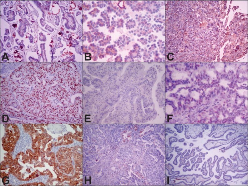
Figure 1. Representative WT1, p53 and p16 immunohistochemical expression in low- (LGSOC) and high-grade serous ovarian carcinoma (HGSOC).
Notes: (A) positive expression of WT1 in LGSOC (10×); (B) negative expression of WT1 in LGSOC (40×); (C) positive expression of WT1 in HGSOC (10×); (D) diffuse nuclear expression of p53 in HGSOC (10×); (E) complete absence of p53 (null type) in HGSOC(10×); (F) focal nuclear expression of p53 (wild type) in LGSOC (40×); (G) positive nuclear and cytoplasmic expression of p16 in HGSOC (10×); (H) negative (focal expression) of p16 in HGSOC (10×); (I) negative (expression in only a few cells) of p16 in LGSOC (10×).

