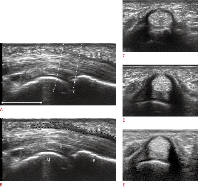
Fig. 1. A 70-year-old man with grade III trigger finger at the left 4th finger.

A. Longitudinal view on ultrasonography over the palmar aspect of the affected metacarpophalangeal joint refers the positional relation of the axial images. Dotted lines indicate the corresponding axial planes shown in C and D. Scale bar=10 mm. B. The same view with A shows the A1 and A2 pulleys (shaded area) over the tendon (T), volar plate (V), metacarpal head (M) and proximal phalanx (P). C, D. Consecutive axial images show the distal, less thick pulley (C) and the thickest pulley (D). Hypoechoic fluid distension was maximal at the level of D, E. Ultrasonography at the same level as D conducted 26 days after the injection shows decreased thickness of the tendon (from 3.78 to 3.41 mm), cross-sectional area of the tendon (from 18.52 to 16.15 mm2), and thickness of the pulley (from 0.922 to 0.533 mm), but no decrease in the transverse diameter of the tendon (from 5.59 to 5.81 mm) in this case.
