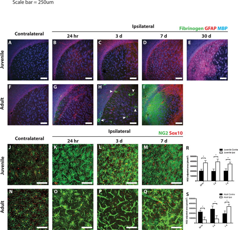
Figure 4. Differential gliotic responses following MCAo in juvenile versus adult mice.

Ischemic and control juvenile (A–E) and adult (F–I) sections were immunostained to detect fibrinogen (green), GFAP (red), and MBP (blue). Scant fibrinogen immunoreactivity was observed in the injured juvenile striatum at any time point examined (A–E). Fibrinogen deposition occurred as early as 24 h after MCAo in the adult striatum (G), with large deposits surrounding blood vessels at 3 days (H, arrowheads). By 7 days, a large sheet of fibrinogen was observed throughout the core of the lesion in adult tissue (I). Astrogliosis was observed surrounding the ischemic core in both juvenile and adult striatum at 3 days post-stroke (C and H, respectively). Peri-infarct astrogliosis was maintained in adults at 7 days post-stroke (I), whereas diffuse astrogliosis throughout the injured striatum was observed in juvenile mice and continued out to 30 days (D,E). Differential NG2-cell response following MCAo in juvenile versus adult mice. Ischemic and control juvenile (J–M) and adult (N–Q) sections were immunostained to detect NG2 (green) and Sox10 (red). Images of juvenile tissue demonstrate hypertrophic NG2-cells (costained with Sox10) and some NG2 immunoreactivity on blood vessels at 24 h post-MCAo (K). Increased density of NG2-cells at 3- and 7-days post-MCAo in juvenile mice (L,M) was seen. Images from adult tissue demonstrate loss of NG2-cells at 24 h (O) and 3 days (P) post-MCAo with high levels of NG2 expression localized to blood vessels. NG2 cells repopulated the adult lesion at 7 days post-MCAo, while blood vessel NG2 immunoreactivity remained prevalent (Q). (R,S) Quantification of non-vascular NG2 immunostaining in the contralateral (dark bars) and ipsilateral (open bars) striatum from juveile (R) and adult (S) mice. White scale bars: 250 lm (A–I), 100 lm (J–Q).
