Abstract
Hadal trenches, oceanic locations deeper than 6,000 m, are thought to have distinct microbial communities compared to those at shallower depths due to high hydrostatic pressures, topographical funneling of organic matter, and biogeographical isolation. Here we evaluate the hypothesis that hadal trenches contain unique microbial biodiversity through analyses of the communities present in the bottom waters of the Kermadec and Mariana trenches. Estimates of microbial protein production indicate active populations under in situ hydrostatic pressures and increasing adaptation to pressure with depth. Depth, trench of collection, and size fraction are important drivers of microbial community structure. Many putative hadal bathytypes, such as members related to the Marinimicrobia, Rhodobacteraceae, Rhodospirilliceae, and Aquibacter, are similar to members identified in other trenches. Most of the differences between the two trench microbiomes consists of taxa belonging to the Gammaproteobacteria whose distributions extend throughout the water column. Growth and survival estimates of representative isolates of these taxa under deep-sea conditions suggest that some members may descend from shallower depths and exist as a potentially inactive fraction of the hadal zone. We conclude that the distinct pelagic communities residing in these two trenches, and perhaps by extension other trenches, reflect both cosmopolitan hadal bathytypes and ubiquitous genera found throughout the water column.
Introduction
The deep sea is one of the largest biomes on Earth, containing over half of the microbial cells in the ocean [1,2]. Pelagic deep-ocean microbial communities are distinct from those above them [3,4,5,6] and in many cases display higher activities under in situ hydrostatic pressures and low temperatures when compared to atmospheric pressure conditions [7]. However, deep-sea environments also contain allochthonous members that descend from above, such as in association with sinking particulate organic matter [8]. These communities can differ from one another, varying by water mass or ocean basin and showing metabolic rates ranging over six orders of magnitude [9]. These variations may reflect resource availability [10,11] and dispersal limitation [12].
More is known about the bathy- and abyssopelagic than the hadopelagic zone, which exists at depths greater than 6,000 m and represents 41% of the oceanic depth continuum [13]. Most hadopelagic sites are associated with trenches, tectonically-active steep-walled depressions that form via subduction. The current study focuses on the Mariana and Kermadec trenches, two hadal sites approximately 6,000 km apart in the Pacific Ocean. The Kermadec Trench in the Southern Hemisphere begins about 120 km off the northeastern coast of New Zealand and reaches its greatest depth at 10,047 m, making it the 5th deepest trench [14]. The Mariana Trench, located in the Northern Hemisphere near the Mariana Islands, extends to 10,984 m at its greatest depth in the Challenger Deep near its southwestern terminus [15], making this the deepest location in the global ocean. Trenches have been proposed to contain unique biodiversity and endemic megafauna due to their geographic isolation [13,16,17,18], but some taxa show a more cosmopolitan distribution, suggesting potential for between-trench dispersal [19,20]. However, studies comparing microbial communities within trenches have not been conducted.
Until recently our understanding of hadal microbial communities has been restricted to highly selective culture-based analyses and small sample size 16S rRNA gene sequence studies [21,22,23]. Distinct microbial taxa adapted to high hydrostatic pressures have been cultured from hadal zones [24,25,26,27,28], of which many are piezophiles, microbes that show optimum growth at pressures greater than 0.1 and as high as 140 Megapascals (MPa; [29]). Recently, increased sample collection and the use of next generation sequencing approaches to hadopelagic communities from the Mariana, Japan, and Puerto Rico trenches have shown that hadal microbial communities are distinct from those above them [30,31,32,33]. At the greatest depths of the Mariana Trench, Gammaproteobacteria, including Pseudomonas and Pseudoalteromonas, are present as major constituents [31,33], findings attributed to trench topography funneling sinking organic matter downward and thereby fueling greater heterotrophic activity [34,35,36,37]. To address microbial community structure, potential endemism, and high-pressure adaptation within hadal trenches, we analyzed the microbial communities inhabiting abyssal and hadal bottom waters within the Kermadec and Mariana trenches using culture-independent high-throughput sequencing, culture-dependent taxonomic characterization, and estimates of microbial activity and abundance.
Methods
Sites and sample collection
Kermadec Trench samples were collected aboard the R/V Thompson from April to May 2014 using HROV Nereus [38], CTD casts, and free-falling/ascending landers (Elevator Lander; [39]). Samples were collected from the Mariana Trench, including within the deepest point of the Sirena Deep [40], aboard the R/V Falkor from November to December 2014 using CTD casts and free-falling/ascending landers (Rock Grabber (RG), Schmidt Ocean Institute, https://schmidtocean.org/technology/elevators-landers/; Leggo, Scripps Institution of Oceanography (SIO), https://scripps.ucsd.edu/labs/dbartlett/contact/challenger-deep-cruise-2014/). One sample was also collected from the Challenger Deep in the Mariana Trench. Lander-based seawater samples were recovered from vertically-positioned Niskin bottles 2 m above the sediment-water interface within the benthic boundary layer, and closed ~12 hours after landing to avoid capturing resuspended sediment material. Recovered samples were immediately transferred to a 4°C cold room and their temperature taken. Sampling within the Kermadec Trench was approved under a NIWA Special Permit issued by the New Zealand Ministry for Primary Industries and within the Mariana Trench by the NOAA Monuments office.
Environmental data
Bathymetry [41] was plotted using the R package marmap [42]. Seawater used for inorganic nutrient analyses was frozen at -20°C and processed at the Oceanographic Data Facility at SIO (S1 Text; https://scripps.ucsd.edu/ships/shipboard-technical-support/odf/documentation/nutrient-analysis). Replicates that varied by over two standard deviations from the mean within each trench were removed. Technical replicates were averaged at each collection site. For cell counts seawater was fixed with 1% paraformaldehyde and stored at -800078C. Samples were later thawed, stained with SYBR Green (Thermo Fisher Scientific, Waltham, MA), and cells enumerated using flow cytometry (Attune Acoustic Focusing Flow Cytometer, Applied Biosystems, Foster City, CA).
Microbial activity within the Mariana trench
Activity was evaluated using biorthogonal noncanonical amino acid tagging (BONCAT; [43,44]) with the methionine analog homopropargylglycine (HPG; Thermo Fisher Scientific). Seawater was placed into KAPAK bags (Komplete Packaging, Grand Prairie, TX) in 50 mL aliquots in duplicate. Bags were amended with 5 μM HPG, heat sealed, and incubated at 4°C in pressure vessels [45] at 0.1 MPa, in situ pressure at collection depth, and 110 MPa to mimic full trench depth. Negative controls were amended with 3% formaldehyde prior to incubation. After 48 hours, samples were fixed with formaldehyde, filtered onto 25 mm, 0.2 μm pore-size GTTP filters (EMD Millipore, Billerica, MA), and stored at -20°C. Active cells were detected as previously described (S1 Text; [43]). The percentage of active cells in each sample was calculated by dividing the number of active cells (DAPI + HPG active) by the total number of cells (DAPI) in each sample. Values from duplicate incubations from each location and pressure condition were averaged and the percent of active cells at one pressure was divided by that at another pressure to determine the effect of pressurization.
DNA extraction and sequencing
Seawater (40–120 L per sample) was serially filtered through 3.0 (47 mm diameter), 0.2 (47 mm or Sterivex), and 0.1 μm (142 mm) polycarbonate filters using a peristaltic pump. Filters were then placed into a sucrose buffer [46] and frozen at -80°C. DNA was extracted from whole filters using a protocol previously described [33,47]. Negative controls using blank filters were extracted in concomitance with every extraction performed.
The 16S rRNA gene region between 515f-926R was amplified in triplicate for 30 cycles and pooled [48]. Samples were tagged with sample-specific Illumina barcodes during a secondary PCR step, combined at equimolar concentrations, and sent for sequencing on an Illumina Miseq (S1 Text). Overlapping paired reads were merged using FLASH [49] and discarded if they fell below a q score of 33 within a 50 bp sliding window [48] using Trimmomatic [50]. Primers were removed and operational taxonomic units (OTUs) picked at 97% similarity using UCLUST in QIIME 1.9.1 [51]. Chimeras were identified with the Ribosomal Database Project gold database (training database v9) using VSEARCH [52] and removed. Taxonomy was assigned against the SILVA [53] 123 database and sequences identified as contaminants were discarded (S1 Text). Finally, OTUs with fewer than 3 reads in at least 4 samples across the entire dataset were excluded. Sequence data have been submitted to the SRA database under accession numbers SRR5643386-SRR5643480.
Statistical analyses
Sequencing reads were processed with the R package phyloseq [54]. Samples were rarefied to even sampling depth to account for differing sequencing depths. Alpha diversity was calculated using vegan [55] and comparisons between samples were performed using the beta-diversity metrics Bray-Curtis and weighted Unifrac [56]. Ordinations based on Bray-Curtis dissimilarity and permutational analysis of variance with adonis in vegan were used to identify statistically significant variables. Samples were classified into depth groups of surface, abyssal, or hadal by UPGMA hierarchical clustering using the command hclust. Samples were classified based on a hard depth cutoff in each trench; samples that clustered with one depth group but belonged to the other based on depth cutoff were grouped by depth of collection. A differential expression enrichment analysis using DESeq2 [57] was used to test the hypotheses that certain taxa are enriched within specific trenches, hadal zones, and certain size fractions using the un-rarefied dataset [58] with low abundance (at least >1000 total reads per OTU or >3000 reads per phylum) taxa removed. For construction of phylogenetic trees, sequences were aligned using the SINA Aligner [59] and trees built using FastTree [60].
Isolation and characterization of microbes
Microbes were cultured at 4°C on agar plates at 0.1 MPa or in transfer bulbs (Samco, Thermo Fisher Scientific) at either 0.1 MPa or high pressure. Enrichments from the Kermadec Trench were conducted using 2216 Marine Medium (2216; BD DifcoTM), A1 Medium, or a seawater minimal medium (S1 Text), while those from the Mariana Trench were conducted in 2216 only. For incubations at high pressure the media was inoculated, mixed with gelatin at a final concentration of 4%, transferred into bulbs, and incubated at the desired pressure [45]. Kermadec Trench samples were incubated at 100 MPa while those from the Mariana Trench were incubated at in situ pressure (40–110 MPa). After ~2 months colony forming units (CFUs) were calculated and representative isolates identified via PCR using the primers 27F and 1492R [61].
Isolate growth and survival
The high-pressure growth and survival characteristics of select strains were evaluated. This included strains from the Mariana Trench isolated at atmospheric pressure; Pseudomonas sp. 28, Pseudoalteromonas sp. 164, Psychrobacter sp. 151, and Halomonas sp. 73, and additional strains collected elsewhere; Pseudomonas pelagia [62], Pseudoalteromonas sp. TW7 [63], Psychrobacter aquimaris [64], Alteromonas mediterranea [65], and Alteromonas sp. SIO [66]. Growth experiments as a function of pressure (0.1, 20, 40, 60, and 90 MPa) and temperature (15°C and 4°C) were set up by inoculating early exponential phase cultures 1:100 into 2216 and either incubated in tubes at 0.1 MPa with shaking at 150 rpm or into bulbs and pressurized. At each time point three bulbs for each strain were sacrificed and their optical density measured (OD600; GENYSIS UV-Vis, Thermo Fisher Scientific).
To estimate survival, early exponential phase cultures were pelleted and reconstituted in 0.2 μm filtered, autoclaved sterile seawater collected from the Scripps Pier. Cultures were then diluted 1:100 into sterile seawater, placed into bulbs, and pressurized at 0.1, 20, 40, 60, and 90 MPa at 4°C. After 30 days, cultures were decompressed and plated on 2216 agar plates at 15°C to estimate surviving CFUs. Cultures were also fixed and total cell counts determined microscopically.
Results and discussion
Hadal microbial communities are adapted to high hydrostatic pressure conditions
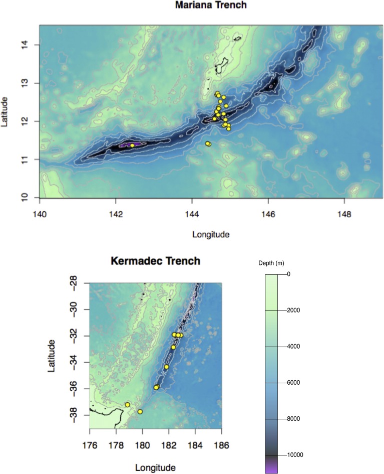
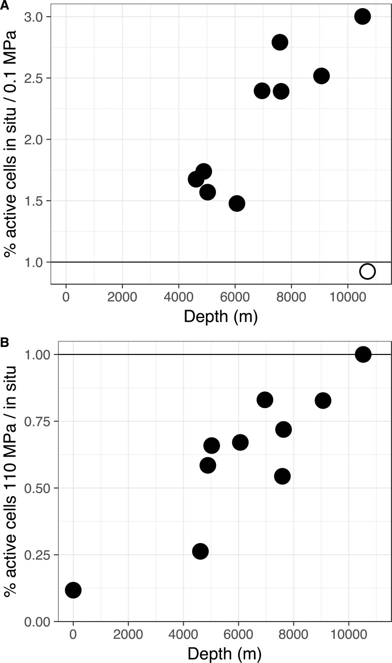
Seawater samples were collected at 38 locations within the Kermadec and Mariana trenches at depths up to 10,004 m and 10,920 m, respectively (Fig 1, S1 Fig, S1 Table). Cell abundances of abyssal and hadal samples were approximately 104 cells mL-1 (S2 Fig). To assess the activity of microbes collected from the Mariana Trench, the fraction of cells engaged in protein synthesis was evaluated using BONCAT. Although many estimates of deep-ocean microbial activity have been conducted under atmospheric pressure conditions, community activity in stratified waters may be highest under in situ hydrostatic pressures [7]. However, such measurements are lacking from hadal locations (e.g. [67,68]). Therefore, the fraction of Mariana Trench cells that were active at atmospheric pressure and following recompression to in situ pressures was determined. In the abyssal and hadal samples ~18% (6.5–34.5%) of the cells were active after recompression to in situ pressures while ~9% (4.7–13.8%) were active under atmospheric pressure conditions (S3 Fig). Low percentages of active cells may be consistent with prior measurements of microbial activity in the bathypelagic, where activity rates drop over two orders of magnitude from those at the surface and turnover times are estimated to be 0.1–30 years [7,9]. In nearly all samples the proportion of active cells was higher under high hydrostatic pressure (Fig 2). The only exception was a sample from the Challenger Deep, which had warmed to above 15°C during retrieval. These results likely reflect the thermal sensitivity and selective inactivation of autochthonous deep-sea residents over allochthonous microbes from shallower depths [69]. Comparisons of communities incubated at full-ocean depth pressures versus in situ pressure showed increasing ratios of full-ocean depth active members with collection depth (Fig 2), indicative of progressive increases in the extent of high pressure adaptation with depth. In contrast to the low percentage of active cells in the deep ocean, over 75% of the community was active in the surface waters at 28°C (S3 Fig). This percentage dropped to ~10% when incubated at 4°C and was further repressed at increasing pressure. We emphasize that bulk community activity was not estimated here, which may exhibit more or less activity as a function of pressure due to the variability of taxa-specific activity rates. Furthermore, benthic boundary layer communities may be more or less active as a function of pressure, relative to the deep pelagic, because these sites are a mixing zone of autochthonous members, allochthonous sinking taxa, and resuspended organic matter from the seafloor [70,71]. Regardless, these findings suggest that hadal communities contain active members adapted to high hydrostatic pressures, even following the stresses imposed by decompression during sample retrieval.
Fig 1. Sample collection locations.
Pelagic sample collection locations within the Kermadec and Mariana trenches.
Fig 2. Microbial activity as a function of pressure using HPG.
A; Ratio of the percentage of active cells under in situ pressure versus atmospheric pressure. B; Ratio of the percentage of active cells at 110 MPa versus in situ pressure. Filled circles, seawater collected at <13°C, excluding the surface sample; open circles, seawater collected at >15°C.
Hadopelagic communities are unique from abyssal assemblages
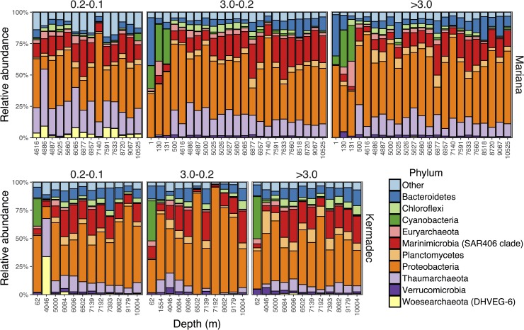
Microbial communities within the Kermadec and Mariana trenches were compared using high-throughput sequencing of the V4-V5 region of the 16S rRNA gene. The dataset consists of 95 samples and 7,169,109 total sequences, with rarefaction resulting in 15,346 sequences per sample and 8,908 total OTUs (S2 Table). The most abundant group in the abyssal and hadal samples was the Gammaproteobacteria within the Proteobacteria (Fig 3), composed primarily of Alteromonas, Idiomarina, Pseudoalteromonas, Psychrobacter, and Shewanella. These taxa represented 10–20% of the community on each size fraction but were sometimes >50%. Other abundant groups included the Marinimicrobia, Thaumarchaeota, Bacteroidetes, and SAR324, consistent with other studies of deep-ocean and hadal communities.
Fig 3. Abundant pelagic phyla.
Relative abundances of the ten most abundant phyla in the pelagic zones of the Kermadec and Mariana trenches, organized by trench and size fraction (>3.0, 3.0–0.2, 0.2–0.1 μm).
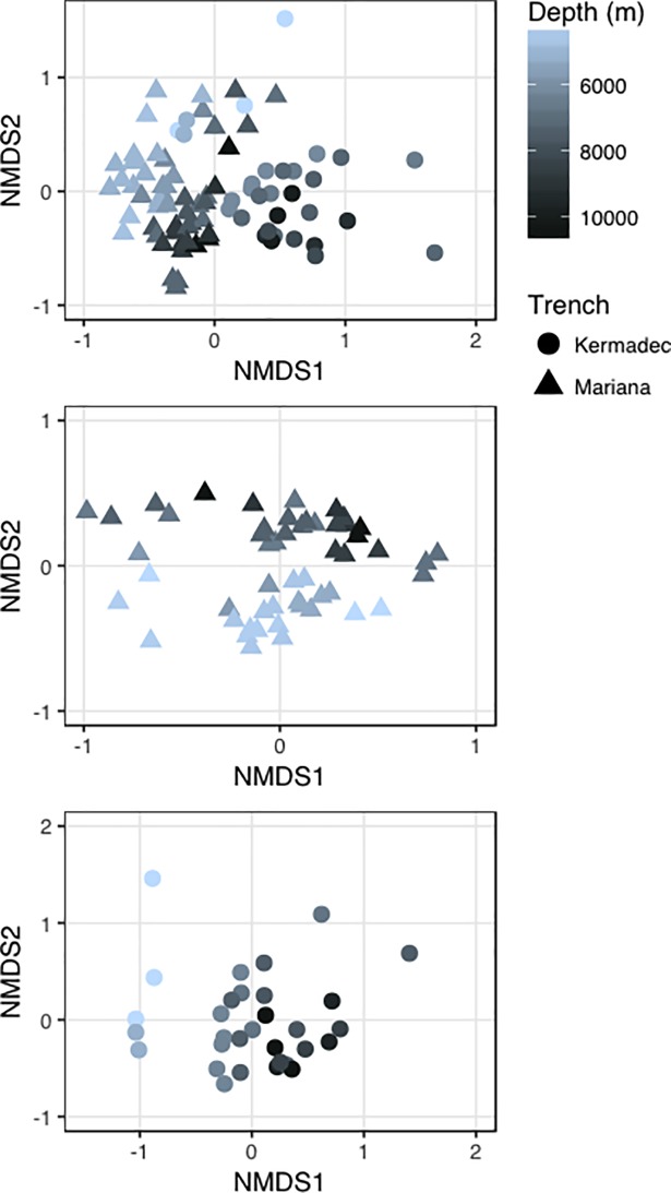
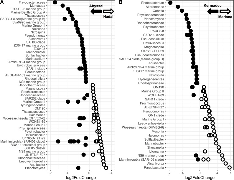
Hadal microbial communities have been shown to be different from those present in shallower locales [30,31,32,33]. Here, the hadal communities in the Kermadec and Mariana trenches were distinct from those in the abyssal zone (Fig 4, S4 Fig). Depth was a significant driver of community composition in both trenches (Both trenches, R2 = 0.13, p<0.001; Mariana, R2 = 0.16, p<0.001; Kermadec R2 = 0.21, p<0.001) when comparing samples deeper than 4,000 m. Distinct communities were identified at depths greater than 6,084 m in the Kermadec Trench (Abyssal, 4,046–5,000 m; Hadal, 6,084–10,004 m) and 6,877 m in the Mariana Trench (Abyssal, 4,616–6,065 m; Hadal, 6,877–10,525 m). These findings are similar to changes in megafaunal demersal fish assemblages, which showed distinct hadal communities starting at 6,750 m in the Kermadec Trench and 6,831 m in the Sirena Deep [72]. The Bacteroidetes and Nitrospira were more abundant in hadal zones and Thaumarchaeota and Chloroflexi were more abundant in abyssal zones (S5 Fig), which may in part be driven by ammonia concentration/flux and higher amounts of organic matter with increasing depth [31,32,73,74]. Many hadal-enriched taxa identified here (Fig 5, S3 Table) shared sequence similarity with microbes previously obtained from trenches, including OTUs belonging to the Marinimicrobia, Planctomycetaceae, Rhodobacteraceae, and Flavobacteriaceae. The Marinimicrobia have been observed as one of the most abundant phyla within the Puerto Rico [75] and Mariana [31,33] trenches, consistent with their high abundances seen here. OTUs related to Aquibacter (Flavobacteriaceae; 96% similar to A. zeaxanthinifaciens) and Defluviicoccus (Rhodospirilliceae; 85% similar to D. vanus) were specifically enriched in the hadal samples and reached abundances of up to 11% and 1.5%, respectively. These OTUs showed high similarity to sequences previously obtained from the pelagic Puerto Rico, Mariana, and Japan trench datasets as well as sediments from the Ogasawara Trench (S6 Fig; [76,77]). These groups may represent bathytypes, taxa adapted to the specific ecological niches associated with depth. Heterotrophic Gammaproteobacteria, including Psychrobacter and Halomonas, were also preferentially enriched within the hadal versus abyssal depths examined, but were found to have widespread, cosmopolitan distributions that include shallow-water locations (e.g. S13 Fig).
Fig 4. Ordinations of sequenced abyssal and hadal communities.
Distances between abyssal and hadal communities visualized via ordinations using Bray-Curtis dissimilarity. A, Mariana Trench; B, Kermadec Trench; C, both trenches.
Fig 5. OTUs enriched in the hado- or abysso- pelagic communities.
A; OTUs showing enrichment within the hadal or abyssal zones of the Mariana and Kermadec trenches labeled by the lowest discernible taxonomic rank. Filled, enriched in the abyssal zone; open, enriched in the hadal zone. B; OTUs showing enrichment within the hadal zone of the Kermadec or Mariana trench. Filled, enriched in the Kermadec Trench; open, enriched in the Mariana Trench.
Kermadec and Mariana trench communities are distinct from each other
Trenches may represent independent zoographic provinces [16,17], each with its own unique signature of biodiversity and endemism due to their extreme depth, topography, nutrient inputs, and in many cases isolation from other trench systems. Comparisons of the Kermadec and Mariana trench communities revealed distinctive memberships in each trench (Fig 4, S4 Fig; R2 = 0.15, p<0.001). When excluding surface samples, 50% of all OTUs were specific to one trench but made up only 5% of the total sequences. Of these unique OTUs 75% belonged to the Mariana Trench, in agreement with increased richness in the Mariana relative to the Kermadec (S7 Fig). When comparing the abyssal and hadal zones 48% of the OTUs were zone-specific but made up only 5% of all sequences, while 13% of all OTUs were found in both trenches and both pelagic zones and accounted for 83% of all sequences. Therefore, these assemblages contain widespread, abundant lineages and a number of rare, potentially endemic taxa that make up a large number of OTUs but a small fraction of the entire community. Similar findings have been reported for the meso- and bathypelagic [12,78], where these communities are composed of both abundant and widely distributed species as well as rare, sample-specific taxa that represent a minor fraction of the total community [4]. The importance of such low-abundance taxa is not well understood, but they may represent a microbial seed bank capable of becoming more abundant under specific environmental conditions [79].
Taxa were identified that showed differential abundances between the two hadal zones (Fig 5, S4 Table). Regardless of abundance, however, they were found in both trenches and showed similarity to other deep-ocean sequences (e.g. S7 Fig). Therefore these taxa are widespread but differentially abundant within the deep-sea communities. Variations in abundance of depth-cosmopolitan taxa, especially members of the Gammaproteobacteria, were also seen. While the Kermadec Trench was enriched in sequences related to Alteromonas, Pseudoalteromonas, Photobacterium, Cobetia, and Psychrobacter, the Mariana Trench was enriched in sequences related to Shewanella, Alcanivorax, Idiomarina, Marinobacter, Halomonas, and Pseudomonas. All of these sequences show similarity to those within surface waters. The differences in community composition between these two trenches may be due to fluxes of organic matter, as annual rates of primary production in the overlying waters of the Kermadec have been estimated as 87 g C m-2 yr-1, compared to lower productivity (59 g C m-2 yr-1) in the waters above the Mariana Trench [13,80]. Water masses, which can have distinct biogeochemical properties, may also affect community composition [81]. Differences in nutrients between the two trenches (S2 Fig) indicate different water mass inputs, with the Kermadec being the first trench in the Pacific Ocean to receive Lower Circumpolar Deep Water [16] while the Mariana receives both Lower Circumpolar Deep Water and North Pacific Deep Water [82]. Site-to-site variations may also be driven by benthic boundary layer nutrient concentrations, perhaps because of differing influences from topographic focusing of settling organic material and/or sediment resuspension. For example, within the Mariana Trench higher relative abundances of nitrogen-cycling Nitrospira and Nitrosomonas correlated with sites containing higher nitrite concentrations (S8 Fig).
Community composition differs between size fractions
Different size fractions may represent distinct niches, with larger size fractions containing particle-attached microbes and smaller size fractions including free-living microbes. We separated the trench microbial communities into >3.0 μm (particle-associated), 3.0–0.2 μm (free-living), and 0.2–0.1 μm (ultra-small free-living) size fractions. Size fraction was a small driver of community composition (both trenches; R2 = 0.05, p<0.001; Mariana, R2 = 0.12, p<0.001; Kermadec, R2 = 0.08, p>0.08). The most well represented taxa associated with the particle fraction included members of the phylum Planctomycetes (S9 Fig), which have a particle-attached lifestyle in bathypelagic settings [83]. The taxa most enriched in the free-living fraction included Alteromonas, SAR324, SAR202, and the Marinimicrobia. Comparisons between the free-living and ultra-small free-living size fractions showed that the ultra-small free-living fraction was enriched in three archaeal phyla, including the Thaumarchaeota, Marine Hydrothermal Vent Group (MHVG), and Woesearchaeota, and two members of the candidate phyla radiation [84], the Parcubacteria and Gracilibacteria (S10 Fig). Many of these taxa were specifically enriched in the Mariana Trench (Fig 5, S4 Table) and show best, albeit low, similarity to sequences from the Mariana, Japan, and Ogasawara trenches, highlighting their potential uniqueness. The Parcubacteria and other members of the candidate phyla radiation represent some of the smallest bacteria known [85]. Our findings support the observation that unique taxa can be exceptionally small and the abundances of many microbes may be misinterpreted when sampling stops at the 0.2 μm pore size.
Culturable microbes are widespread
Piezophiles have been isolated from a variety of deep-ocean trenches, with most belonging to the genera Colwellia, Shewanella, Moritella, or Psychromonas [24,25,26,86,87], r-strategists that grow relatively rapidly in nutrient-rich media. Abundance estimates of these cultured piezophiles indicate that they make up a small fraction of hadal seawater communities despite their consistent isolation [30]. Previously no discernible portion of the community in the Sirena Deep was attributed to known piezophiles, although they made up ~0.5% of the communities in the Challenger Deep [33]. However, distribution and rates of isolation of piezophiles have not been thoroughly investigated.
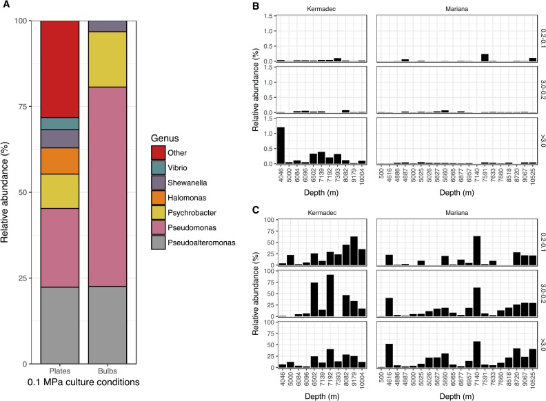
To determine if previously isolated piezophilic species were present in the Mariana and Kermadec trenches, the community sequence data was searched for OTUs with >97% similarity to known piezophiles. Abundances were typically less than 1% within each size fraction and were higher in the Kermadec relative to the Mariana Trench (Fig 6; T-test, p<0.04). Sequences related to piezophilic taxa were preferentially enriched on the particle fraction, indicative of a surface-attached lifestyle associated with the utilization of particulate organic matter (T-test, both trenches, p>0.05; Kermadec Trench, p<0.05). Now three trenches, including the Puerto Rico, Mariana, and Kermadec, have been shown to contain sequences associated with previously isolated and cultured piezophiles at relative abundances of less than 1%. Thus, these piezophiles, along with other putative bathytypes, appear to maintain connectivity between trenches, including within the northern and southern hemispheres and the Pacific and Atlantic oceans. Some obligate piezophiles show growth and activity at pressures as low as 40 MPa, suggesting they can survive at abyssal depths. Therefore many hadal microbes, as with other deep-sea microbes [12], could be transported into other regions of the global ocean in association with water mass circulation at abyssal depths. Curiously, no isolates were obtained at high hydrostatic pressure from the water column in either trench (S5 Table). The inability to culture piezophiles here may be due to low abundances of these taxa, especially in the Mariana Trench, or the small numbers of enrichments performed. Because many piezophilic microbes have been isolated from hosts and sediments [24,26,86,88], they may be more abundant in these niches than in the water column.
Fig 6. Isolates and their abundances.
A; Relative abundances of cultured isolates from seawater in the Kermadec and Mariana trenches at 0.1 MPa and 4°C on plates (n = 170) or in bulbs (n = 31). B; Combined relative abundances of OTUs similar to piezophilic Colwellia, Shewanella, Moritella, or Psychromonas species derived from the community data. C; Combined relative abundances of OTUs related to the genera Pseudoalteromonas, Pseudomonas, Psychrobacter, Halomonas, Shewanella, and Vibrio derived from the community data.
Many microbes have also been isolated from deep-ocean samples when incubated at atmospheric pressure, but the role of these isolates in situ remains unclear. In contrast to our high pressure culturing results, 102–103 CFUs mL-1 were obtained from trench samples on plates and in bulbs incubated at atmospheric pressure (S5 Table). Most isolates were related to the genera Pseudoalteromonas, Pseudomonas, Shewanella, Halomonas, and Psychrobacter (Fig 6, S6 Table). Related taxa have been isolated from other deep-ocean samples [31,89,90,91], suggesting there is selection even for microbes that grow at atmospheric pressure. Interestingly, atmospheric pressure isolates are more representative of deep-ocean communities than microbes isolated under in situ conditions (Fig 6). To assess the ability of deep-sea associated, atmospheric pressure-isolated microbes to grow under deep-sea conditions, growth studies were performed for nine isolates belonging to the genera Pseudoalteromonas, Pseudomonas, Psychrobacter, Alteromonas, and Halomonas, including five deep-sea isolates and four related shallow-water species (S7 Table). None of the isolates were piezophilic or grew at pressures greater than 40 MPa (S11 Fig), consistent with other estimates of high-pressure growth of related strains [89,92,93]. Although growth rates were repressed under the low oxygen conditions that develop in batch high-pressure cultures, the reduced oxygen availability does not explain the pressure sensitivity of these strains. After pressurization for one month at 90 MPa cells remained intact and some strains remained cultivable (S12 Fig), indicating they may remain viable in the deep sea for long periods. Sequences representative of these genera were found in the surface, abyssal, and hadal communities in high abundances (S8 Table, S13 Fig), indicating they may not be obligate bathytypes, and related taxa have been found associated with sinking particles [94,95,96]. Therefore these atmospheric-pressure adapted “deep-sea” isolates could represent cell types which colonize particles, either at the surface or at meso- or bathy- pelagic depths [12,97] and descend to full-ocean depths where they can survive prolonged periods at high pressure. Future studies should evaluate whether the hadal populations of these genera represent a mixture of closely-related strains that show ecotype differentiation (e.g. [98]), some of which are autochthonous to the deep-sea and others that may have sank from shallower depths.
Conclusions
Hadal microbial communities have been proposed to contain distinct taxa adapted to the unique in situ conditions found in trenches. Here, we show that hadal communities within the Kermadec and Mariana trenches are indeed distinct from the abyssal assemblages above them. Hadal communities are enriched in certain taxa that may represent bathytypes, including clades such as the Marinimicrobia and specific genera such as Aquibacter. Sequences related to known piezophiles were identified in both trenches, albeit in higher abundances in the Kermadec Trench, but at <1% of total communities. These findings suggest similar hadal-associated taxa are present in multiple trenches, potentially transported by deep-ocean currents. Such lineages may be responsible for the higher rates of activity under in situ rather than atmospheric pressures determined here. Communities were also distinct between the Mariana and Kermadec trenches, showing varying abundances of cosmopolitan taxa and the presence of unique but rare OTUs. Inter-trench variation was largely driven by differentially abundant heterotrophic Gammaproteobacteria that show a remarkable ability to survive long-term pressurization and may be from bathyal and shallower depths where they colonize particles and sink. Trenches are therefore home to unique microbial communities, comprised of autochthonous, pressure-adapted members and ubiquitous genera found throughout the water column.
Supporting information
(DOCX)
Circles, CTD cast; Triangles, lander.
(TIF)
Red circles, Kermadec Trench; Blue circles, Mariana Trench.
(TIF)
The percentage of active cells at either (A) atmospheric or (B) in situ pressures, where filled circles were obtained at <13°C (excluding the surface sample) and open circles at >15°C. C; The percentage of active cells of a Mariana Trench surface water sample as a function of temperature and pressure.
(TIF)
A; Beta diversity community comparisons between water samples visualized by either weighted Unifrac or Bray-Curtis ordinations. B; Heirarchical clustering of samples based on Bray-Curtis dissimilarity colored broadly by pelagic collection location.
(TIF)
Positive, hadal; negative, abyssal.
(TIF)
A, Aquibacter; B, Defluviicoccus; C, Rhodobacteraceae; D, Planctomyces.
(TIF)
Red, Kermadec Trench; Blue, Mariana Trench.
(TIF)
A; Relative abundances of the nitrogen-cycling bacteria Nitrosomonas, Nitrospina, and Nitrospira in the community Itag data. B; Nitrite concentrations within the Kermadec and Mariana Trenches. C; Relative abundances of Nitrosomonas and Nitrospira within each abyssal or hadal sampling site in either the Mariana (left) or Kermadec (right) trench plotted as a function of nitrite concentrations.
(TIF)
Taxonomic labels represent the lowest discernible taxonomic rank. Positive, >3.0 μm; negative, 3.0–0.2 μm.
(TIF)
Positive, 0.2–0.1 μm; negative, 3.0–0.2 μm.
(TIF)
(TIF)
T0; counts prior to long-term pressurization.
(TIF)
Phylogenetic trees of OTUs related to Pseudoalteromonas (A), Psychrobacter (B), and Halomonas (C) that were abundant in surface, abyssal, and hadal samples.
(TIF)
(XLSX)
(XLSX)
Positive log2FoldChange, hadal; negative abyssal.
(XLSX)
Positive log2FoldChange, Mariana; negative Kermadec.
(XLSX)
(XLSX)
(XLSX)
(XLSX)
Relative abundances shown were determined after combining samples by size fraction and depth zone (Surface (1–131 m), abyssal (500- ~6000 m), hadal (~6000–10525 m)).
(XLSX)
Acknowledgments
We thank Matt Norenberg, David Price, the R/V Falkor, R/V Thompson, and the HADal Ecosystem Studies (HADES) team for help at sea, Mackenzie Gerringer and Than Kyaw for helpful advice, and Bethany Kolody for artistic expertise
Data Availability
Sequence data are available at the SRA database under accession numbers SRR5643386-SRR5643480.
Funding Statement
LMP and DHB were supported by the National Science Foundation (0801973, 0827051, 1536776), National Aeronautics and Space Administration (NNX11AG10G, NNX14AP13H), the Prince Albert II Foundation (Project 1265), and a gift from Earthship Productions. MJC and AN were supported by the Simons Foundation via the Simons Collaboration on Ocean Processes and Ecology (SCOPE; grant ID 329108). The funders had no role in study design, data collection and analysis, decision to publish, or preparation of the manuscript.
References
- 1.Whitman WB, Coleman DC, Wiebe WJ. Prokaryotes: The unseen majority. Proc Natl Acad Sci. 1998;95: 6578–6583. [DOI] [PMC free article] [PubMed] [Google Scholar]
- 2.Kallmeyer J, Pockalny R, Adhikari RR, Smith DC, D’Hondt S. Global distribution of microbial abundance and biomass in subseafloor sediment. Proc Natl Acad Sci. 2012;109: 16213–16216. doi: 10.1073/pnas.1203849109 [DOI] [PMC free article] [PubMed] [Google Scholar]
- 3.DeLong EF, Preston CM, Mincer T, Rich V, Hallam SJ, Frigaard N, et al. Community genomics among stratified microbial assemblages in the ocean’s interior. Science. 2006;311: 496–503. doi: 10.1126/science.1120250 [DOI] [PubMed] [Google Scholar]
- 4.Sogin ML, Morrison HG, Huber JA, Welch DM, Huse SM, Neal PR, et al. Microbial diversity in the deep sea and the underexplored “rare biosphere.” Proc Natl Acad Sci. 2006; 103: 12115–12120. doi: 10.1073/pnas.0605127103 [DOI] [PMC free article] [PubMed] [Google Scholar]
- 5.Brown MV, Philip GK, Bunge JA, Smith MC, Bissett A, Lauro FM, et al. Microbial community structure in the North Pacific ocean. ISME J. 2009;3: 1374–1386. doi: 10.1038/ismej.2009.86 [DOI] [PubMed] [Google Scholar]
- 6.Smedile F, Messina E, la Cono V, Tsoy O, Monticelli LS, Borghini M, et al. Metagenomic analysis of hadopelagic microbial assemblages thriving at the deepest part of Mediterranean Sea, Matapan-Vavilov Deep. Environ Microbiol. 2013;15: 167–182. doi: 10.1111/j.1462-2920.2012.02827.x [DOI] [PubMed] [Google Scholar]
- 7.Tamburini C, Boutrif M, Garel M, Colwell RR, Deming JW. Prokaryotic responses to hydrostatic pressure in the ocean—a review. Environ Microbiol. 2013;15: 1262–1274. doi: 10.1111/1462-2920.12084 [DOI] [PubMed] [Google Scholar]
- 8.Agusti S, Gonzalez-Gordillo JI, Vaque D, Estrada M, Cerezo MI, Salazar G, et al. Ubiquitous healthy diatoms in the deep sea confirm deep carbon injection by the biological pump. Nat Commun. 2015;6: 7608 doi: 10.1038/ncomms8608 [DOI] [PMC free article] [PubMed] [Google Scholar]
- 9.Nagata T, Tamburini C, Arístegui J, Baltar F, Bochdansky AB, Fonda-Umani S, et al. Emerging concepts on microbial processes in the bathypelagic ocean–ecology, biogeochemistry, and genomics. Deep-Sea Res Pt II. 2010;57: 1519–1536. [Google Scholar]
- 10.Nagata T, Fukuda H, Fukuda R, Koike I. Bacterioplankton distribution and production in deep Pacific waters: Large-scale geographic variations and possible coupling with sinking particle fluxes. Limnol Ocean. 2000;45: 426–435. [Google Scholar]
- 11.Hansell DA, Ducklow HW. Bacterioplankton distribution and production in the bathypelagic ocean: Directly coupled to particulate organic carbon export? Limnol Ocean. 2003;48: 150–156. [Google Scholar]
- 12.Salazar G, Cornejo-Castillo FM, Benítez-Barrios V, Fraile-Nuez E, Alvarez-Salgado XA, Duarte CM, et al. Global diversity and biogeography of deep-sea pelagic prokaryotes. ISME J. 2015a;10: 596–608. [DOI] [PMC free article] [PubMed] [Google Scholar]
- 13.Jamieson AJ. The Hadal Zone: Life in the Deepest Oceans. London: Cambridge University Press; 2015. [Google Scholar]
- 14.Angel MV. Ocean trench conservation. The Environmentalist. 1982;2: 1–17. [Google Scholar]
- 15.Gardner JV, Armstrong AA, Calder BR, Beaudoin J. So, how deep is the Mariana Trench? Marine Geod. 2014;37: 1–13. [Google Scholar]
- 16.Belyaev GM. Deep-sea ocean trenches and their fauna Nauka Publishing House: Moscow: 1989. [Google Scholar]
- 17.Vinogradova NG. Zoogeography of the abyssal and hadal zones. Adv Mar Biol. 1997;32: 325–387. [Google Scholar]
- 18.Linley TD, Gerringer ME, Yancey PH, Drazen JC, Weinstock CL, Jamieson AJ. Fishes of the hadal zone including new species, in situ observations and depth records of Liparidae. Deep-Sea Res Pt I. 2016;114: 99–110. [Google Scholar]
- 19.Jamieson AJ, Lacey NC, Lorz AN, Rowden AA, Piertney SB. The supergiant amphipod Alicella gigantean (Crustacea: Alicellidae) from hadal depths in the Kermadec Trench, SW Pacific Ocean. Deep Sea Res Pt II. 2013;92: 107–113. [Google Scholar]
- 20.Ritchie H, Jamieson AJ, Piertney SB. Population genetic structure of two congeneric deep-sea amphipod species from geographically isolated hadal trenches in the Pacific Ocean. Deep-Sea Res Pt I. 2017;119: 50–57. [Google Scholar]
- 21.Kato C, Li L, Tamaoka J, Horikoshi K. Molecular analyses of the sediment of the 11000-m deep Mariana Trench. Extremophiles. 1997;1: 117–123. [DOI] [PubMed] [Google Scholar]
- 22.Li L, Kato C, Horikoshi K. Microbial diversity in sediments collected from the deepest cold-seep area, the Japan Trench. Mar Biotechnol. 1999;1: 391–400. [DOI] [PubMed] [Google Scholar]
- 23.Yanagibayashi M, Nogi Y, Li L, Kato C. Changes in the microbial community in Japan Trench sediment from a depth of 6292m during cultivation without decompression. FEMS Microbiol Lett. 1999;170: 271–279. [DOI] [PubMed] [Google Scholar]
- 24.Yayanos AA, Dietz AS, Van Boxtel R. Obligately barophilic bacterium from the Mariana Trench. Proc Natl Acad Sci. 1981;78: 5212–5215. [DOI] [PMC free article] [PubMed] [Google Scholar]
- 25.Nogi Y, Kato C, Horikoshi K. Psychromonas kaikoae sp. nov., a novel piezophilic bacterium from the deepest cold-seep sediments in the Japan Trench. Int J Syst Evol Microbiol. 2002;52: 1527–1532. doi: 10.1099/00207713-52-5-1527 [DOI] [PubMed] [Google Scholar]
- 26.Nogi Y, Hosoya S, Kato C, Horikoshi K. Colwellia piezophila sp. nov., a novel piezophilic species from deep-sea sediments of the Japan Trench. Int J Syst Evol Microbiol. 2004;54: 1627–1631. doi: 10.1099/ijs.0.03049-0 [DOI] [PubMed] [Google Scholar]
- 27.Eloe EA, Malfatti F, Gutierrez J, Hardy K, Schmidt WE, Pogliano K, et al. Isolation and characterization of a psychropiezophilic alphaproteobacterium. Appl Environ Microbiol. 2011c;77: 8145–8153. [DOI] [PMC free article] [PubMed] [Google Scholar]
- 28.Cao Y, Chastain RA, Eloe EA, Nogi Y, Kato C, Bartlett DH. Novel psychropiezophilic Oceanospirillales species Profundimonas piezophila gen. nov., sp. nov., isolated from the deep-sea environment of the Puerto Rico Trench. Appl Environ Microbiol 2014;80: 54–60. doi: 10.1128/AEM.02288-13 [DOI] [PMC free article] [PubMed] [Google Scholar]
- 29.Kusube M, Kyaw TS, Tanikawa K, Chastain RA, Hardy K, Cameron J, et al. Colwellia marinimaniae sp nov., a hyperpiezophilic species isolated from an amphipod within the Challenger Deep, Mariana Trench. Int J Syst Evol Microbiol. 2016;67: 824–831. [DOI] [PubMed] [Google Scholar]
- 30.Eloe EA, Fadrosh DW, Novotny M, Allen LZ, Kim M, Lombardo MJ, et al. Going deeper: metagenome of a hadopelagic microbial community. PLoS ONE. 2011a;6: 1–15. [DOI] [PMC free article] [PubMed] [Google Scholar]
- 31.Nunoura T, Takaki Y, Hirai M, Shimamura S, Makabe A, Koide O, et al. Hadal biosphere: Insight into the microbial ecosystem in the deepest ocean on Earth. Proc Natl Acad Sci. 2015;112: E1230–E1236. doi: 10.1073/pnas.1421816112 [DOI] [PMC free article] [PubMed] [Google Scholar]
- 32.Nunoura T, Hirai M, Yoshida-Takashima Y, Nishizawa M, Kawagucci S, Yokokawa T, et al. Distribution and niche separation of planktonic microbial communities in the water columns from the surface to the hadal waters of the Japan Trench under the eutrophic ocean. Front Microbiol. 2016;7: 1261 doi: 10.3389/fmicb.2016.01261 [DOI] [PMC free article] [PubMed] [Google Scholar]
- 33.Tarn J, Peoples LM, Hardy K, Cameron J, Bartlett DH. Identification of free-living and particle-associated microbial communities present in hadal regions of the Mariana Trench. Front Microbiol. 2016;7: 665 doi: 10.3389/fmicb.2016.00665 [DOI] [PMC free article] [PubMed] [Google Scholar]
- 34.Danovaro R, Croce ND, Dell’Anno A, Pusceddu A. A depocenter of organic matter at 7800 m depth in the SE Pacific Ocean. Deep-Sea Res Pt I. 2003;50: 1411–1420. [Google Scholar]
- 35.Glud RN, Wenzhöfer F, Middelboe M, Oguri K, Turnewitsch R, Canfield DE, et al. High rates of microbial carbon turnover in sediments in the deepest oceanic trench on Earth. Nat Geosci. 2013;6: 284–288. [Google Scholar]
- 36.Ichino MC, Clark MR, Drazen JC, Jamieson A, Jones DOB, Martin AP, et al. The distribution of benthic biomass in hadal trenches: A modeling approach to investigate the effect of vertical and lateral organic matter transport to the seafloor. Deep-Sea Res Pt I. 2015;100: 21–33. [Google Scholar]
- 37.Wenzhofer F, Oguri K, Middelboe M, Turnewitsch R, Toyofuku T, Kitazato H, et al. Benthic carbon remineralization in hadal trenches: Assessment by in situ O2 microprofile measurements. Deep-Sea Res Pt I. 2016;116: 276–286. [Google Scholar]
- 38.Fletcher B, Bowen A, Yoerger DR, Whitcomb LL. Journey to the Challenger Deep: 50 Years Later with the Nereus Hybrid Remotely Operated Vehicle. Marine Technology Society Journal. 2009;43: 65–76. [Google Scholar]
- 39.Nunnally CC, Friedman JR, Drazen JC. In situ respiration measurements of megafauna in the Kermadec Trench. Deep-Sea Res Pt I. 2016;118: 30–36. [Google Scholar]
- 40.Fryer P, Becker N, Appelgate B, Martinez F, Edwards M, Fryer G. Why is the Challenger Deep so deep? Earth Planet Sci Lett. 2003;211: 259–269. [Google Scholar]
- 41.Amante C, Eakins BW. ETOPO1 1 arc-minute global relief model: procedures, data sources and analysis Colorado: US Department of Commerce, National Oceanic and Atmospheric Administration, National Environmental Satellite, Data, and Information Service, National Geophysical Data Center, Marine Geology and Geophysics Division; 2009. [Google Scholar]
- 42.Pante E, Simon-Bouhet B. marmap: A package for importing, plotting, and analyzing bathymetric and topographic data in R. PLoS ONE. 2013;8: e73051 doi: 10.1371/journal.pone.0073051 [DOI] [PMC free article] [PubMed] [Google Scholar]
- 43.Samo TJ, Smriga S, Malfatti F, Sherwood BP, Azam F. Broad distribution and high proportion of protein synthesis active marine bacteria revealed by click chemistry at the single cell level. Front Mar Sci. 2014;1: 48. [Google Scholar]
- 44.Hatzenpichler R, Connon SA, Goudeau D, Malmstrom RR, Woyke T, Orphan VJ. Visualizing in situ translational activity for identifying and sorting slow-growing archaeal-bacterial consortia. Proc Natl Acad Sci. 2016;113: E4069–E4078. doi: 10.1073/pnas.1603757113 [DOI] [PMC free article] [PubMed] [Google Scholar]
- 45.Yayanos AA. Deep-sea piezophilic bacteria. Method Microbiol. 2001;30: 615–637. [Google Scholar]
- 46.Rusch DB, Halpern AL, Sutton G, Heidelberg KB, Williamson S, Yooseph S, et al. The Sorcerer II Global Ocean Sampling Expedition: northwest Atlantic through eastern tropical Pacific. PLoS Biology. 2007;5: 0398–0431. [DOI] [PMC free article] [PubMed] [Google Scholar]
- 47.Fuhrman JA, Comeau DE, Hagstrom A, Chan AM. Extraction from natural plankton microorganisms of DNA suitable for molecular biological studies. Appl Environ Microbiol. 1988;54: 1426–1429. [DOI] [PMC free article] [PubMed] [Google Scholar]
- 48.Parada AE, Needham DM, Fuhrman JA. Every base matters: assessing small subunit rRNA primers for marine microbiomes with mock communities, time series and global field samples. Environ Microbiol. 2015;18: 1403–1414. doi: 10.1111/1462-2920.13023 [DOI] [PubMed] [Google Scholar]
- 49.Magoc T, Salzberg SL. FlASH: fast length adjustment of short reads to improve genome assemblies. Bioinformatics. 2011;27: 2957–2963. doi: 10.1093/bioinformatics/btr507 [DOI] [PMC free article] [PubMed] [Google Scholar]
- 50.Bolger AM, Lohse M, Usadel B. Trimmomatic: a flexible trimmer for Illumina sequence data. Bioinformatics. 2014;30: 2114–2120. doi: 10.1093/bioinformatics/btu170 [DOI] [PMC free article] [PubMed] [Google Scholar]
- 51.Caporaso JG, Kuczynski J, Stombaugh J, Bittinger K, Bushman FD, Costello EK, et al. QIIME allows analysis of high-throughput community sequencing data. Nature Methods. 2010;7: 335–336. doi: 10.1038/nmeth.f.303 [DOI] [PMC free article] [PubMed] [Google Scholar]
- 52.Rognes T, Flouri T, Nichols B, Quince C, Mahe F. VSEARCH: a versatile open source tool for metagenomics. PeerJ. 2016;4: e2584 doi: 10.7717/peerj.2584 [DOI] [PMC free article] [PubMed] [Google Scholar]
- 53.Pruesse E, Quast C, Knittel K, Fuchs BM, Ludwig W, Peplies J, et al. SILVA: a comprehensive online resource for quality checked and aligned ribosomal RNA sequence data compatible with ARB. Nucleic Acids Res. 2007;35: 7199–7196. [DOI] [PMC free article] [PubMed] [Google Scholar]
- 54.McMurdie PJ, Holmes S. phyloseq: An R package for reproducible interactive analysis and graphics of microbiome census data. PLoS ONE. 2013;8: e61217 doi: 10.1371/journal.pone.0061217 [DOI] [PMC free article] [PubMed] [Google Scholar]
- 55.Oksanen J, Blanchet FG, Friendly M, Kindt R, Legendre P, McGlinn D, et al. vegan: Community Ecology Package. https://cran.r-project.org/web/packages/vegan/vegan.pdf. 2017.
- 56.Lozupone C, Lladser ME, Knights D, Stombaugh J, Knight R. UniFrac: an effective distance metric for microbial community comparison. ISME J. 2011;5: 169–172. doi: 10.1038/ismej.2010.133 [DOI] [PMC free article] [PubMed] [Google Scholar]
- 57.Love MI, Huber W, Anders S. Moderated estimation of fold change and dispersion for RNA-seq data with DESeq2. Genome Biol. 2014;15: 550 doi: 10.1186/s13059-014-0550-8 [DOI] [PMC free article] [PubMed] [Google Scholar]
- 58.McMurdie PJ, Holmes S. Waste not, want not: Why rarefying microbiome data is inadmissible. PLoS Comput Biol. 2014;10: e1003531 doi: 10.1371/journal.pcbi.1003531 [DOI] [PMC free article] [PubMed] [Google Scholar]
- 59.Pruesse E, Peplies J, Glockner FO. SINA: Accurate high-throughput multiple sequence alignment of ribosomal RNA genes. Bioinformatics. 2012;28: 1823–1829. doi: 10.1093/bioinformatics/bts252 [DOI] [PMC free article] [PubMed] [Google Scholar]
- 60.Price MN, Dehal PS, Arkin AP. FastTree 2 –Approximately Maximum-Likelihood Trees for Large Alignments. PLoS ONE. 2010;5: 1–10. [DOI] [PMC free article] [PubMed] [Google Scholar]
- 61.Lane DJ. 16S/23S rRNA sequencing In: Stackebrandt E Goodfellow M (eds). Nucleic Acid Techniques in Bacterial Systematics. New York: John Wiley and Sons; 1991. [Google Scholar]
- 62.Hwang CY, Zhang GI, Kang SH, Kim HJ, Cho BC. Pseudomonas pelagia sp. nov., isolated from a culture of the Antarctic green alga Pyramimonas gelidicola. Int J Syst Evol Microbiol. 2009;59: 3019–3024. doi: 10.1099/ijs.0.008102-0 [DOI] [PubMed] [Google Scholar]
- 63.Bidle KD, Azam F. Bacterial control of silicon regeneration from diatom detritus: Significance of bacterial ectohydrolases and species identity. Limnol Ocean. 2001;46: 1606–1623. [Google Scholar]
- 64.Yoon JH, Lee CH, Yeo SH, Oh TK. Psychrobacter aquimaris sp. nov. and Psychrobacter namhaensis sp. nov., isolated from sea water of the South Sea in Korea. Int J Syst Evol Microbiol. 2005;55: 1007–1013. doi: 10.1099/ijs.0.63464-0 [DOI] [PubMed] [Google Scholar]
- 65.Ivanova EP, Lopez-Lopez M, Zabalos M, Nguyen SH, Webb HK, Ryan J, et al. Ecophysiological diversity of a novel member of the genus Alteromonas, and description of Alteromonas mediterranea sp. nov. Antonie van Leeuwenhoek. 2015;107: 119–132. doi: 10.1007/s10482-014-0309-y [DOI] [PubMed] [Google Scholar]
- 66.Pedler BE, Aluwihare LI, Azam F. Single bacterial strain capable of significant contribution to carbon cycling in the surface ocean. Proc Natl Acad Sci. 2014;111: 7202–7207. doi: 10.1073/pnas.1401887111 [DOI] [PMC free article] [PubMed] [Google Scholar]
- 67.Seki H, ZoBell CE. Microbial assimilation of carbon dioxide in the Japan Trench. J. Oceanogr. Soc. Japan. 1976;23: 182–188. [Google Scholar]
- 68.Schwarz JR, Yayanos AA, Colwell RR. Metabolic activities of the intestinal microflora of a deep-sea invertebrate. Appl Environ Microbiol. 1976;31: 46–48. [DOI] [PMC free article] [PubMed] [Google Scholar]
- 69.Yayanos AA, Dietz AS. Thermal inactivation of a deep-sea barophilic bacterium, isolate CNPT-3. Appl Environ Microbiol. 1982;43: 1481–1489. [DOI] [PMC free article] [PubMed] [Google Scholar]
- 70.Turley C. Bacteria in the cold deep-sea benthic boundary layer and sediment-water interface of the NE Atlantic. FEMS Microbiol Ecol. 2000;33: 89–99. [DOI] [PubMed] [Google Scholar]
- 71.Kawagucci S, Yoshida YT, Noguchi T, Honda MC, Uchida H, Ishibashi H, et al. Disturbance of deep-sea environments induced by the M9.0 Tohoku Earthquake. Scientific Reports. 2012;2: 1–7. [DOI] [PMC free article] [PubMed] [Google Scholar]
- 72.Linley TD, Stewart AL, McMillan PJ, Clark MR, Gerringer ME, Drazen JC, et al. Bait attending fishes of the abyssal zone and hadal boundary: Community structure, functional groups and species distribution in the Kermadec, New Hebrides and Mariana trenches. Deep-Sea Res Pt I. 2017;21: 38–53. [Google Scholar]
- 73.Sintes E, Bergauer K, De Corte D, Yokokawa T, Herndl GJ. Archaeal amoA diversity points to distinct biogeography of ammonia-oxidizing Crenarchaeota in the ocean. Environ Microbiol. 2013;15: 1647–1658. doi: 10.1111/j.1462-2920.2012.02801.x [DOI] [PMC free article] [PubMed] [Google Scholar]
- 74.Sintes E, De Corte D, Haberleitner E, Herndl GJ. Geographic distribution of archaeal ammonia oxidizing ecotypes in the Atlantic Ocean. Front Microbiol 2016;7: 77 doi: 10.3389/fmicb.2016.00077 [DOI] [PMC free article] [PubMed] [Google Scholar]
- 75.Eloe EA, Shulse CN, Fadrosh DW, Williamson SJ, Allen EE, Bartlett DH. Compositional differences in particle-associated and free-living microbial assemblages from an extreme deep-ocean environment. Environ Microbiol Rep. 2011b;3: 449–458. [DOI] [PubMed] [Google Scholar]
- 76.Nunoura T, Nishizawa M, Kikuchi T, Tsubouchi T, Hirai M, Koide O, et al. Molecular biological and isotopic biogeochemical prognoses of the nitrification-driven dynamic microbial nitrogen cycle in hadopelagic sediments. Environ Microbiol. 2013;15: 3087–107. doi: 10.1111/1462-2920.12152 [DOI] [PubMed] [Google Scholar]
- 77.Yoshida M, Takaki Y, Eitoku M, Nunoura T, Takai K. Metagenomic analysis of viral communities in hado(pelagic) sediments. PLoS ONE. 2013;8: e57271 doi: 10.1371/journal.pone.0057271 [DOI] [PMC free article] [PubMed] [Google Scholar]
- 78.Galand PE, Casamayor EO, Kirchman DL, Lovejoy C. Ecology of the rare microbial biosphere of the Arctic Ocean. Proc Natl Acad Sci. 2009;106: 22427–22432. doi: 10.1073/pnas.0908284106 [DOI] [PMC free article] [PubMed] [Google Scholar]
- 79.Fuhrman JA. Microbial community structure and its functional implications. Nature. 2009;459: 193–199. doi: 10.1038/nature08058 [DOI] [PubMed] [Google Scholar]
- 80.Longhurst A, Sathyendranath S, Platt T, Caverhill C. An estimate of global primary production in the ocean from satellite radiometer data. J Plankton Res. 1995;17: 1245–1271. [Google Scholar]
- 81.Galand PE, Potvin M, Casamayor EO, Lovejoy C. Hydrography shapes bacterial biogeography of the deep Arctic Ocean. ISME J. 2010;4: 564–576. doi: 10.1038/ismej.2009.134 [DOI] [PubMed] [Google Scholar]
- 82.Siedler G, Holfort J, Zenk W, Muller TJ, Csernok T. Deep-water flow in the Mariana and Caroline Basins. J Phys Oceanogr. 2003;34: 566–581. [Google Scholar]
- 83.Salazar G, Cornejo-Castillo FM, Borrull E, Díez-Vives C, Lara E, Vaqué D, et al. Particle-association lifestyle is a phylogenetically conserved trait in bathypelagic prokaryotes. Mol Ecol. 2015b;24: 5692–5706. [DOI] [PubMed] [Google Scholar]
- 84.Brown CT, Hug LA, Thomas BC, Sharon I, Castelle CJ, Singh A, et al. Unusual biology across a group comprising more than 15% of domain bacteria. Nature. 2015;523: 208–211. doi: 10.1038/nature14486 [DOI] [PubMed] [Google Scholar]
- 85.Luef B, Frischkorn KR, Wrighton KC, Holman HYN, Birarda G, Thomas BC, et al. Diverse uncultivated ultra-small bacterial cells in groundwater. Nat Commun. 2015;6: 6372 doi: 10.1038/ncomms7372 [DOI] [PubMed] [Google Scholar]
- 86.Kato C, Li L, Nogi Y, Nakamura Y, Tamaoka J, Horikoshi K. Extremely barophilic bacteria isolated from the Mariana Trench, Challenger Deep, at a depth of 11,000 meters. Appl Environ Microbiol. 1998;64: 1510–1513. [DOI] [PMC free article] [PubMed] [Google Scholar]
- 87.Nogi Y, Kato C, and Horikoshi K. Taxonomic studies of deep-sea barophilic Shewanella strains and description of Shewanella violacea sp. nov. Arch Microbiol. 1998;170: 331–338. [DOI] [PubMed] [Google Scholar]
- 88.Nakayama A, Saito R, Matsuzaki M, Yano Y, Yoshida K. Phylogenetic analysis based on 16S rRNA gene sequences of deep-sea bacteria isolated from intestinal contents of deep-sea fishes retrieved from the abyssal zone. J Gen Appl Microbiol. 2005;51: 385–394. [DOI] [PubMed] [Google Scholar]
- 89.Tamegai H, Li L, Masui N, Kato C. A denitrifying bacterium from the deep sea at 11000-m depth. Extremophiles. 1997;1: 207–211. [DOI] [PubMed] [Google Scholar]
- 90.Maruyama A, Honda D, Yamamoto H, Kitamura K, Higashihara T. Phylogenetic analysis of psychrophilic bacteria isolated from the Japan Trench, including a description of the deep-sea species Psychrobacter pacificensis sp. nov.. Int J Syst Evol Microbiol. 2000;50: 835–846. doi: 10.1099/00207713-50-2-835 [DOI] [PubMed] [Google Scholar]
- 91.Tamegai H, Furukawa T, Nitta J, Chikuma S, Miyazaki M, Nogi Y, et al. Halomonas sp. strain DT-W, a halophile from the 11,000 m-depth of the Mariana Trench. Journal of Japanese Society for Extremophiles. 2006;5: 27–33. [Google Scholar]
- 92.Kaneko H, Takami H, Inoue A, Horikoshi K. Effects of hydrostatic pressure and temperature on growth and lipid composition of the inner membrane of barotolerant Pseudomonas sp. BT1 isolated from the deep-sea. Biosci Biotechnol Biochem. 2000;64: 72–79. doi: 10.1271/bbb.64.72 [DOI] [PubMed] [Google Scholar]
- 93.Kaye JZ, Baross JA. Synchronous effects of temperature, hydrostatic pressure, and salinity on growth, phospholipid profiles, and protein patterns of four Halomonas species isolated from deep-sea hydrothermal-vent and sea surface environments. Appl Environ Microbiol. 2004;70: 6220–6229. doi: 10.1128/AEM.70.10.6220-6229.2004 [DOI] [PMC free article] [PubMed] [Google Scholar]
- 94.Ivars-Martínez E, Martin-Cuadrado AB, D’Auria G, Mira A, Ferriera S, Johnson J, et al. Comparative genomics of two ecotypes of the marine planktonic copiotroph Alteromonas macleodii suggests alternative lifestyles associated with different kinds of particulate organic matter. ISME J. 2008;2: 1194–1212. doi: 10.1038/ismej.2008.74 [DOI] [PubMed] [Google Scholar]
- 95.Tamburini C, Goutx M, Guigue C, Garel M, Lefevre D, Charriere B, et al. Effects of hydrostatic pressure on microbial alteration of sinking fecal pellets. Deep-Sea Res Pt II. 2009;56: 1533–1546. [Google Scholar]
- 96.Fontanez KM, Eppley JM, Samo TJ, Karl DM, DeLong EF. Microbial community structure and function on sinking particles in the North Pacific Subtropical Gyre. Front Microbiol. 2015;6: 1–14. [DOI] [PMC free article] [PubMed] [Google Scholar]
- 97.Herndl GJ, Reinthaler T. Microbial control of the dark end of the biological pump. Nature Geosci. 2013;6: 718–724. [DOI] [PMC free article] [PubMed] [Google Scholar]
- 98.Kaye JZ, Sylvan JB, Edwards KJ, Baross JA. Halomonas and Marinobacter ecotypes from hydrothermal vent, subseafloor and deep-sea environments. FEMS Microbiol Ecol. 2011;75: 123–133. doi: 10.1111/j.1574-6941.2010.00984.x [DOI] [PubMed] [Google Scholar]
Associated Data
This section collects any data citations, data availability statements, or supplementary materials included in this article.
Supplementary Materials
(DOCX)
Circles, CTD cast; Triangles, lander.
(TIF)
Red circles, Kermadec Trench; Blue circles, Mariana Trench.
(TIF)
The percentage of active cells at either (A) atmospheric or (B) in situ pressures, where filled circles were obtained at <13°C (excluding the surface sample) and open circles at >15°C. C; The percentage of active cells of a Mariana Trench surface water sample as a function of temperature and pressure.
(TIF)
A; Beta diversity community comparisons between water samples visualized by either weighted Unifrac or Bray-Curtis ordinations. B; Heirarchical clustering of samples based on Bray-Curtis dissimilarity colored broadly by pelagic collection location.
(TIF)
Positive, hadal; negative, abyssal.
(TIF)
A, Aquibacter; B, Defluviicoccus; C, Rhodobacteraceae; D, Planctomyces.
(TIF)
Red, Kermadec Trench; Blue, Mariana Trench.
(TIF)
A; Relative abundances of the nitrogen-cycling bacteria Nitrosomonas, Nitrospina, and Nitrospira in the community Itag data. B; Nitrite concentrations within the Kermadec and Mariana Trenches. C; Relative abundances of Nitrosomonas and Nitrospira within each abyssal or hadal sampling site in either the Mariana (left) or Kermadec (right) trench plotted as a function of nitrite concentrations.
(TIF)
Taxonomic labels represent the lowest discernible taxonomic rank. Positive, >3.0 μm; negative, 3.0–0.2 μm.
(TIF)
Positive, 0.2–0.1 μm; negative, 3.0–0.2 μm.
(TIF)
(TIF)
T0; counts prior to long-term pressurization.
(TIF)
Phylogenetic trees of OTUs related to Pseudoalteromonas (A), Psychrobacter (B), and Halomonas (C) that were abundant in surface, abyssal, and hadal samples.
(TIF)
(XLSX)
(XLSX)
Positive log2FoldChange, hadal; negative abyssal.
(XLSX)
Positive log2FoldChange, Mariana; negative Kermadec.
(XLSX)
(XLSX)
(XLSX)
(XLSX)
Relative abundances shown were determined after combining samples by size fraction and depth zone (Surface (1–131 m), abyssal (500- ~6000 m), hadal (~6000–10525 m)).
(XLSX)
Data Availability Statement
Sequence data are available at the SRA database under accession numbers SRR5643386-SRR5643480.


