Figure 8.
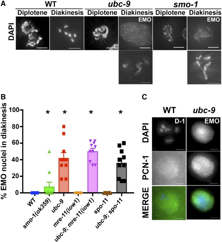
SUMOylation mutants have aberrant chromosomal morphology and EMO nuclei at diakinesis. (A) Representative images of diplotene and diakinesis nuclei in wild-type (WT), ubc-9(tm2610), and smo-1(ok359) strains. Both EMO and non-EMO phenotypes are shown for mutants. Mutant non-EMO oocytes still had abnormal morphology compared to WT. Points on the graph indicate individual data points, with the bar indicating the mean of all data points. (B) Percentage of EMO nuclei counted from DAPI-stained whole worms. At least 10 worms from each genotype were counted. Error bars signify the mean ± SEM. (C) Representative EMO oocyte from a ubc-9(tm2610) gonad staining strongly for PCN-1. WT oocytes have low levels of background PCN-1 staining not associated with chromatin. Ten WT D-1 oocytes and 11 EMO oocytes were counted for PCN-1 staining. Points on the graph indicate individual data points, with the bar indicating the mean of all data points.

