The classic article by Riedl and Frey1 [Plast Reconstr Surg.131(4): 802–810, 2013] is the most exhaustive study on the categorization of the sural nerve (SN). The authors reviewed 221 of 6,237 relevant contributions to the literature; all findings were analyzed and categorized into patterns I-V (p. 804, Fig. 1), and 4 patterns were confirmed by their own cadaver study (p. 805, Fig. 2). Just as they pointed out, their study covered all the details about the SN anatomy published internationally.
Fig. 1.

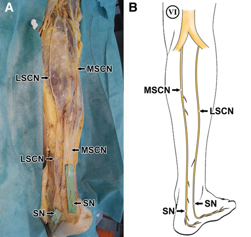
A, The LSCN and MSCN coursed in parallel, passed the retromalleolar region, and formed the SN separately in left leg. B, The new SN pattern drawing in the right leg, which was consistent with “Figure. 2” of the literature in the study by Riedl et al.
Fig. 2.

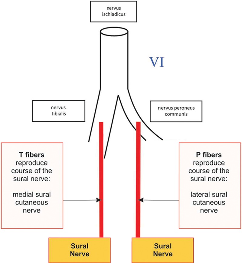
Illustrating figure of pattern VI, added to “Figure. 1” of the literature in the study by Riedl et al. (Adapted with permission from Wolters Kluwer).
The SN is commonly used for nerve grafting, sural flap’s reinnervation, and biopsy for disease diagnosis; therefore, our research group has a long-lasting interest in this topic.
We identified a new pattern that differed from the above SN categorization recently in our dissection: Both the lateral sural cutaneous nerve (LSCN) and the medial sural cutaneous nerve (MSCN) coursed in parallel, passed the retromalleolar region separately, then gave off sensory sub-branches to the lateral dorsal surface of the foot (Fig. 1A). Considering the criteria that the sural cutaneous nerve could be defined as the SN was that the sural cutaneous nerve passed the retromalleolar region1 or the level of the lateral malleolus,2 rather than terminating subcutaneously in the middle and distal calf. Our anatomical image indicated that both the LSCN and the MSCN became the SN separately (Fig. 1B). Therefore, we categorize this distinct case as pattern VI (Fig. 2), which was not covered in the existing patterns I–V of Riedl and Frey1. Clearly, it is different from pattern II or IV, but it is similar to pattern III plus V of the Riedl and Frey1 classification.
We have not found the same exact pattern in other publications so far, though there were a few similar types of case reports hinting pattern VI previously, where there was no anatomical panorama photograph of the whole SN3,4—the key evidence in morphology. Authors of these reports were unable to exclude the possibility of the LSCN and MSCN union under lateral malleolus5 (pattern I under Riedl and Frey1) or existence of a communicative branch between the LSCN and MSCN.4
The reason why Riedl and Frey1 didn’t list pattern VI from enormous previous literature may be that they took the possibility of imaging deficiency of previous reports into consideration. Furthermore, the limited cadaver number in the majority of anatomy studies and very low rate of pattern VI may have contributed to its omission of inclusion as a separate pattern.
This supplementary type—pattern VI found by our anatomical study--does exist and, it is hoped, completes the range of categorization by Riedl and Frey1. This letter provides further understanding of the SN classification of patterns.
Footnotes
Disclosure: The authors have no financial interest to declare in relation to the content of this article. The Article Processing Charge was paid for by the authors.
REFERENCES
- 1.Riedl O, Frey M. Anatomy of the sural nerve: cadaver study and literature review. Plast Reconstr Surg. 2013;131:802–810.. [DOI] [PubMed] [Google Scholar]
- 2.Strauch B, Goldberg N, Herman CK. Sural nerve harvest: anatomy and technique. J Reconstr Microsurg. 2005;21:133–136.. [DOI] [PubMed] [Google Scholar]
- 3.Pyun SB, Kwon HK. The effect of anatomical variation of the sural nerve on nerve conduction studies. Am J Phys Med Rehabil. 2008;87:438–442.. [DOI] [PubMed] [Google Scholar]
- 4.Vuksanovic-Bozaric A, Radunovic M, Radojevic N, et al. The bilateral anatomical variation of the sural nerve and a review of relevant literature. Anat Sci Int. 2014;89:57–61.. [DOI] [PubMed] [Google Scholar]
- 5.Mahakkanukrauh P, Chomsung R. Anatomical variations of the sural nerve. Clin Anat. 2002;15:263–266.. [DOI] [PubMed] [Google Scholar]


