GENETICS Correction for “MicroRNA-373 induces expression of genes with complementary promoter sequences,” by Robert F. Place, Long-Cheng Li, Deepa Pookot, Emily J. Noonan, and Rajvir Dahiya, which was first published January 28, 2008; 10.1073/pnas.0707594105 (Proc Natl Acad Sci USA 105:1608–1613).
The authors wish to note the following: “The corresponding authors were made aware of errors in Fig. 2D that required further investigation. The University of California, San Francisco (UCSF) and VA Medical Center, San Francisco, conducted a joint investigation into the cause of the errors. The investigation was overseen by the VA Office of Research Oversight (ORO) and, in part, by the UCSF Office of Research Integrity (ORI) independent of the authors.
Fig. 2.
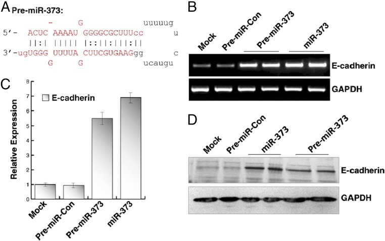
PremiR-373 induces E-cadherin expression. (A) Sequence of the miR-373 precursor hairpin RNA (premiR-373). Bases in red indicate the mature sequence of the miR-373 duplex. (B) PC-3 cells were transfected at 50 nM premiR-Con, premiR-373, or miR-373 for 72 h. E-cadherin and GAPDH mRNA expression levels were assessed by standard RT-PCR. (C) Relative expression was determined by real-time PCR (mean ± SE from four independent experiments). Values of E-cadherin were normalized to GAPDH. (D) E-cadherin and GAPDH protein levels were detected by immunoblot analysis. GAPDH served as a loading control.
“The Investigation Committee reviewed Fig. 2D including the E-cadherin panel (lanes 5 and 6) and GAPDH panel (lanes 2 and 5) and concluded that these images were derived from the same source of data despite representing different experimental conditions. Further analysis also led to the finding that a portion of the GAPDH panel, encompassing approximately 1.5 lanes, was mirrored and added to the panel. The Committee concluded that the manipulation of the images in Fig. 2D could only have occurred intentionally, representing instances of scientific misconduct. The Committee could not definitively attribute the research misconduct to any individual.
“One author then was able to identify an image copy of the original GAPDH immunoblot and two exposures of a corresponding E-cadherin immunoblot in their digital archives. The data were then submitted to the Committee and PNAS for evaluation. After review, it was determined by the Committee that a correction to Fig. 2D would be appropriate. The correction does not impact interpretation of the data.”
The corrected figure has been evaluated by the editor and approved for publication. The corrected figure and legend appear below.


