Abstract
[Purpose] This study aimed to examine the effects of pelvic movements on the back function of patients with lumbar disc herniation when lumbar stabilization exercise was applied to the patients, suggest an intervention method that can be used in future clinical therapies. [Subjects and Methods] Thirty patients with lumbar disc herniation underwent the intervention 30 minutes per day, three times per week for 4 weeks. Of them, 15 patients were assigned to the balance center stabilization resistance exercise group (experimental group I) and the other 15 were assigned to the three-dimensional stabilization exercise group (experimental group II). Before the intervention, disc herniation index was measured using magnetic resonance imaging, sacral angle was measured using X-ray, and back function was measured using the KODI. Four weeks later, these three factors were re-measured and analyzed. [Results] There was a significant pre- versus post-intervention difference in disc herniation index, sacral angle, and KODI in experimental group I and a significant difference in disc herniation index and KODI in experimental group II, and each group of disc herniation index and sacral angle had a significant difference. In experimental group I, each disc herniation index and sacral angle had a negative correlation. [Conclusion] The lumbar stabilization exercise, which controls balance using pelvic movements, improves mobility and stability of the sacroiliac joint; therefore, it increases pelvic and back movements. These kinds of movements not only improved proprioception sense, they also had positive effects on lumbar disc function recovery.
Key words: Disc herniation index, Lumbar stabilization exercise, Sacral angle
INTRODUCTION
Back pain is one of the most common diseases in modern society since 60–80% of the population has experienced it at least once in their lifetime1). Among back pain diseases, lumbar disc herniation is the most common disease2), decreasing the normal lumbar curve and creating muscular stiffness. It also causes sacroiliac joint instability since the facet joints and ligament flava are degenerated as the pelvis is warped and the sacral angle of inclination is increased3, 4). It also causes constant back pain and nerve root injection between the legs since it makes sitting properly difficult and warps the posture, and problems occur in balance and walking because of the weakened muscles surrounding the spine3). These symptoms become severe due to disability of the disc function caused by the herniation, which makes it difficult to maintain sitting or standing postures since it is not able to disperse the impact applied to the back5, 6). Constant health care and functional recovery are needed since these symptoms can cause disease recurrence in the same area and herniation in other segments. Therefore, it is necessary to perform early ambulation, exercise, and training7, 8).
Exercise in particular aims to actively strengthen the multifidus muscles and transverse abdominis muscles, deep muscles of the back; hence, lumbar stability exercises are needed to stabilize the spine segments and vitalize the local muscles around the back9). However, lumbar stability exercises are difficult to perform in the acute phase when back pain is severe and do not address pelvis problems such as sacral joint instability since they focus only on the local trunk muscles. To compensate for these deficiencies, exercises are performed by patients using equipment that can be used in early parts, which stimulates not only the deep muscles but also the whole trunk muscles and creates trunk stability10,11,12).
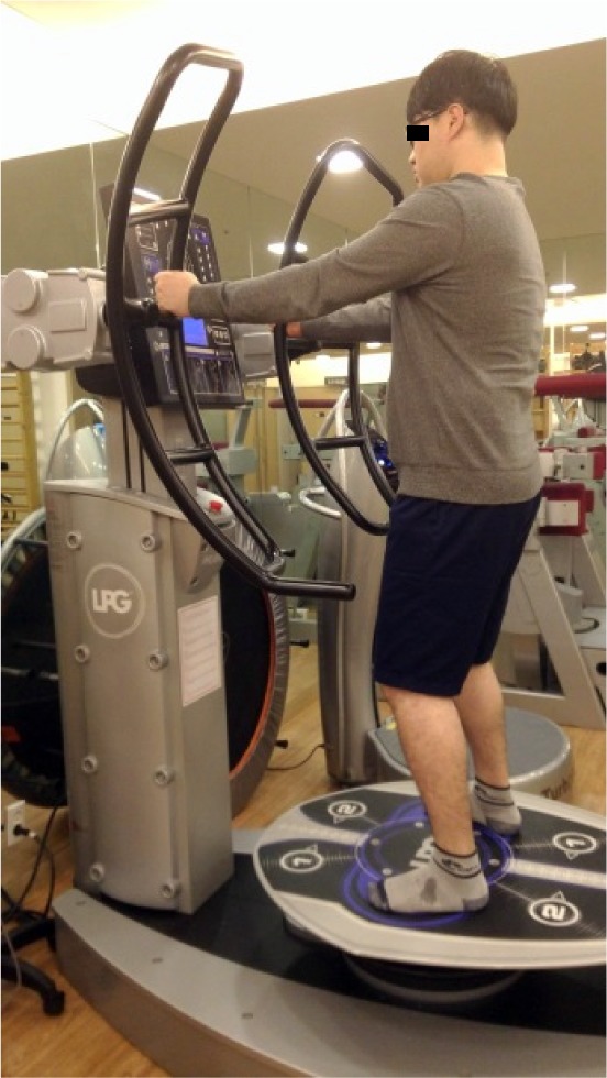
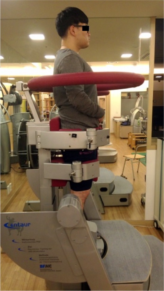
With this principle, the balance center stabilization resistance exercise (Fig. 1), which uses the pelvic movements on moving disc, and three-dimensional (3D) stabilization exercises (Fig. 2), which can exercise on diverse degree. However, it remains to be seen how accompanying symmetrical pelvis movements through these types of quantitative exercises positively affect one’s back. Therefore, this study aimed to examine the effects of pelvis movements on back function when the balance center stabilization resistance and 3D stabilization exercises are applied to patients via the quantitative measurement of patients with lumbar disc herniation and investigated the correlation between the effects; analyze the effects of back stabilization through pelvis movements on disc herniation index, sacral angle, and KODI; and make recommendations about how to manage lumbar disc herniation patients.
Fig 1.

The balance center stabilization resistance exercise.
Fig 2.

The 3D stabilization exercise.
SUBJECTS AND METHODS
This study was conducted for about 6 weeks from April 14 to May 22, 2017 after receiving approval from the Institutional Review Board of Sehan University (approval No. SH-IRB 2017–03). A total of 30 patients aged 25–50 years who were diagnosed with lumbar disc herniation (below the protrusion) and visited the medical institutions in Seoul were included. The included patients had no other diseases such as spondylolisthesis, a surgical history, and no other surgical diseases or neurological disorders; each was able to walk independently without a walker; and each understood the purpose of the study and participated voluntarily (Table 1).
Table 1. Subjects’ general characteristics.
| Item | Experimental group I (n=15) |
Experimental group II (n=15) |
|---|---|---|
| Age (years) | 32 ± 7.9 | 35.5 ± 10.9 |
| Height (cm) | 168 ± 9.1 | 164.3 ± 10.3 |
| Weight (kg) | 60.93 ± 13.6 | 67.38 ± 15.1 |
| SMM (kg) | 24.8 ± 7.1 | 22.68 ± 7.0 |
| BFM (kg) | 16.5 ± 4.0 | 21.19 ± 8.0 |
| BMI (kg/m2) | 21.7 ± 2.2 | 25.29 ± 3.7 |
Data are presented as mean ± SD obtained using the Shapiro-Wilk test. SMM: skeletal muscle mass; BFM: body fat mass; BMI: body mass index.
The diagnoses of the 30 patients were verified using magnetic resonance imaging (MRI), after which point they were randomly assigned to the perform the balance center stabilization resistance exercise (experimental group I; n=15) (Fig. 1) or the 3D back stabilization exercise group (experimental group II; n=15) (Fig. 2). The intervention consisted of performing the prescribed exercise for 30 minutes three times a week for 4 weeks. Before the intervention, herniation index was measured using MRI, sacral angle was measured using standing right posture X-rays, and back function was measured using the Korean Oswestry Disability Index (KODI). After 4 weeks, these variables were re-examined and analyzed.
The disc herniation index measurement consists of measurement of the disc and intervertebral foramen at the intercept of the axial direction13). The sagittal distance of the herniated disc was recorded as the maximum anteroposterior disc height (AB), while length of the vertebral foramen was the maximum anteroposterior canal length (EF). The width of the herniated disc was measured by drawing a line in the middle of the anteroposterior disc length (CD), while the width of the spinal canal was calculated at the same level (GH). The formula of the herniated disc index is as follows: [(AB × CD)/(EF × GH)] ×1,00014).
Sacral angle is the angle formed by the line drawn along the bottom surface of the sacrum with the horizontal line in the lateral lumbar spine view using PACS (Picture Archiving and Communications System) after the X-ray were taken and the normal range of 40°3).
The KODI has nine questions, each of which is scored at a maximum of 6 points. The reliability of the Korean version of the back pain disability index is high (r=0.92). The back pain disability index is evaluated as maximum 45 points; the higher the total score, the greater the degree of disability and the lower the functional performance15, 16).
The balance center stabilization resistance exercise was conducted in the patients for about 15–30 minutes using programs consisting of exercise mixed with core and balance. All participants controlled the handle checking out whether the proper force was delivered through the monitor while exercising on the moving platform12, 17).
The 3D back stabilization exercise required patients to stand upright and contract the transverse abdominis muscle, and tilted patients at each angles using Centaur after fixing pelvic and femoral and making chins be “Chin in.” The exercise was conducted with the patients regulating the turning time, pausing time, and angles gradually and the exercise time was varied at 15–30 minutes (3–5 sets)18).
The normality of the general characteristics of the subjects both groups were tested by using the Shapiro-Wilk test and paired t-test was used to compare the changes in each group of disc herniation index, sacral angle, and KODI. Analysis of covariance was used to compare intergroup changes, while SPSS 20.0 for Windows was used to examine changes in herniated index, sacral angle, and KODI. In addition, to investigate the correlation between disc herniation index, sacral angle, and KODI, Pearson correlation analysis was used at a significance level of α=0.05.
RESULTS
No special variables related to the general characteristics of the subject were found between the group, thus indicating normality (Table 1). There were significant differences in changes of both groups in disc herniation index and KODI (Table 2); after application of the sacral angle experiment, there was a significant difference in experimental group I only (Table 2). There were also significant differences in the comparison of changes between herniated index and sacral angle (Table 2). Also, in experimental group I, only herniated index and sacral angle showed a negative correlation, and there was no correlation among herniated index, sacral angle, and back function in experimental group II (Table 3).
Table 2. Comparisons of changes between groups on disc herniation index, sacral angle, and KODI.
| Item | Group | Pre-test (M ± SD) |
Post-test (M ± SD) |
|
|---|---|---|---|---|
| Disc herniation index (points) | Experimental group I | 256.5 ± 23.6 | 186.7 ± 19.8** | * |
| Experimental group II | 265.6 ± 21.2 | 206.8 ± 18.2* | ||
| Sacral angle (°) | Experimental group I | 37.2 ± 6.2 | 31.52 ± 5.2** | |
| Experimental group II | 36.2 ± 7.2 | 34.52 ± 6.5 | ** | |
| KODI (points) | Experimental group I | 26.4 ± 4.2 | 16.3 ± 6.3** | |
| Experimental group II | 26.1 ± 2.3 | 17.6 ± 6.2** | ||
*p<0.05, **p<0.01.
Data are presented as mean ± SD, paired t-test, analysis of covariance.
KODI: Korean Oswestry Disability Index.
Table 3. Correlation between groups on disc herniation index, sacral angle, and KODI.
| Experimental group I | Disc herniation index |
Sacral angle |
KODI | Experimental group II | Disc herniation index |
Sacral angle |
KODI |
|---|---|---|---|---|---|---|---|
| Disc herniation index | Disc herniation index | ||||||
| Sacral angle | −0.771* | Sacral angle | −0.152 | ||||
| KODI | 0.352 | −0.141 | KODI | 0.324 | −0.233 |
*p<0.05.
Data are presented as mean ± SD, Pearson correlation.
KODI: Korean Oswestry Disability Index.
DISCUSSION
Performing quantitative exercise through the exercise equipment, patients who have lumbar disc herniation are stabilized in various activities. This lumbar stabilization exercise is used in patients in clinics to retrain the proprioceptive senses of the tissues surrounding the joints10, 12, 19).
Letafatkar et al.19) reported that balance center stabilization resistance exercise positively affects back function since back pain was reduced and proprioceptive sense, lumbar movement control, and quality of life increased. There were some significant differences in each disc herniation index group and KODI group in this study. However, there was a significant difference only in the experimental group I in sacral angle test. The reason for this is that the 3D stabilization exercise can lead to functional improvement due to the improvement of stabilization since it is used by patients when their lower limbs and pelvises are fixed, but it led to different results from those of the balance center stabilization resistance exercise since couple motion of pelvic and back were not occurred. Also, the improvement of disk herniation index was evident as inflammatory exudation was eliminated after increase of the spinal nerves and blood flow rate of the intervertebral foramen by light exercise20) through quantitative movements applied to the patients in the range without pain and shown through the increased diameter of the intervertebral foramen and the improved circulation around the discs.
Han et al.21) reported that the lumbar stabilization exercise, which eases the mental effort, decreases spinal movements, and improves pelvic tilt, had positive effects on body balance and decreased weight and gravity on the lumbosacral area. Couillandre et al.17) reported that balance center stabilization resistance exercise improves coordination as well as the muscular strength of back and the legs and improves posture as the center of pressure is upgraded. In this study, there was a significant intergroup difference in disc herniation index and sacral angle. The result of this is as follows. The balance center stabilization resistance exercise that patients performed on the moving plate by balancing through pelvic movement improved pelvis tilts more than the 3D stabilization exercise, which does not have a joint movement but creates changes in proprioceptive sense19) and has positive effects on the sacroiliac joint. The conclusion of Kim et al.22) also supports this study since they stated that the proprioceptive sense can be improved when the dynamic balance sense exercise was applied to the patients using tools such as exercise balls.
Chu et al.23) analyzed the correlation between lumbar disc herniation and intercostal line height in 445 back pain patients. They concluded that the iliolumbar ligament brings instability to the lumbosacral joint and increases the spinning movement when the iliolumbar ligament loses its function, so it increases the possibility of L5/S1 lumbar disc herniation24). On the other hand, the iliolumbar ligament becomes short and thin and L4/L5 lumbar disc degeneration occurs more rapidly and becomes susceptible to herniation25). In this study, there was a negative correlation only in experimental group I as disc herniation index increased, decreasing the sacral angle. The reason for this is that a decreasing sacral angle increases stability while decreasing mobility but improves stability and mobility at the same time through the pelvic movement. This result improves the stability of the sacroiliac joint and the physiological effects of the areas surrounding the lumbar disc20) since it results in functional improvement of the back by reducing stress on the spine and sacroiliac joint16). Thus, this study demonstrated that the balance center stabilization resistance exercise, which increases sacroiliac joint stability by controlling balance through pelvic movements, can be clinically effective in patients with lumbar disc herniation causing postural and spinal imbalances.
The limitation of this study is that it is difficult to generalize our findings to every lumbar disc herniation patient since it was conducted at one medical institution and it was not able to control confounding variables such as drugs taken by the patients.
Acknowledgments
This paper was supported by the Sehan University Research Fund in 2017.
REFERENCES
- 1.Krekoukias G, Gelalis ID, Xenakis T, et al. : Spinal mobilization vs. conventional physiotherapy in the management of chronic low back pain due to spinal disk degeneration: a randomized controlled trial. J Manual Manip Ther, 2017, 25: 66–73. [DOI] [PMC free article] [PubMed] [Google Scholar]
- 2.Deyo RA, Walsh NE, Martin DC, et al. : A controlled trial of transcutaneous electrical nerve stimulation (TENS) and exercise for chronic low back pain. N Engl J Med, 1990, 322: 1627–1634. [DOI] [PubMed] [Google Scholar]
- 3.Neumann DA: Kinesiology of the musculoskeletal system: foundations for rehabilitation. Elsevier Health Sciences, 2013, 357–379. [Google Scholar]
- 4.Gay RE, Bronfort G, Evans RL: Distraction manipulation of the lumbar spine: a review of the literature. J Manipulative Physiol Ther, 2005, 28: 266–273. [DOI] [PubMed] [Google Scholar]
- 5.Adams MA, Roughley PJ: What is intervertebral disc degeneration, and what causes it? Spine, 2006, 31: 2151–2161. [DOI] [PubMed] [Google Scholar]
- 6.Raj PP: Intervertebral disc: anatomy-physiology-pathophysiology-treatment. Pain Pract, 2008, 8: 18–44. [DOI] [PubMed] [Google Scholar]
- 7.Chou R, Huffman LH, American Pain SocietyAmerican College of Physicians: Nonpharmacologic therapies for acute and chronic low back pain: a review of the evidence for an American Pain Society/American College of Physicians clinical practice guideline. Ann Intern Med, 2007, 147: 492–504. [DOI] [PubMed] [Google Scholar]
- 8.Wheeler AH, Hanley EN, Jr: Nonoperative treatment for low back pain. Rest to restoration. Spine, 1995, 20: 375–378. [DOI] [PubMed] [Google Scholar]
- 9.Wirth K, Hartmann H, Mickel C, et al. : Core stability in athletes: a critical analysis of current guidelines. Sports Med, 2017, 47: 401–414. [DOI] [PubMed] [Google Scholar]
- 10.Kim GY, Ahn CS, Kim SS: The effects of 3-dimensional lumbar stabilization exercise have an effect on the improvement of pain and static or dynamic balance ability in 20’s age group with low back pain. J Korean Soc Phys Med, 2011, 6: 235–246. [Google Scholar]
- 11.Anders C, Brose G, Hofmann GO, et al. : Evaluation of the EMG-force relationship of trunk muscles during whole body tilt. J Biomech, 2008, 41: 333–339. [DOI] [PubMed] [Google Scholar]
- 12.Markovic G, Sarabon N, Greblo Z, et al. : Effects of feedback-based balance and core resistance training vs. Pilates training on balance and muscle function in older women: a randomized-controlled trial. Arch Gerontol Geriatr, 2015, 61: 117–123. [DOI] [PubMed] [Google Scholar]
- 13.Fagerlund MK, Thelander U, Friberg S: Size of lumbar disc hernias measured using computed tomography and related to sciatic symptoms. Acta Radiol, 1990, 31: 555–558. [PubMed] [Google Scholar]
- 14.Kang JI, Jeong DK, Choi H: Effect of spinal decompression on the lumbar muscle activity and disk height in patients with herniated intervertebral disk. J Phys Ther Sci, 2016, 28: 3125–3130. [DOI] [PMC free article] [PubMed] [Google Scholar]
- 15.Kim DY, Lee SH, Lee HY, et al. : Validation of the Korean version of the oswestry disability index. Spine, 2005, 30: E123–E127. [DOI] [PubMed] [Google Scholar]
- 16.Kang JI, Jeong DK: The effect of lumbosacral stabilization exercise on Oswestry Disability Index and gait velocity of patients with chronic low back pain. J Digit Converg, 2013, 11: 243–250. [Google Scholar]
- 17.Couillandre A, Duque Ribeiro MJ, Thoumie P, et al. : Changes in balance and strength parameters induced by training on a motorised rotating platform: a study on healthy subjects. Ann Readapt Med Phys, 2008, 51: 59–73. [DOI] [PubMed] [Google Scholar]
- 18.Kwon WA, Yang KH, Lee JH: The effects on 3-dimensional exercise of lumber stabilization for chronic low back pain. J Kor Soc Phys Ther, 2006, 18: 25–34. [Google Scholar]
- 19.Letafatkar A, Nazarzadeh M, Hadadnezhad M, et al. : The efficacy of a HUBER exercise system mediated sensorimotor training protocol on proprioceptive system, lumbar movement control and quality of life in patients with chronic non-specific low back pain. J Back Musculoskeletal Rehabil, 2017, 30: 767–778. [DOI] [PubMed] [Google Scholar]
- 20.Onel D, Tuzlaci M, Sari H, et al. : Computed tomographic investigation of the effect of traction on lumbar disc herniations. Spine, 1989, 14: 82–90. [DOI] [PubMed] [Google Scholar]
- 21.Han JE, Kim JH, Lee WJ, et al. : Effects of body balance and trunk temperature on modes of waist-hip exercise stability after operated by percutaneous endoscopic discectomy. Korean J Sports Sci, 2010, 19: 1173–1187. [Google Scholar]
- 22.Kim CH, Kim WM, Lee HW: Effects of 8 weeks lumbar stabilization exercise program on the muscle’s cross-sectional area, pain and dynamic balancing capability of patients with lumbar disk herniation. Korean J Phys Educ, 2012, 51: 451–461. [Google Scholar]
- 23.Choo WJ, Kim MY, Seo MS, et al. : Radiological findings of relation between intercrestal line and HIVD of lumbar spine. J Korea CHUNA Man Med Spine Nerves, 2011, 2: 53–60. [Google Scholar]
- 24.Pool-Goudzwaard A, Hoek van Dijke G, Mulder P, et al. : The iliolumbar ligament: its influence on stability of the sacroiliac joint. Clin Biomech (Bristol, Avon), 2003, 18: 99–105. [DOI] [PubMed] [Google Scholar]
- 25.Aihara T, Takahashi K, Ono Y, et al. : Does the morphology of the iliolumbar ligament affect lumbosacral disc degeneration? Spine, 2002, 27: 1499–1503. [DOI] [PubMed] [Google Scholar]


