Abstract
Background
Patients with annuloaortic ectasia may be surgically treated with modified Bentall or David I valve-sparing procedures. Here, we compared the long-term results of these procedures.
Methods
A total of 181 patients with annuloaortic ectasia underwent modified Bentall (102 patients, Group 1) or David I (79 patients, Group 2) procedures from 1994 to 2015. Mean age was 62 ± 11 years in Group 1 and 64 ± 16 years in Group 2. Group 1 patients were in poorer health, with a lower ejection fraction and higher functional class.
Results
Early mortality was 3% in Group 1 and 2.5% in Group 2. Patients undergoing a modified Bentall procedure had a higher incidence of thromboembolism and hemorrhage, whereas those undergoing a David I procedure had a higher incidence of endocarditis. Actuarial survival was 70 ± 6% at 15 years in Group 1 and 84 ± 7% at 10 years in Group 2. Actuarial freedom from reoperation was 97 ± 2% at 15 years in Group 1 and 84 ± 7% at 10 years in Group 2. In Group 2, freedom from procedure-related reoperations was 98 ± 2% at 10 years. At last follow-up, no cases of moderate or severe aortic regurgitation were observed.
Conclusions
The modified Bentall and David I procedures showed excellent early and late results. The modified Bentall procedure with a mechanical conduit was associated with thromboembolic and hemorrhagic complications, whereas the David I procedure was associated with unexplained occurrences of endocarditis. Thus, the David I procedure appears to be safe, reproducible, and capable of achieving stable aortic valve repair and is therefore our currently preferred solution for patients with annuloaortic ectasia. However, the much shorter follow-up for David I patients limits the strength of our comparison between the two techniques.
Keywords: Aorta, Aortic valve, Aortic aneurysm
Introduction
Patients presenting with aortic root dilatation and aortic valve regurgitation have been traditionally treated with simultaneous replacement of the aortic valve and ascending aorta with a composite conduit using the Bentall and De Bono technique or its modification [1, 2]. To treat these patients in the early 1990’s, David and Feindel introduced an aortic valve-sparing technique [3], and Sarsam and Yacoub demonstrated the feasibility of remodeling the aortic root and thus replacing only the ascending aorta without replacing the aortic valve [4]. Such techniques rapidly became extremely popular and were extensively employed in patients with annuloaortic ectasia, as they have been demonstrated to be reproducible and stable and have excellent long-term results [5, 6]. Although the David technique appears to be associated with a better quality of life [7], some controversy remains regarding whether valve sparing is superior to the Bentall operation in terms of overall late results [8-12]. Here, we present the results of a long-term comparison of the two techniques in patients with annuloaortic ectasia.
Materials and Methods
This study was approved by our local ethical committee without the need for patient informed consent due to its retrospective nature.
Patient Data
Most preoperative patient characteristics are summarized in Table 1. A total of 181 patients with annuloaortic ectasia received surgical treatment at our institution. Of these, 102 (57%) underwent a modified Bentall operation (MB; Group 1) from January 1994 to June 2015, and 79 (43%) underwent a David I procedure (DP; Group 2) from January 2001 to June 2015. During the same time period, 41 patients underwent MB with a biological conduit but were excluded from this report. Patients in Group 1 were mostly male (87%) and had a mean age of 62 ± 11 years, New York Heart Association (NYHA) functional class of 2.4 ± 0.9, and ejection fraction of 52 ± 10%; 15% had a bicuspid valve, and 3% had Marfan syndrome. Mean aortic annulus diameter was 27.5 ± 3 mm, with a mean aortic root diameter of 45 ± 1 mm. Patients in Group 2 were mostly male (89%) and had a mean age of 64 ± 14 years, NYHA class of 1.8 ± 0.8, and ejection fraction of 55 ± 6%; 9% had a bicuspid valve, and 2.5% had Marfan syndrome. Mean aortic annulus diameter was 26.5 ± 2 mm, with a mean aortic root diameter of 44.6 ± 2 mm. There was a significant difference between groups in preoperative status (p = 0.001), with Group 1 patients being in poorer health.table 2
Table 1.
Summary of clinical and surgical data.
| Group 1 (Bentall) N (%) | Group 2 (David I) N (%) | p | |
|---|---|---|---|
| Number (N) of patients | 102 | 79 | |
| Gender | |||
| - males | 89 (87) | 70 (89) | n.s. |
| - females | 13 (13) | 9 (11) | n.s. |
| Mean age (years) | 62 ± 11 | 64 ± 14 | n.s. |
| NYHA class | |||
| Mean | 2.4 ± 0.9 | 1.8 ± 0.8 | 0.001 |
| - I | 23 (23) | 35 (44) | |
| - II | 22 (21) | 26 (33) | |
| - III | 47 (46) | 15 (19) | |
| - IV | 10 (10) | 3 (4) | |
| Rhythm | |||
| - sinus | 92 (90) | 77 (97.5) | n.s. |
| - atrial fibrillation | 10 (10) | 2 (2.5) | n.s. |
| LV ejection fraction | 52 ± 10 | 55 ± 6 | 0.001 |
| Associated pathology | |||
| - Coronary artery disease | 7(7) | 7 (9) | n.s. |
| - Marfan syndrome | 3 (3) | 2 (2.5) | n.s. |
| - Bicuspid aortic valve | 15 (15) | 7 (9) | n.s. |
| Diabetes | 8 (8) | 10 (12) | n.s. |
| EuroSCORE (%) | 7 ± 2 | 10 ± 7 | n.s. |
| Associated procedures | |||
| - CABG | 5 (5) | 7 (9) | n.s. |
| - arch or hemiarch replacement | 2 (2) | 4 (5) | n.s. |
| - mitral valve surgery | 2 (2) | 2 (2.5) | n.s. |
| Mean CPB time (minutes) | 122 ± 51 | 154 ± 30 | 0.001 |
| Mean aortic cross clamp (minutes) | 98 ± 29 | 129 ± 7 | 0.001 |
NYHA = New York Heart Association; LV = Left ventricular; EuroSCORE = European System for Cardiac Operative Risk Evaluation; CABG = Coronary artery bypass grafting; CPB = Cardiopulmonary bypass; n.s. = Not significant.
Table 2.
Actuarial freedom from major postoperative complications.
| Group 1 (Bentall) % ± SE |
Group 2 (David I) % ± SE |
||||||
|---|---|---|---|---|---|---|---|
| N. | 10 years | 15 years | N. | 5 years | 10 years | p | |
| Late deaths | 18 | 82 ± 4 | 70 ± 6 | 4 | 91 ± 5 | 84 ± 8 | |
| Thromboembolism | 10 | 91 ± 4 | 79 ± 7 | - | 100 | 100 | |
| Hemorrhages | 7 | 92 ± 3 | 92 ± 3 | - | 100 | 100 | |
| Endocarditis | - | 100 | 100 | 5 | 92 ± 4 | 92 ± 4 | |
| Reoperation | 3 | 97 ± 2 | 97 ± 2 | 7 | 90 ± 5 | 84 ± 7 | |
| Major complications | 22 | 80 ± 5 | 68 ± 7 | 7 | 90 ± 4 | 83 ± 7 | |
SE = Standard error.
Data of Group 1 and 2 are not congruent due to different follow-up intervals.
Surgical Techniques
All patients were operated through a full sternotomy and standard cardiopulmonary bypass (CPB) with moderate hypothermia (32°C nasopharyngeal temperature). In most cases, the distal ascending aorta or arch was cannulated together with the right atrium. In patients requiring hemiarch or arch replacement, the femoral or right axillary artery was cannulated, and distal open repair was performed during deep hypothermic arrest or more recently with direct antegrade cerebral perfusion with hypothermia to 24°C. In patients requiring mitral valve procedures, bicaval cannulation was used. Myocardial protection was generally achieved with antegrade cold blood cardioplegia and external cooling with ice slush. The technique used for MB was previously described [13]. DP was initially performed following David’s original technique using a straight tube [3]. However, since we have more recently adopted the use of the Valsalva graft (Vascutek-Terumo, Scotland, UK), we employed the technique described by El Khoury’s group [14, 15]. Specifically, the aortic root was extensively dissected, and the aortic valve was carefully examined. Graft size was determined by measuring the height of the non/left commissure from a line connecting the nadir of the two adjacent cusps to the top of the commissure. After graft insertion, the aortic valve was re-implanted using three running sutures with the same technique used to implant a pericardial stentless bioprosthesis [16]. After coronary reimplantation, valve competence was tested, and any cusp prolapse or abnormality was corrected. Finally, valve function was again evaluated in the pressurized graft through the cardioplegia line before performing distal graft anastomosis. At the end of surgery and before release of the aortic cross-clamp, all external sutures were covered with fibrin glue (Tisseel, formerly Tissucol; Baxter BioScience, Deerfield, IL). After weaning the patient from CPB, final valve assessment was obtained by transesophageal 2- or 3-D echo, mainly aiming to obtain a coaptation length of at least 0.8 cm.
After surgery, Group 1 patients were kept on life-long oral anticoagulants with a target international normalized ratio of 2.5, whereas Group 2 patients were routinely administered only life-long antiplatelet medications.
Patient Evaluation and Follow-Up
Patients were followed in our outpatient clinic 1 and 6 months after surgery and on a yearly basis thereafter. Preoperative and intraoperative data were recovered from our institutional database. Information on patient status was obtained from direct visit or phone interviews for those unable to return to our center, and additional information was provided by relatives or referring physicians when needed. Clinical evaluation aimed to elicit all major postoperative complications, which were defined according to standard guidelines [17], and periodic echocardiographic studies were planned for all long-term survivors to assess prosthetic valve performance or stability of aortic valve repair. In Group 2 patients, procedure-related failures were considered only those directly related to recurrent aortic regurgitation requiring reoperation, and residual aortic insufficiency (AI) at last follow-up was graded as 0 (absent), 1+ (trivial), 2+ (mild), 3+ (moderate), or 4+ (severe). Mean follow-up was 9.5 ± 5.6 years (range, 0.02-20.8) for Group 1 patients and 4.0 ± 3.3 years (range, 0.02-14.4) for Group 2 patients. Follow-up was 100% complete for Group 2, whereas one patient was lost to follow-up in Group 1.
Statistical Analysis
Continuous variables are expressed as mean ± standard deviation and were compared using Student’s t-test for paired data. Categorical variables, expressed as percentages, were analyzed using χ2 test. The Kaplan-Meier method was used to obtain curves for survival and freedom from major postoperative complications, which were compared using log-rank tests. Due to different follow-up durations between groups, actuarial estimates are reported at 10 and 15 years for Group 1 and 5 and 10 years for Group 2. All variables with a p < 0.05 in univariate analysis were considered statistically significant. Statistical analysis was performed using SPSS 20.0 software.
Results
Surgical Data
In Group 1, only mechanical conduits were employed. Composite grafts (St. Jude Medical Inc; St. Paul, MN) were used in 24 patients, and CarboSeal conduits (Sorin Biomedica, Saluggia, Italy) were used in 30 patients. Since 2006, St. Jude Medical Valsalva grafts were exclusively employed (48 patients). Associated procedures were performed in 9 patients (9%): coronary artery bypass grafting in 5, mitral valve operation in 2, and hemiarch replacement in 2. In Group 2, a straight tube was used in 8 patients, whereas all others received a Valsalva graft. The most frequently used graft sizes were 28 and 30 mm. Aortic cusp repair with central cusp plication for cusp prolapse was performed in 10 (13%) patients, and subcommissural plication of the interleaflet triangles was performed in 18 (23%) patients. Associated procedures were performed in 13 (16%) patients: coronary artery bypass in 7, hemiarch or total arch replacement in 4, and mitral valve surgery in 2. Mean duration of CPB was 122 ± 51 min in Group 1 versus 154 ± 30 min in Group 2 (p = 0.001), and mean aortic cross-clamp time was 98 ± 29 min in Group 1 and 129 ± 7 min in Group 2 (p = 0.001; Table 1).
Early Results
There were 3 operative deaths in Group 1 (3%) and 2 in Group 2 (2.5%); 2 patients in Group 1 died of septic shock and 1 of uncontrolled bleeding, and 2 patients in Group 2 died of low output after DP associated with myocardial revascularization. Major postoperative complications included postoperative bleeding requiring chest re-exploration in 16 (16%) Group 1 patients and 8 (10%) Group 2 patients, occurrence of transient atrial fibrillation in 40 (40%) Group 1 patients and 42 (54%) Group 2 patients, and prolonged mechanical ventilation in 8 (8%) Group 1 patients and 10 (13%) Group 2 patients. A mean of 1.8 ± 2.1 and 1.5 ± 2.2 blood units were transfused in Group 1 and 2 patients, respectively.
Late Results
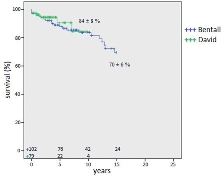
There were 18 late deaths in Group 1 and 4 in Group 2. In Group 1, 11 patients died of non-cardiac causes (8 neoplasms, 2 senectus, and 1 pulmonary failure), 3 of cardiac failure, and 4 of valve-related causes (2 stroke, 1 valve thrombosis, and 1 sudden death). In Group 2, 3 patients died of cardiac failure and 1 of bowel ischemia. Actuarial survival was 82 ± 4% at 10 years and 70 ± 6% at 15 years in Group 1 and 91 ± 5% at 5 years and 84 ± 8% at 10 years in Group 2 (Figure 1). At last follow-up, mean NYHA class was 1.2 ± 0.6 and 1.1 ± 0.4 in Group 1 and 2 patients, respectively.
Figure 1.
Actuarial survival after the modified Bentall and David I aortic valve-sparing procedures. Numbers on the horizontal axis indicate patients at risk.
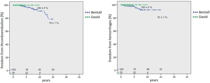
Thromboembolic episodes occurred in 10 patients in Group 1 at a mean delay of 8.5 ± 3.9 years from MB (range, 2.6-12.9). Nine patients had a cerebral embolism, 1 of whom died and 1 had permanent sequelae; 1 patient died due to prosthetic thrombosis without reoperation. Actuarial freedom from thromboembolism was 91 ± 4% at 10 years and 79 ± 7% at 15 years (Figure 2). Seven patients in Group 1 experienced anticoagulant-related hemorrhage at a mean delay of 6.2 ± 5.1 years (range, 0.6-16.2); 6 had minor bleeding, while 1 patient had fatal cerebral hemorrhage. Actuarial freedom from bleeding was 92 ± 3% at both 10 and 15 years. No embolic or hemorrhagic episodes were observed in Group 2.
Figure 2.
Actuarial freedom from thromboembolic complications (left panel) and anticoagulant-related hemorrhage (right panel).
Endocarditis occurred in 5 patients in Group 2 at a mean delay of 0.9 ± 1.3 years from DP (range, 0.3-3.2), involving the aortic valve in 3 patients and the mitral valve in 2 patients. All patients were successfully reoperated, with 3 patients requiring aortic valve replacement and 2 requiring mitral valve repair. Actuarial freedom from endocarditis in Group 2 was 92 ± 4% at both 5 and 10 years. No cases of endocarditis occurred in Group 1.
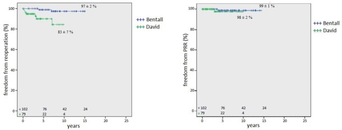
Reoperation was performed in 3 patients in Group 1 at a mean delay of 9.2 ± 6 years (range, 3.7-16.5 years) and 7 patients in Group 2 at a mean delay of 2.2 ± 0.2 years (range, 0.2-7.6 years). In Group 1, causes of reoperation were 1 valve thrombosis, 1 aortic pseudoaneurysm, and 1 mitral regurgitation, with no deaths. In Group 2, reoperation was required due to endocarditis in 5 patients, and 2 were reoperated due to aortic regurgitation and aortic stenosis 3 and 7 years after DP, respectively. Actuarial freedom from reoperation was 97 ± 2% at both 10 and 15 years in Group 1 and 90 ± 5% at 5 years and 84 ± 7% at 10 years in Group 2 (Figure 3). There were two procedure-related reoperations in Group 1 (valve thrombosis and pseudoaneurysm formation) and one in Group 2 (aortic regurgitation) with an actuarial freedom of 99 ± 1% at both 10 and 15 years in Group 1 and 98 ± 2% at both 5 and 10 years in Group 2.
Figure 3.
Actuarial freedom from reoperation for all causes (left panel) and procedure-related reoperations (PRR, right panel).
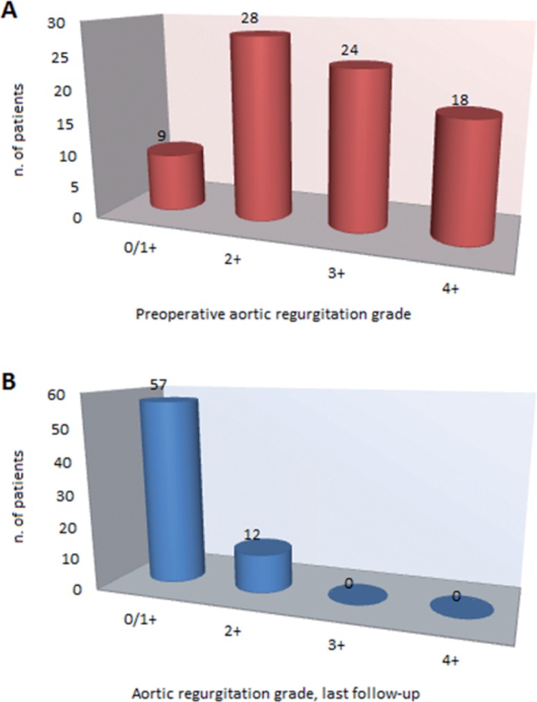
In Group 2, complete echocardiographic data were available for 69 long-term survivors at a mean delay of 4.3 ± 3.3 years. Variation in AI grades is shown in Figure 4; at last follow-up, 57 (83%) patients showed absent or trivial AI and 12 (17%) showed mild AI, whereas no cases of moderate or severe AI were observed.
Figure 4.
Graphic representation of preoperative aortic incompetence grade (Panel A) and aortic incompetence grade at last post-repair follow-up (Panel B).
Discussion
In patients with aortic valve disease and aneurysms of the aortic root, combined replacement of the aortic valve and ascending aorta with a composite graft, as proposed by Bentall and De Bono and later modified by Kouchoukos et al. [1, 2], has been the gold standard procedure for many years. This is true also for patients with annuloaortic ectasia and various degrees of AI, where the aortic valve may be substantially preserved without structural anomalies. Demonstrations that these patients can be treated conservatively by aortic root replacement with an adequately fashioned graft [4] or aortic valve resuspension within a graft tube [3] by valve-sparing procedures restoring adequate aortic valve function have significantly changed the outlook of patients with annuloaortic ectasia. However, given the favorable long-term results of MB [18-20], whether valve-sparing operations are superior to composite graft replacement of the aortic root is still a matter of debate [8-12].
In the present study, we compared outcomes between MB and DP for patients with annuloaortic ectasia. Our finding of better survival in patients undergoing DP can in part be explained by a difference in preoperative clinical status, as MB patients were in poorer health as indicated by a lower ejection fraction and higher NYHA class. As expected, thromboembolic and hemorrhagic complications were observed in the MB group, with a 79% freedom from embolism and 92% freedom from anticoagulant-related bleeding at 15 years; indeed, all patients received a mechanical composite graft requiring life-long anticoagulation. However, in a recent report, long-term freedom from bleeding and thromboembolism in patients undergoing composite graft replacement of the aortic root was 94% at 15 years despite that 76% of patients received a mechanical conduit [21]. On the other hand, patients undergoing DP, who are routinely not given oral anticoagulants, did not experience such complications in this series, with a 100% freedom from both events at 10 years.
In the DP group, we observed an unusual incidence of endocarditis involving the aortic valve in 3 out of 5 patients, 2 of whom had mitral valve infection. A 92% freedom from aortic valve infection 10 years after DP is disturbing, considering that avoidance of prosthetic material should limit this complication, and is not clearly explained by our analysis, particularly as no cases of infection were observed in the MB group despite the use of a mechanical conduit. In a recent systematic review of aortic valve preservation and repair, postoperative endocarditis was reported with a rate of up to 0.78% patient-years [22]. Kallenback et al. [23] observed four cases of endocarditis at a mean follow-up of 41 months after DP in a series of 284 patients. Svensson et al. [24] reported three reoperations due to infection out of 313 patients undergoing DP, whereas David et al. [5] observed two cases of endocarditis requiring reoperation out of 371 consecutive patients. Probably because this complication seems quite rare, no specific comments on this event are present in the literature; however, as infection may compromise the results of aortic valve repair, we believe it merits further and more detailed analysis.
Although our experience with aortic root reconstruction is limited, we can confirm what others have reported about valve-sparing procedures in larger populations at medium- and long-term intervals, showing that patients with annuloaortic ectasia can be safely treated with DP. Following technical indications and lessons from leaders in the field [5, 14, 15], this operation appears to be reproducible and stable over time, even in less experienced hands. In fact, in the present series, only one patient required reoperation due to recurrent AI after DP, with an actuarial freedom from procedure-related reoperation of 98% at 10 years. At last follow-up, the vast majority of patients showed absent or trivial AI, with no cases of moderate or severe AI. If we exclude the experience of David et al. [5], others report excellent long-term results with DP [23-25], which is currently our procedure of choice in the treatment of patients with AI and aortic root dilatation. In our series, most patients had a tricuspid aortic valve, but DP can also be employed with similar favorable outcomes in patients with Marfan syndrome or bicuspid aortic valves [11, 12, 24, 25]. For these reasons, we believe that DP should always be planned and attempted in patients with annuloaortic ectasia, whereas MB should be reserved for patients with structural aortic valve disease associated with root dilatation or in cases of early or late DP failure.
Finally, in most of our patients, we employed a Valsalva graft for DP, which is a specifically designed Dacron graft incorporating pseudosinuses of Valsalva. This graft has been considered to facilitate aortic valve reimplantation procedures and is associated with satisfactory results for up to 10 years [26].
Major limitations of this report are its retrospective nature and the fact that the two series were not concurrent but belonged to different, partially overlapping time intervals; this is because earlier patients were typically treated with MB, whereas more recent patients tended to be treated with DP. Therefore, the two groups had different follow-up durations that were shorter for the DP group, which makes meaningful comparisons between groups difficult. Nevertheless, a careful patient re-evaluation with nearly 100% follow-up may at least partially mitigate this limitation. Furthermore, as previously described, all MBs were performed using a mechanical conduit due to the relatively young age of our patient population. Patients receiving MB with a biological conduit were excluded from this report due to their small number; however, we would currently favor the use of biological conduits for MB in an elderly population.
In conclusion, our experience suggests that both MB and DP produce overall excellent early- and late-term results. MB using a mechanical conduit was associated with the unavoidable occurrence of thromboembolic and hemorrhagic complications, whereas DP was associated with an unexplained occurrence of endocarditis. DP appears to be safe, reproducible, and able to achieve stable aortic valve repair over time, suggesting that it is currently an ideal solution for patients with annuloaortic ectasia. Thus, we currently reserve MB only for patients with a severely damaged aortic valve. In elderly patients requiring MB, the use of biological conduits should probably be expanded. Finally, the much shorter follow-up of DP patients considerably limits the strength of our comparison between the two techniques; therefore, longer follow-up is necessary to provide evidence regarding effectiveness of the DP technique.
Conflict of Interest
The authors have no conflict of interest relevant to this publication.
References
- 1.Bentall H, De Bono A. A technique for complete replacement of the ascending aorta. Thorax. 1968;23:338-339. PMID: [DOI] [PMC free article] [PubMed] [Google Scholar]
- 2.Kouchoukos NT, Marshall WG Jr, Wedige-Stecher TA. Eleven-year experience with composite graft replacement of the ascending aorta and aortic valve. J Thorac Cardiovasc Surg. 1986;92:691-705. PMID: [PubMed] [Google Scholar]
- 3.David TE, Feindel M. An aortic valve-sparing operation for patients with aortic incompetence and aneurysm of the ascending aorta. J Thorac Cardiovasc Surg. 1992;103: 617-622. PMID: [PubMed] [Google Scholar]
- 4.Sarsam MA, Yacoub M. Remodeling of the aortic valve annulus. J Thorac Cardiovasc Surg. 1993;105:435-438. PMID: [PubMed] [Google Scholar]
- 5.David TE, Feindel CM, David CM, Manlhiot C. A quarter of a century of experience with aortic valve-sparing operations. J Thorac Cardiovasc Surg. 2014;148:872-879. DOI: 10.1016/j.jtcvs.2014.04.048 [DOI] [PubMed] [Google Scholar]
- 6.David TE. Aortic valve sparing in different aortic valve and aortic root conditions. J Am Coll Cardiol. 2016;68:654-664. DOI: 10.1016/j.jacc.2016.04.062 [DOI] [PubMed] [Google Scholar]
- 7.Franke UFW, Isecke A, Nagib R, Breuer M, Wippermann J, Tigges-Limmer K, et al. Quality of life after aortic root surgery: reimplantation technique versus composite replacement. Ann Thorac Surg. 2010;90:1869-1875. DOI: 10.1016/j.athoracsur.2010.07.067 [DOI] [PubMed] [Google Scholar]
- 8.Tourmousoglou C, Rokkas C. Is aortic valve-sparing operation or replacement with a composite graft the best option for aortic root and ascending aortic aneurysms? Interact Cardiovasc Thorac Surg. 2009;8:134-147. DOI: 10.1510/icvts.2008.186544 [DOI] [PubMed] [Google Scholar]
- 9.Lim JY, Kim JB, Jung SH, Choo SJ, Chung CH, Lee JW. Surgical management of aortic root dilatation with advanced aortic regurgitation: Bentall operation versus valve-sparing procedure. Korean J Thorac Cardiovasc Surg. 2012;45:141-147. DOI: 10.5090/kjtcs.2012.45.3.141 [DOI] [PMC free article] [PubMed] [Google Scholar]
- 10.Kellenbach K, Kojic D, Oezsoez M, Bruckner T, Sandrio S, Arif R, et al. Treatment of ascending aortic aneurysms using different surgical techniques: a single-centre experience with 548 patients. Eur J Cardiothorac Surg. 2013;44:337-345. DOI: 10.1093/ejcts/ezs661 [DOI] [PubMed] [Google Scholar]
- 11.Price J, Magruder JT, Young A, Grimm JC, Patel ND, Alejo D, et al. Long-term outcomes of aortic root operations for Marfan syndrome: a comparison of Bentall versus aortic valve-sparing procedures. J Thorac Cardiovasc Surg. 2016;151:330-338. DOI: 10.1016/j.jtcvs.2015.10.068 [DOI] [PubMed] [Google Scholar]
- 12.Vallabhajosyula P, Szebo WY, Habertheuer F, Komlo C, Milewski RK, McCarthy F, et al. Bicuspid aortic insufficiency withy aortic root aneurysm: root reimplantation versus Bentall root replacement. Ann Thorac Surg. 2016;102:1221-1228. DOI: 10.1016/j.athoracsur.2016.03.087 [DOI] [PubMed] [Google Scholar]
- 13.Pratali S, Milano A, Codecasa R, De Carlo M, Borzoni G, Bortolotti U. Improving hemostasis during replacement of the ascending aorta and aortic valve with a composite graft. Texas Heart Inst J. 2000;27:246-249. PMID: [PMC free article] [PubMed] [Google Scholar]
- 14.de Kerchove L, Nezhad ZM, Boodhwani M, El Khoury G. How to perform valve sparing reimplantation in a tricuspid aortic valve. Ann Cardiothorac Surg. 2013;2:105-112. DOI: 10.3978/j.issn.2225-319X.2013.01.16 [DOI] [PMC free article] [PubMed] [Google Scholar]
- 15.Boodhwani M, de Kerchove L, Gilneur D, Rubay J, Vanoverschelde JL, Noirhomme P, et al. Repair of regurgitant bicuspid aortic valves: a systematic approach. J Thorac Cardiovasc Surg. 2010;140:276-284. DOI: 10.1016/j.jtcvs.2009.11.058 [DOI] [PubMed] [Google Scholar]
- 16.Stanger O, Tevaerarai H, Carrel T. The Freedom SOLO bovine pericardial stentless valve. Res Rep Clin Cardiol. 2014;5:1-13. DOI: 10.2147/RRCC.S72978 [DOI] [Google Scholar]
- 17.Akins CW, Miller DC, Turina MI, Kouchoukos NT, Blackstone EH, Grunkemejer GL, et al. Guidelines for reporting mortality and morbidity after cardiac valve interventions. J Thorac Cardiovasc Surg. 2008;135:732-738. DOI: 10.1016/j.athoracsur.2007.12.082 [DOI] [PubMed] [Google Scholar]
- 18.Etz CD, Bischoff MS, Bodian C, Roder F, Brenner R, Griepp RB, et al. The Bentall procedure: is it the gold standard? A series of 597 consecutive cases. J Thorac Cardiovasc Surg. 2010:140:S64-S70. DOI: 10.1016/j.jtcvs.2010.07.033 [DOI] [PubMed] [Google Scholar]
- 19.van Putte BP, Ozturk S, Siddiqi S, Schepens MAAM, Heijmen RH, Morshuis WJ. Early and late outcome after aortic root replacement with a mechanical prosthesis in a series of 528 patients. Ann Thorac Surg. 2012;93:503-509. DOI: 10.1016/j.athoracsur.2011.07.089 [DOI] [PubMed] [Google Scholar]
- 20.Varrica A, Satriano A, de Vincentiis C, Biondi A, Trimarchi S, Ranucci M, et al. Bentall operation in 375 patients: long-term results and predictors of death. J Heart Valve Dis. 2014;23:127-134. PMID: [PubMed] [Google Scholar]
- 21.Mok SCM, Ma WG, Mansour A, Charilaou P, Chou AS, Peterss S, et al. Twenty-five year outcomes following composite graft aortic root replacement. J Cardiac Surg. 2017;32:99-109. DOI: 10.1111/jocs.12875 [DOI] [PubMed] [Google Scholar]
- 22.Saczkowski R, Malas T, de Kerchove L, El Khoury G, Boodhwani M. Systematic review of aortic valve preservation and repair. Ann Cardiothorac Surg. 2013;2:3-9. DOI: 10.3978/j.issn.2225-319X.2013.01.07 [DOI] [PMC free article] [PubMed] [Google Scholar]
- 23.Kallenbach K, Kark M, Pak D, Salcher R, Khaladj N, Leyh R, et al. Decade of aortic valve sparing reimplantation. Are we pushing the limits too far? Circulation. 2005;112(Suppl. 1):I253-I259. DOI: 10.1161/01.CIRCULATIONAHA.104.525907 [DOI] [PubMed] [Google Scholar]
- 24.Svensson LG, Blackstone EH, Alsalihi M, Batizy LH, Roselli EE, McCullogh R, et al. Midterm results of David reimplantation in patients with connective tissue disorder. Ann Thorac Surg. 2013;95;555-562. DOI: 10.1016/j.athoracsur.2012.08.043 [DOI] [PubMed] [Google Scholar]
- 25.Kvitting JP, Kari FA, Fischbein MP, Liang DH, Beraud AS, Stephens EH, et al. David valve-sparing aortic root replacement: equivalent mid-term outcome for different valve types with or without connective tissue disorders. J Thorac Cardiovasc Surg. 2013;145:117-126. DOI: 10.1016/j.jtcvs.2012.09.013 [DOI] [PMC free article] [PubMed] [Google Scholar]
- 26.De Paulis R, Scaffa R, Nardella S, Maselli D, Welter L, Bertoldo F, et al. Use of the Valsalva graft and long-term follow-up. J Thorac Cardiovasc Surg. 2010;140:S23-S27. DOI: 10.1016/j.jtcvs.2010.07.060 [DOI] [PubMed] [Google Scholar]


