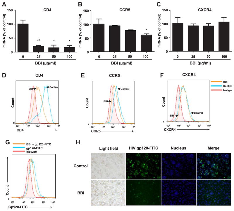
Fig. 2. BBI decreases CD4 and CCR5.
Macrophages were treated with/without BBI at indicated concentrations for 6 h (mRNA) or BBI (100 μg/ml) for 24 h (protein). Cellular RNA was collected and subjected to real time PCR for the genes indicated and GAPDH RNA (A–C). The data are expressed as RNA levels percentage (%) for CD4, CCR5, CXCR4 to untreated control, which is defined as 100%. Data are shown as mean±SD for three independent experiments. (D–F) CD4 in the surface of human macrophages stained with PE-cy7 anti-human CD4 (clone: SK3) antibody, CCR5 stained with FITC anti-human CD195 (CCR5) antibody, CXCR4 stained with PE anti-human CXCR4 antibody and all of them were detected by flow cytometer (FACSCantoII), the arrows indicated macrophages stimulated with/without BBI for 24 h. Representative data from three independent experiments are shown. (*P<0.05, **P<0.01, when performing Student’s t-test). Macrophages were stimulated with BBI (100 μg/ml) for 24 h. After washing 3 times with PBS, Macrophages treated with/without BBI were incubated with gp120-FITC (15 μg/ml) for additional 2 h, the binding of gp120-FITC to CD4 was detected by flow cytometer (G) and fluorescence microscope (magnification, 200) (H). The data from flow cytometer were analyzed with Flow-Jo software. Representative data from three independent experiments are shown.

