



Abstract
Owing to its complex structure and highly diverse cell populations, the study of hypothalamic development has historically lagged behind that of other brain regions. However, in recent years, a greatly expanded understanding of hypothalamic gene expression during development has opened up new avenues of investigation. In this review, we synthesize existing work to present a holistic picture of hypothalamic development from early induction and patterning through nuclear specification and differentiation, with a particular emphasis on determination of cell fate. We will also touch on special topics in the field including the prosomere model, adult neurogenesis, and integration of migratory cells originating outside the hypothalamic neuroepithelium, and how these topics relate to our broader theme.
INTRODUCTION
The hypothalamus controls a wide range of homeostatic processes essential for life, including feeding, thirst, sleep, circadian rhythms, reproductive behavior, and more. This brain region, derived from the rostral diencephalon of the forebrain, is enormously anatomically complex. Where other brain regions such as the cortex or hippocampus have a clear overall structure of columns and parallel feed-forward loops, respectively, the hypothalamus has a densely interconnected patchwork of often quite ill-defined nuclei. Partially for this reason, the study of hypothalamic development has lagged behind that of many other brain regions, whose simpler anatomy facilitated the identification of factors that pattern them. However, as determinants of hypothalamic identity were studied in recent years, it has become increasingly clear that many of the same factors implicated in development of the cortex, retina, and other CNS regions play similar roles in the hypothalamus.
Shortly after the neural plate forms following gastrulation, diffusible morphogens begin patterning the developing nervous system, including the hypothalamus. In the beginning, these factors are generated outside of neural tissues, in mesodermal domains including the axial notochord and more anterior prechordal plate (PCP), as well as other more lateral tissues. Often, production of these same cues is later induced in neural tissue near or within the nascent hypothalamus, where dynamically shifting sources of morphogen production create ever-finer concentration gradients within the region. Sharp boundaries are translated from these gradients through induction of distinct and often mutually repressive sets of cell-autonomous transcription factors, progressively defining the fate of neural progenitors and emerging hypothalamic nuclei.1,2
In this review, we will first outline the contribution of several major growth and differentiation factor families in inducing, patterning, and sometimes specifying subdomains of the hypothalamus. We will then discuss how the pathways guiding neurogenesis, specification, and differentiation diverge in the heterogeneous hypothalamus to produce its variegated adult nuclei, with a focus on the transcriptional networks that control this process.
HYPOTHALAMIC INDUCTION AND SPATIAL PATTERNING
Wingless Family (Wnt) Signaling
The Wnts are a large family of secreted, lipid-modified glycoproteins long known to be involved in patterning during development, having originally been implicated in developmental segmentation by their homology to the Drosophila Wingless gene. Anterior-posterior (AP) patterning of newly induced neural plate is initiated by the posteriorizing morphogen Wnt8, secreted by lateral mesodermal precursors. Its effect is enhanced and sustained by nodal signaling in the posterior neural plate. Conversely, Wnt inhibition is necessary for anterior patterning, including the region that eventually gives rise to the hypothalamus. A variety of Wnt inhibitors are expressed in spatially and temporally dynamic patterns, first in the PCP (Figure 1) and later in the developing anterior brain. These inhibitors include sFRP1, sFRP2, sFRP3/Frzb1, Crescent/Frzb2, Dickkopf1, and Cerberus.4,5
FIGURE 1.

Schematic showing the inductive signals that act on the ventral neural plate. The posterior neural plate receives inductive signals from the notochord to become floor plate, whereas the more anterior neural plate that goes on to become the hypothalamus receives a different set of inductive signals from the prechordal plate. Circles represent signaling from the prechordal plate/notochord, whereas the diagonal bars in the neural plate represent local gene expression. It should be noted that the juxtaposition of the prechordal plate and the hypothalamic neural plate is transient. These structures migrate out of register with each other. The prechordal plate reaches its final rostral position in advance of the hypothalamic neural plate, which migrates over and rostrally past the prechordal plate. Thus by the time the floor plate and the hypothalamic neural plate are specified, the prechordal plate actually resides caudal to the hypothalamic neural plate.3 A, anterior; P, posterior
After initial AP patterning, Wnt signaling in turn subdivides the forebrain. The domain receiving lower Wnt signaling gives rise to the telencephalon, which includes the cerebral cortex, hippocampus, striatum, amygdala, and associated structures. Higher Wnt signaling induces development of the diencephalon, including a more rostral domain that gives rise to the hypothalamus and prethalamus, and a more caudal domain that gives rise to the thalamus.6 Telencephalic and rostral diencephalic neuroepithelium are hereafter distinguished by expression of the forkhead domain transcription factors FOXG1 and FOXD1, respectively.7,8
However, despite relatively high Wnt signaling in the diencephalon early on, inhibition of canonical Wnt signaling by Axin1 is essential for initial hypothalamic specification. In Axin1−/− mice, the ventral midline cells of the hypothalamus fail to develop, instead assuming a caudal floor plate identity.9 Later roles of Wnts in hypothalamic development explain this seeming paradox. During the late gastrula stage in zebrafish, Wnt8b and Wnt1 secreted from the midbrain–hindbrain boundary pattern the forebrain and induce specification of posterior diencephalon, including the sensory thalamus and habenula. Overexpression of these Wnts or their receptors, or loss of the Wnt inhibitor Axin1, induce expanded expression of posterior hypothalamic markers such as Emx2 at the expense of anterotuberal hypothalamic markers such as Rx3, suggesting a posteriorization of hypothalamic neuroepithelium.9,10
Consistent with this finding, Wnt8b is expressed in the mouse posterior hypothalamus (PH) beginning at ~ E8.5,8,11 consistent with a role in patterning, but its function has not been directly explored in this context (Figure 2(e)). By E12.5, Wnt8b becomes restricted to the mammillary region, suggesting it may act to impart posterior hypothalamic identity in mammals as it does in zebrafish8 (Figure 2(f)). WNT7a/7b become expressed selectively in prethalamic and hypothalamic GABAergic neuronal progenitors around the same time, suggesting a role in interneuron development, but their function is even less well-characterized than WNT8b8 (Figure 2(F)).
FIGURE 2.

Sagittal view of morphogen expression over the course of development in the mouse forebrain, emphasizing the hypothalamus. (a–c). Forebrain Shh expression from E8.5 through E11.5. (d). Hypothalamic Shh expression at E12.5. White bars represent areas of diffuse Shh expression. (e) Wnt expression in the forebrain at E9.5. (f). Wnt expression in the hypothalamus at E12.5. Wnt 7a/7b is expressed in interneuron progenitors only while Wnt 3/3a and Wnt 8a/8b are expressed more broadly. BP, basal plate; ZLI, zona limitans intrathalamica; POA, preoptic area; Tel, telencephalon; PrTh, prethalamus; Hy, hypothalamus.
Meanwhile, a gradient of WNT3 signaling further patterns the developing forebrain, including the hypothalamus (Figure 2(f)). High levels of WNT3 induce Irx3 expression in the sensory thalamus, whereas low WNT3 signaling is permissive for more anterior Six3 expression in the hypothalamus and prethalamus.12 Mutual cross-repression of SIX3 and IRX3 demarcates the border between these regions, an effect that is reinforced by SIX3-mediated dampening of WNT responsiveness. Confirming the importance of these interactions, Six3−/− mice show a rostral expansion of caudal diencephalic markers at the expense of the hypothalamus and telencephalon, largely due to an anterior expansion of WNT1 expression in the roof plate of the forebrain.1,13
Sonic Hedgehog (SHH) Signaling
Concurrent with Wnts’ central role in early AP patterning of the developing neural tube, the lipid-linked polypeptide signal SHH is a key secreted morphogen controlling neural dorso-ventral (DV) patterning. SHH is first expressed in the PCP (Figure 1), and is necessary for initial induction of the hypothalamus.14,15 The ventral midline cells initially have a quasi-caudal floor plate identity, including expression of HNF3β but not the floor plate markers FP3 and FP416 (Figure 1). PCP-derived SHH goes on to induce transient Shh expression in the ventral diencephalic midline (and much of the rest of the developing hypothalamus) through a GLI-mediated signaling cascade that activates expression of transcription factors including SIX3, SOX2, and SOX3.12,17 SOX2 and SOX3, members of the SOXB1 transcription factor family, directly activate and maintain Shh transcription in hypothalamic neuroepithelium.17 SIX3 likewise targets the Shh brain enhancer-2 to directly activate Shh transcription there.18,19
A subsequent downregulation of Shh expression is important for hypothalamic patterning. Indeed, early patterning of the ventral hypothalamus along the AP axis is achieved in part through downregulation of Shh expression in ventral tuberomammillary hypothalamus15,20,21 (Figure 2(b)). For instance, studies in chick demonstrate cells giving rise to this region must downregulate Shh to adopt their proper fate through induction of Tbx2 expression, which in turn triggers Shh downregulation by upregulation of Bmp7 in the tuberomammillary hypothalamic neuroepithelium (see next section).
In contrast to the ventral tuberomammillary hypothalamus, Shh expression in the basal plate (BP), which is initially induced by PCP Shh and additional feed-forward Shh signaling from the ventral midline, is maintained through E14.522,23 (Figure 2(b)–(d)). This more dorsally located hypothalamic region begins immediately posterior and ventral to the presumptive suprachiasmatic nucleus (SCN) and terminates in the mammillary region8,24 (Figure 2(b)–(d)). Shh(+) cells of the BP originate at the diencephalic/mesencephalic border, then migrate to their final destination in the hypothalamus.21 BP SHH plays a critical role in hypothalamic patterning. Several studies have looked at loss of function mutants in which Shh is deleted from the BP. In these studies, the hypothalamus is still specified, but neurons fail to undergo further differentiation, resulting in the loss or reduction of several nuclear markers. In all mutant lines examined, both AP and DV patterning are disrupted, although the reported phenotypes are not identical among these studies, most likely because they used Cre-drivers with different spatial and temporal patterns of activity.8,17,25,26
BP Shh is also necessary to initiate sustained Shh expression in the zona limitans incerta (ZLI), a secondary Shh(+) organizer dividing the prethalamus and thalamus.27,28 ZLI-derived Shh plays a central role in patterning the sensory thalamus, and is required for proliferation and differentiation of prethalamic progenitors29,22,23 (Figure 2(b)–(d)). Importantly, ZLI Shh expression is preserved in Cre/lox studies where Shh was deleted in the BP, showing that the hypothalamic patterning roles of BP- and ZLI-derived Shh are distinct.
Bone-Morphogenetic Protein (Bmp) Signaling
BMP7 is the primary member of this family of TGF-β superfamily members that has been studied in the context of hypothalamic development. Surprisingly given the classical physical separation and antagonistic effects of BMPs and SHH in spinal DV patterning, many known actions of BMP7 in the hypothalamus revolve around cooperative interactions with SHH. For instance, during gastrulation PCP-derived BMP7 and SHH cooperate to induce expression of early general hypothalamic markers such as Nkx2.1 in the ventral diencephalic midline, distinct from more caudal midline gene expression induced by notochord-derived SHH alone16 (Figure 1).
During later stages of hypothalamic patterning, expression of Bmp7 and Shh are transiently coexpressed in ventral tuberomammillary hypothalamus,21 (Figure 1). BMP7 swiftly downregulates Shh expression here by activating expression of first Tbx2 and subsequently Gli3, although it is unclear if TBX2 directly activates Gli3 expression.20,21 In chick this process begins around Hamburger and Hamilton (HH) stage 10, inducing complete elimination of Shh by HH stage 16 (roughly equivalent to E9.5–10.5 in mouse).24 Tbx2 itself is in turn downregulated during stages 16–22; toward the end of this period, ventral tuberomammillary markers such as Fgf10 and Emx2 begin to be expressed in these now Shh(−) subdomains of the developing hypothalamus.
BMP7 signaling is also required for induction of Pax7 expression in more dorsally located tuberomammillary progenitors, which occurs by HH stage 30. BMP7 accomplishes this by both downregulating Shh expression in ventral tuberomammillary hypothalamus, and by directly activating Pax7 transcription via induction of Smad5 phosphorylation.20 Interestingly, widespread expression of the Bmp antagonist Chrdl1 is detected in chick hypothalamus at HH stage 13, with expression restricted to the anterioventral hypothalamic neuroepithelium by HH15 as Bmp expression becomes restricted to posterioventral hypothalamus. Chrdl1 expression is upregulated in response to inhibition of Notch signaling, suggesting that Notch may play a role in early hypothalamic development by modulating BMP signaling,30 although this has not yet been shown directly.
Nodal Signaling
Nodal proteins are other members of the TGF-β superfamily that play an important role in hypothalamic patterning. PCP-derived nodal signaling, most notably by cyclops, plays an essential role in initial hypothalamic specification in zebrafish31 (Figure 1). Together with Shh and Bmp7, nodal signaling is essential for induction of early hypothalamic markers such as Nkx2.1; however, unlike in the telencephalon, Shh is not able to restore initial expression of the Nkx2.1 homologue in zebrafish diencephalon in the absence of nodal, suggesting a separable role for nodal in early hypothalamic induction.32
On the other hand, cells defective in both Nodal and Shh signaling are unable to give rise to hypothalamic cells following transplantation into wild-type zebrafish, and Shh can restore some nkx2.1 expression in the zebrafish ventral posterior hypothalamus (vPH) during early stages of hypothalamic development.15,32 Together, these data suggest a cooperative role for Nodal and Shh in later stages of hypothalamic development, with Nodal enhancing the ability of cells to respond to Shh.
There also appears to be cross-talk between Nodals and Wnts in hypothalamic development, as the Wnt inhibitor Axin1 facilitates Nodal signaling.9 Axin1 may thus alter the competence of hypothalamic progenitors to respond to both Nodal and indirectly, to Shh signaling, likely explaining the non-cell-autonomous requirement for Nodal signaling in patterning of the dorsal anterior hypothalamus (dAH) by Shh.15 Conversely, cells of the zebrafish vPH require Nodal signaling cell-autonomously in order to activate emx2 expression, whereas excessive Shh signaling inhibits emx2 expression. Thus, Nodal must cooperate with Bmp7 to induce emx2 in vPH—while Nodal acts as a cell-autonomous factor conferring competence to activate emx2 expression, Bmp7 inhibits local shh to prevent Shh signaling from becoming excessive. Thus, Axin1 sits at the nexus of Wnt, Shh, Nodal, and Bmp signaling, from which it serves as a critical, integrative determinant of AP identity.
Fibroblast Growth Factor (FGF) Signaling
FGF family members do not appear to play a critical role in early stages of hypothalamic patterning. Irx3 is required for competence to respond to Fgf8 signaling, but is excluded from the hypothalamus during this time by its cross-repressive interaction with Six3. The ZLI lies between the Six3(+) rostral diencephalon and Irx3(+) caudal diencephalon, demarcating the boundary of competence to Fgf8 signaling from the isthmic organizer and telencephalic roof plate.33 That said, FGF family members do regulate cell proliferation at later stages of hypothalamic development, as discussed in later sections.
HYPOTHALAMIC NEUROGENESIS AND LATE PATTERNING
Hypothalamic neurogenesis occurs from ~ E10.5–16.5 in mice, with the great majority of hypothalamic neurons being generated between E12.5 and E14.5.34,30,35 Birthdating studies using tritiated thymidine and BrdU indicated that neurons in lateral hypothalamic nuclei are typically born before medial nuclei, resulting in an ‘outside-in’ pattern of birth in the hypothalamus.35 However, later research has contradicted this model. Looking at different parvocellular neuronal cell types within the neurosecretory dorsolateral anterior hypothalamus (dlAH), all were found to be born during the same approximate interval, despite occupying different medial-lateral locations within the hypothalamus.36 Similar results have been reported for different neuronal subtypes in the arcuate nucleus (ARC),37 suggesting that for progenitors in at least some regions of the hypothalamus, this ‘outside-in’ order of cell generation may not hold.
Growth and differentiation factors that pattern the developing hypothalamus also play important roles in later stages of hypothalamic cell fate specification. For example, while SHH is required for both early and late neurogenesis in much of the hypothalamus,8,38 Shh downregulation is necessary for proliferation and expansion of the ventral tuberomammillary progenitor pool.21 Meanwhile, canonical Wnt signaling, through its effector Lef1, plays an important role in both progenitor maintenance and in driving progenitor differentiation in the PH.39,40 Notch signaling is also involved in progenitor maintenance in the hypothalamus, as it is elsewhere in the developing nervous system, but the specifics of its role in different parts of the hypothalamus has only recently begun to be understood.30,41 In the rostral hypothalamus, Notch signaling works through lateral inhibition to regulate the progenitor population and generate new neurons. High levels of Notch signaling in AH upregulate Hes and Hey family genes while downregulating proneural genes such as Ascl1 and Nhlh1. The genes Tagln3, Chga, Robo2, Slit1, and Chrdl1 are then needed for terminal neural differentiation once precursor cells have exited mitosis.
Expression of a diverse assortment of transcription factors are induced by the intricate spatiotemporal code of exposure to morphogens discussed previously, which in turn confer intrinsic spatial identity upon hypothalamic progenitor cells. Patterning processes establish the anatomical DV and AP axes within the hypothalamus, limiting which nuclei can develop at any given set of coordinates. Consequently, many markers ubiquitously expressed in early hypothalamic progenitors become more restricted once neurogenesis begins. For instance, Six3 and Lhx2 are expressed in the anterior and tuberal hypothalamus (AH and TH), but not the PH. Meanwhile, Rax and Nkx2.1 are expressed in the TH and PH, but only in limited regions of the AH.42
However, these still relatively broad domains of transcription factor expression become even more regionalized as neurogenesis progresses. Continuing with the previous examples, Lhx2 becomes restricted to the dorsolateral aspects of AH (dlAH) while Six3 becomes restricted to the ventral AH (vAH). Meanwhile, Rax expression is rapidly downregulated everywhere except the ventral TH, though Nkx2.1 remains comparatively broadly expressed in the TH and PH.8,42–45 By mouse E12.5, progressive restriction of expression of these and assorted other genes has already defined many nuclei found in the adult hypothalamus (Figure 3).
FIGURE 3.
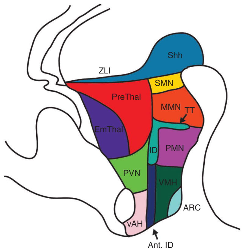
Sagittal map of nuclei in the mouse hypothalamus at E12.5 at the level of the third ventricle. ZLI, zona limitans intrathalamica; PreThal, prethalamus; EmThal, thalamic eminence; PVN, paraventricular nucleus; vAH, ventral anterior hypothalamus; ant. ID, anterior intrahypothalamic diagonal; ID, intrahypothalamic diagonal; ARC, arcuate nucleus; VMH, ventromedial hypothalamus; PMN, premammillary nucleus; TT, tuberomammillary terminal; MMN, mammillary nucleus; SMN, supramammillary nucleus.
Developmental expression of over a thousand such mouse genes were documented in an extensive screen published by Shimogori et al in 2010. Among other findings, this study identified the intrahypothalamic diagonal/tuberomammillary terminal (ID/TT) complex, an excellent example of progressive regionalization in the hypothalamus. This region traverses most of the hypothalamus in the sagittal plane starting at E12.5 (Figure 3) and is characterized by uniform expression of Arx and patterned expression of Lim homeodomain factors Lhx1, Lhx6, Lhx8, and Lhx9. This largely nonoverlapping pattern of LIM homeodomain factor expression appears to define a number of hypothalamic nuclei involved in control of circadian rhythms and sleep homeostasis, such as the SCN, dorsomedial hypothalamic (DMH), and parts of the lateral hypothalamus (LH) (Figure 4). In at least two cases, these genes are required for proper development of the hypothalamic cell types in which they are expressed.46,47 Such findings have already begun to facilitate the study of the development of individual hypothalamic nuclei, the topic we shall focus on for the remainder of the review.
FIGURE 4.

(a–d) Coronal diagrams of the nuclei of the developing hypothalamus at ~E15.5. Insets in the upper right-hand corners indicate the approximate position of these coronal sections in the sagittal plane. Note that the angle of the cut shown in the developing hypothalamus sometimes places nuclei normally present on different coronal planes in typical adult brain sections on the same plane in our diagram (e.g.: SCN and DMH in panel b). Abbreviations indicating divisions of the hypothalamus larger than single nuclei as described in the text (e.g.: AH and PH) here show the disposition of poorly defined and/or less well-studied nuclei within those regions. For example, though the PVN and SCN are derived from the broader AH, in this figure they are shown separately and the region labeled AH only encompasses less-studied regions we do not discuss in detail, such as the periventricular, subparaventricular, and retrochiasmatic nuclei. SCN, suprachiasmatic nucleus; DMH, dorsomedial hypothalamic; PVN, paraventricular nucleus; AH, anterior hypothalamus; PH, posterior hypothalamus.
DEVELOPMENT OF SPECIFIC HYPOTHALAMIC NUCLEI AND NEURONAL SUBTYPES
Dorsolateral Anterior Hypothalamus (dlAH): Paraventricular Nucleus (PVN), Supraoptic Nucleus (SON), and Anterior Periventricular Nucleus (APV)
The dlAH, or neuroendocrine hypothalamus, plays a central role in controlling systemic hormone secretion, with important implications for feeding and other homeostatic processes.48,49 Although Rax expression in dlAH has been controversial,43 fate mapping using Rax-CreER knock-in mice confirmed its previously reported low level expression beginning at E10.5.8,50 Six3, Six6, and Foxd1 also label the entire AH, including the dlAH, around this time.8 In zebrafish dlAH, fezf2 and olig2 are respectively required for induction of orthologs for the early specific dlAH markers otp and sim151,52 (Figure 5(a)). Fezf2 and Olig2 are expressed in mouse dlAH by E11.5, suggesting that these functions may be preserved in mammals.8,42 Furthermore, in zebrafish, Pac1 receptor signaling positively regulates levels of Otp protein, but not otp transcript.51
FIGURE 5.

Factors controlling specification and differentiation within specific nuclei or regions of the developing hypothalamus. Regions depicted include the dlAH (a), SCN (b), DMH (c), VMH (d), ARC (e), and PH (f). Blue arrows indicate factors that direct cells within the region toward the fate pointed at by its arrowhead, whereas red arrows indicate factors that inhibit the indicated fate. Serial arrows show that the downstream arrow is dependent upon the upstream arrow for its expression, directly or indirectly. Parallel arrows represent factors that act in concert to direct cells toward a common fate, but are not known to be directly dependent on one another for their expression. dlAH, dorsolateral anterior hypothalamus; SCN, suprachiasmatic nucleus; DMH, dorsomedial hypothalamic; VMH, ventromedial hypothalamus; ARC, arcuate nucleus; PH, posterior hypothalamus.
In contrast, early Lhx2 expression suppresses neurosecretory specification through inhibition of Otp and Sim1 (Figure 5(a)); the expression domains of these factors are expanded in Lhx2−/− AH.44 Lhx2 is also required to repress dlAH-specific genes in retina, even after retinal neurogenesis has begun. This implies that LHX2 acts as a fairly general repressor of dlAH identity in structures like the eye field that are derived from vAH hypothalamic neuroepithelium.44
Otp, Sim1, and the SIM1 binding partner Arnt2 are expressed in mouse dlAH by E12.5, concurrent with downregulation of Six3 and Six6 in PVN and SON, but not APV.8,45,53 SIM1/ARNT2 and OTP function in parallel to drive expression of several dlAH neuropeptides via their downstream factors Brn2 and Sim2, as well as specifying A11 dopaminergic neurons of the dlAH52,54–59 (Figure 5(a)). Fezf2, previously discussed as an upstream regulator of Otp expression, is similarly required for the formation of dopaminergic and neuropeptidergic lineages in zebrafish.51,60
However, in Otp−/− mice the dlAH exhibits additional developmental defects not observed in Sim1−/− mice, including ectopic expression of Six3 and Dlx1, loss of calbindin expression, and defects in radial migration of neural progenitors.54,57,59 This suggests that Otp is required at earlier stages of dlAH specification than Sim1. Conversely, SIM1 has additional later roles in dlAH, such as regulating longitudinal innervation of the spinal cord by A11 dopamine neurons in zebrafish and maintaining AH-specific gene expression in the postnatal mouse.61,62 Accordingly, both early and late Sim1 expression is essential for PVN function; both Sim1+/− and CaMKII-Cre;Sim1lox/lox mice are hyperphagic and obese.62,63
Acting downstream of Sim1 and Otp, Brn2 is required for expression of Avp and Oxt in PVN/SON, and Crh in PVN, whereas Sim2 is required for expression of Trh in PVN/APV and Sst in APV55,64,65 (Figure 5(a)). Sampling tissue from distinct Sim2 and Brn2 expressing subdomains of the developing Sim1(+) dlAH, a microarray-based screen identified additional Sim1-controlled genes, many of which are specifically expressed in cells expressing Sim2 or Brn2.66 This screen is thus also a starting point for identifying genes that act downstream of Brn2 and Sim2 in developing dlAH.
One of the few Sim1-regulated genes expressed throughout the entire domain of Sim1 expression is Uncx4.1.66 In Uncx4.1−/− mice, gross morphology of the PVN and expression of Otp and Avp is normal, although neuronal connectivity in the PVN is disrupted. No further gene expression analysis was reported in this study, but Uncx4.1 is known to promote dopaminergic cell fate at the expense of glutamatergic cells in the midbrain.67,68 Thus, given Sim1’s role in specifying hypothalamic dopamine neurons in zebrafish,52 it is tempting to speculate that UNCX4.1 may act downstream of SIM1 to promote A11 neuron specification, in a pathway parallel to Brn2- and Sim2-dependent determination of dlAH neuropeptidergic cell fate.
Both extracellular and intracellular factors influence dlAH development downstream of Sim2 and Brn2. Notch signaling plays a central role in this process. APV SST(+) cells are selectively depleted in Hes1−/− mice, and those that remain innervate the pituitary aberrantly, implicating Hes1 in specification of this lineage69 (Figure 5(a)). PVN and SON AVP(+) neurons also migrate improperly and ectopically express Gad67 in Hes1−/− mice, suggesting a role in maturation.69 Fgf8 hypomorphs show a more specific defect, with processed OXT peptide expression lost in PVN and SON, OXT propeptide expression lost only in SON, and Oxt mRNA unaffected in both regions, suggesting a role for FGFs in promoting maturation of these cells.70 AVP protein is also reduced in Fgf8 hypomorphic PVN, possibly because of similar deficits in maturation.71 Finally, Uncx4.1 is required for proper pituitary innervation by dlAH nuclei, whereas Nhlh2 is required for Pcsk expression and production of mature peptide in PVN Trh(+) neurons.67,72
Unlike in most of the hypothalamus, the transcriptional network guiding specification of various PVN, SON, and APV lineages is sufficiently well understood to provide a solid starting point for building on our understanding of dlAH development. Identifying the factors that specify individual neuropeptidergic populations from hypothalamic progenitor pools delineated by Sim2 and Brn2 expression is a topic of particular interest, particularly given recent findings suggesting that the fate of dopamine and SST neurons in adult rat dlAH can be bidirectionally changed by manipulating the circadian cycle the animal experiences, with significant effects on stress behavior.73 This suggests that developmental pathways regulating cell fate in the dlAH may remain active into adulthood, imparting plasticity to this system that can profoundly alter behavior in response to environmental changes.
Ventral Anterior Hypothalamus (vAH): The Suprachiasmatic Nucleus (SCN)
The best-characterized nucleus of the vAH is the SCN, which functions as the body’s central light-entrained circadian clock.74,75 In mouse, the ventral hypothalamic markers Rax and Lhx2, and anterior markers Foxd1 and Nkx2.2, are all expressed early in SCN, but downregulated as development proceeds. Other anterior markers, such as Six3 and Six6, remain expressed throughout development as they become progressively more restricted to the vAH. Fzd5 is expressed early in the Rax/Six3(+) region, possibly implicating Wnts in regulating initial SCN specification. Fzd5 is later downregulated coincident with induction of Dlx2 (specifying GABAergic fate), Lhx1 (the earliest specific marker of developing SCN), and Lhx8 in the anterior ID domain (Figure 3) that gives rise to the SCN8,42,45,50 (Figure 4). Induction of other selective SCN markers follows initial expression of the LIM and pro-GABAergic factors, including Rora, Id4, and Igfbp5 at E14.5 and Rorb, Nr1d1, Vipr2, and Sema6d at E16.5. By E16.5, Lhx8 expression is downregulated in the vAH, but persists in the presumptive DMH nucleus.8,45
In addition to the SCN, the anterior domain of the ID likely gives rise to other regions of the vAH, such as the subparaventricular, retrochiasmatic, and anterior nuclei (Figure 4). For example, the prominent ID marker Arx is present throughout most of the vAH despite being largely excluded from the SCN for most of the region’s development.8,42 However, the vAH is poorly characterized both developmentally and functionally outside of the SCN, and will not be discussed further in this review.
Lhx2 is required for specification of SCN and other vAH cell fates (Figure 5(b)); Lhx2−/− mice lack expression of multiple ventral anterior markers at E12.5, including Lhx1 and Arx.44 Six3 and Six6 are also required for SCN specification (Figure 5(b)), as Nestin-Cre;Six3lox/lox and Six6−/− mice both fail to form a morphologically recognizable SCN or express SCN-specific markers.45,76 Corresponding to this morphological defect, behavioral rhythms are also profoundly disorganized in Six6−/− mice, the only one of the three mutants that survive long enough to assess behavior.76 Although Nestin-Cre;Six3lox/lox mice likely have some disruption of the BP domain of Shh expression that is required for expression of AH markers,8,18,19 mosaic loss of Six3 function in the SCN of a subset of Nestin-Cre;Six3lox/lox mice led to a specific loss of Lhx1 expression only within the Six3-deficient region. This suggests that Six3 has a cell-autonomous role in SCN specification separable from its effects on control of Shh expression.45
Downstream of Six3 and Six6, Lhx1 is a master regulator of mammalian SCN terminal differentiation46 (Figure 5(b)). Six3-Cre;Lhx1lox/lox SCN retains expression and proper regionalization of most markers initially expressed prior to E16.5, but neuropeptides with important roles in adult circadian function including Vip, Grp, Avp, Prok2, Enk, and Nms are lost.46 The relatively normal development of Lhx1-deficient SCN from E11.5–E16.5 suggests possible compensation by one or more factors during this period; Lhx8 is one possible candidate. Lhx1 directly regulates Vip and likely Nms, Prok2, and Enk, but perhaps not Grp and Avp, as they lack conserved Lhx1 binding sites in their proximal enhancers.46,77 Creb3l1, another Lhx1-regulated gene, is a more likely direct regulator of Avp downstream of Lhx1, though the role of CREB3L1 in SCN development has not yet been assessed.77,78 Although its precise composition remains to be worked out, this LHX1-dependent transcriptional network is essential for adult SCN function, as Six3-Cre;Lhx1lox/lox mice show assorted defects in circadian entrainment of activity rhythms, including fragmentation or total arrhythmicity in the absence of external light cues.46
Further downstream, the role of Rora in SCN development has also been examined; Rorasg/sg loss-of-function mice surprisingly have a morphologically wild-type SCN with normal Vip and Avp expression, suggesting Rora may be dispensable for SCN development. However, the molecular characterization of Rorasg/sg SCN tested only these two molecular markers, and it is possible that subtle deficits in SCN development went undetected.45 Currently, the only factor known to control SCN differentiation downstream of LHX1 is FGF8, which is required for SCN-specific expression of Avp. This may reflect a role for FGF8 in promoting maturation of this population, as FGF8 appears to do for AVP+, PVN neurons.70,71 Looking ahead, dissecting the transcriptional networks upstream and downstream of Lhx1 is a promising approach for better understanding not only SCN differentiation, but also the adult function of the nucleus. Already, the Six3-Cre;Lhx1lox/lox mouse presents a unique opportunity to study interactions among SCN signals by disrupting many of them simultaneously with a single gene deletion, and it has been found that Lhx1 expression is directly regulated by light in adulthood, suggesting a possible role for LHX1 in mediating environmental control of neuropeptide signaling.46,77
DORSOMEDIAL HYPOTHALAMUS (DMH)
The DMH is reciprocally interconnected to many different hypothalamic regions, and functions at least in part as an association area that integrates information from many different modalities. A well-studied but highly controversial example is its contribution to food entrainment of circadian rhythms.79–82 The DMH arises from a Rax(−) domain of the Nkx2.1(+) hypothalamic ventricular zone (HVZ), a region also characterized by low Six3 and expression of Otp and Nkx2.2.8,42,43 By E12.5, the ID domain that gives rise to the DMH expresses Dlx2, Arx, Lhx1, and Lhx8, much like developing SCN (Figures 3 and 4). But complementarily to SCN, by E16.5 Lhx8 is preserved and Lhx1 is downregulated in DMH, with weak Lhx8 expression preserved through adulthood in the dense ventromedial core subdomain of DMH.8
No studies have focused solely on DMH cell fate, though some broader studies included characterization of DMH phenotypes. NKX2.1 is crucial for early specification of the entire TH and PH (Figure 5(c)and (e)), and Nkx2.1−/− DMH is hypocellular and fused with ventromedial hypothalamus (VMH).83 Conversely, RAX may suppress selection of DMH fate within TH (Figure 5(c)), as Shh-Cre;Raxlox/lox mice ectopically express the DMH markers Dlx2, Gad67, and Lhx1 in a portion of the VMH at E12.5.43
Otp may be necessary to specify certain DMH lineages (Figure 5(c)); for instance, Otp−/− DMH appears to lack Sst expression,54 though this was not specifically discussed in the original manuscript. Meanwhile, DLX1 is necessary for normal development of A12 dopamine neurons, some of which are located in the DMH84 (Figure 5(c) and (d)). The transcription factors that specify DMH from tuberal hypothalamic neuroepithelium and guide its terminal differentiation are currently unknown. In fact, it is unclear whether DMH’s dense ventromedial core and diffuse dorsolateral shell subdivisions even represent unitary or separable developmental compartments. Lhx8 is a factor of particular interest in this context.
Ventromedial Hypothalamus (VMH)
The VMH is a multifunctional nucleus, including major roles regulating feeding and reproductive behavior.85,86 VMH is derived from the Rax(+)/Nkx2.1(+)/Six3-high HVZ, which is also Otp(−) and Nkx2.2(+).8,42,43 As discussed previously for the DMH, Nkx2.1 is necessary for specification of VMH, whereas Rax prevents the VMH lineage from assuming a DMH-like fate43,83 (Figure 5(d)). Ascl1 (also known as Mash1) is broadly expressed here, and Ngn3 is expressed within a more restricted region. Basic helix-loop-helix transcription (bHLH) factor activity, but not these specific factors per se, are required for VMH neurogenesis (Figure 5(d)); in Ascl1−/− mice, most VMH neurons are lost excepting a small pool derived from a residual Ngn3(+) region, but a Ngn2 knock-in at the Ascl1 locus (Ascl1Ngn2/Ngn2) rescues this defect.87 Notch signaling may regulate Ascl1 expression as it does in ARC, but this has not been directly examined.41 Nr5a1 (also known as Sf-1) is the earliest selective VMH marker, expressed in all VMH neurons exiting neurogenesis; however, Nr5a1 becomes restricted to dorsal-medial (dm)VMH by E14.5, when a distinct Nr5a1(−) cell cluster emerges in ventro-lateral (vl)VMH.8,88 By adulthood, VMH is even further subdivided, between Nr5a1(+) dmVMH, Nkx2.1(+) vlVMH, and Isl1/ERα(+) vmVMH.89
Both Ascl1 and Ngn3 are required for specification of VMH Nr5a1(+) neurons (Figure 5(d)); their numbers are dramatically decreased in both Ascl1−/− and Ngn3−/− mice. This effect is specifically mediated by Ascl1, as Ascl1Ngn2/Ngn2 knock-in mice show only very modest rescue of Nr5a1 expression.87,90 A screen of factors upregulated in neonatal mouse VMH and downregulated in adulthood uncovered a number of differentially expressed genes, including Vgll2, Nr5a1, Sox14, Satb2, Fezf1, Dax1, Nkx2.2, and Nr2f2. The roles of most of these factors in regulating VMH development largely remain unclear, though in zebrafish hypothalamus, morpholinos targeting a2bp1, fezf1, satb2, or sox14 modestly downregulate expression of nr5a1.91
Nr5a1 is a master regulator of VMH terminal differentiation (Figure 5(d)). Although animals lacking Nr5a1 show unchanged VMH cell numbers, all of its subdivisions are grossly disorganized.92,93 The pan-VMH marker Bdnf and subdomain-specific markers including Nkx2.1, Isl1, ERα, Npy, and Gal are all selectively downregulated and/or aberrantly distributed in mutant VMH, and GABAergic markers that are normally excluded from VMH are ectopically expressed.94–96 Furthermore, development of VMH efferent projections to targets both inside and outside of the hypothalamus are compromised.96,97 Consistent with these developmental defects, Nr5a1−/− mice have profound behavioral defects; these mutants are hyperphagic, obese, anxious, and infertile.98–100
For the future, identifying the molecular cues that functionally subdivide the VMH downstream of Nr5a1 is a topic of particular interest. One promising avenue is the study of genes that directly regulate NR5A1 function, such as DAX1, which is coexpressed with Nr5a1 during VMH development. DAX1 inhibits NR5A1 function through direct protein–protein interaction, and DAX1 mutations cause defects in human sexual maturation that likely partially result from disrupted VMH differentiation.101–103 Another negative regulator of NR5A1 is FOXO1; while the developmental expression pattern of this factor is unclear and its effects on VMH gene expression have not been explored, Nr5a1-Cre;Foxo1lox/lox mice are lean, with heightened leptin and insulin sensitivity, the polar opposite of the phenotype seen in Nr5a1 mutants.98,104 Genes whose expression delineates Nr5a1(−) domains of the VMH in adulthood, such as Isl1 and Nkx2.1, are also likely candidates for specifying these respective subdomains from Nr5a1(+) VMH progenitors.89
Arcuate Nucleus (ARC)
The ARC serves as a crucial regulator of feeding homeostasis, as well as dictating the release of most systemic hormones not controlled by the dlAH.105,106 The ARC is derived from the same Rax(+)/Nkx2.1(+)/Six3-high zone as VMH at E12.5, and both Nkx2.1 and Rax are required for ARC specification43,83 (Figure 5(e)). However, Otp and Dlx family genes are also expressed early in ARC development, though Nkx2.2 and Arx are not.8,42,84
As in VMH, Ascl1 and to a lesser extent Ngn3 are present in ARC progenitors and required as general bHLH factors for neurogenesis43,83,87 (Figure 5(e)). Notch signaling inhibits ARC neurogenesis, as conditional deletion of the transcription factor Rbpj, which mediates Notch-dependent gene expression through interaction with the Notch intracellular domain, increases Ascl1 expression and Pomc(+) neuron number at E13.5, leading to a corresponding increase in the number of multiple ARC neuropeptidergic populations at E18.5.41
It has been reported that Ascl1 (but not other bHLH factors) drives not only expression of the early marker Pomc in ARC progenitors at E10.587,37 (Figure 5(e)), but also transient expression of Nr5a1, which is downregulated in the ARC and becomes specific to the VMH shortly thereafter.87 However, other studies do not detect Nr5a1 expression in ARC even very early in development,8 and fate mapping with a knock-in Nr5a1-Cre driver does not label ARC neurons.107 One possible explanation for these divergent results comes from the finding that the Nr5a1 paralog Nr5a2 is expressed at high levels in adult ARC kisspeptin(+) (KISS) neurons108 and more broadly in ARC as late as E14.5.109 It is thus possible that Nr5a2 expression may instead have been detected with the anti-NR5A1 antibody used in the McNay et al, 2006 study, a possibility which could be readily tested by fate mapping using an Nr5a2-Cre driver. In any case, it is uncontroversial that Ascl1, which as a general neurogenic bHLH factor is required for specification of many neuronal subtypes, is also required for specification of the Pomc(+) lineage (Figure 5(e)). Ascl1−/− mice lack Pomc expression at all time points examined, whereas Ascl1Ngn2/Ngn2 knock-in mice recover normal Pomc expression by E12.5.87
Partial loss of Ngn3 expression in Ascl1−/− ARC, and prominent Ngn3 expression in both ARC progenitors and postmitotic neurons, suggest that Ngn3 acts downstream of Ascl1 in its regulation of ARC development87,90 (Figure 5(e)). Much like Ascl1, Ngn3 is also required for the early Pomc/Nr5a expression in many ARC neurons at E10.5, but is partially dispensable for initial specification of the adult Pomc(+) lineage, as cell number partially recovers by E15.5. The very different Pomc specification phenotypes observed early and late in the development of both Ascl1 and Ngn3 mutants are consistent with developmentally phased control of ARC Pomc expression by two independent, conserved enhancers,110 only one of which contains target sites for early-expressed transcription factors such as NKX2.1.111,112 The factors required for specification of the adult Pomc(+) lineage remain relatively poorly understood, despite the central anorexigenic role played by these neurons in feeding homeostasis,105,113,114 though maternal dietary and hormonal signals are known to influence Pomc(+) neuronal specification.115 Thoroughly understanding this process is especially important in light of the highly plastic fate of early Pomc(+) cells; fate mapping studies using the mouse Pomc-Cre allele showed that Pomc is expressed early in the development of not only adult Pomc(+) neurons, but also other ARC lineages,37 including the orexigenic Npy/Agrp(+) neurons that directly oppose the action of adult POMC neurons on feeding.105,116
Downstream of the early bHLH transcription factors, Nhlh2 and NeuroD1 remain downregulated in most Ngn3−/− Pomc(+) neurons past E15.5, despite the partial recovery in Pomc(+) cell number in Ngn3−/− mice seen at this stage. This suggests that Ngn3 may also control terminal differentiation of ARC neurons, as Nhlh2 directly drives ARC expression of Pcsk1/2, enzymes required for proteolytic processing of αMSH from the POMC propeptide.72,117,118 Terminal differentiation of these cells is crucial for maintenance of normal metabolic homeostasis; Nhlh2−/− mice exhibit a rare adult-onset obesity phenotype, likely caused by incomplete differentiation of Pomc(+) ARC neurons.119 Finally, although Neurod1 has not been functionally studied in the ARC, it is able to drive Pomc expression in concert with the pituitary transcription factor Ptx1 in cultured cells,120 and thus may also play a role in these neurons’ differentiation in the ARC. The discovery of functionally distinct Pomc neurons that respond differentially to metabolic cues such as leptin, insulin, and serotonin makes the terminal differentiation of these cells particularly interesting.121,122 Whether the later-acting bHLH factors Nhlh2 and NeuroD1, or other factors, are responsible for this functional divergence is currently unclear.
In contrast to their effects on Pomc(+) neuron differentiation, Ascl1 and Ngn3 suppress terminal differentiation of ARC Npy(+) and A12 dopamine cells (Figure 5(e)), even though Ascl1 is required for their initial generation.87,90 Bsx is crucial for terminal differentiation of Npy/Agrp(+) neurons (Figure 5(e)); while the Bsx(+) lineage is preserved after Bsx deletion, ARC Npy and Agrp expression are selectively and severely downregulated.123 Dlx1 promotes specification of the A12 dopamine lineage in ARC, similar to its role in DMH84 (Figure 5(d) and (e)). In addition, BMP signaling is required specifically for ARC A12 neuron development (Figure 5(e)), as these cells are selectively lost in the ARC but not DMH of Olig1-Cre;Bmpr1alox/lox mice that lack the receptor in ARC progenitors.124
The development of GHRH neurons is perhaps the best understood of all ARC cell types. ASCL1 promotes development of Ghrh(+) neurons in ARC87 (Figure 5(e)). Unlike most Ascl1-regulated ARC lineages, NGN3 does not appear to regulate development of the Ghrh(+) lineage; instead, ASCL1 specifies these neurons by regulating Gsh1, an essential factor for ARC Ghrh expression90,125 (Figure 5(e)). Hmx2 and Hmx3 are also redundantly required for specification of GHRH neurons126 (Figure 5(e)). The requirement of Gsh1 and Hmx2/3 for proper GHRH neuron development is ARC-specific; VMH Ghrh expression is unaffected by loss of these factors, and only ARC GHRH neurons contribute to neuroendocrine deficiencies that cause the dwarfism observed in Gsh1−/− and Hmx2−/−;Hmx3−/− mice.125,126 Meanwhile, ARC expression of Sst requires Otp, ARC expression of Gal requires either Hmx2 or Hmx3, and ARC Kiss expression requires Nr5a254,108,126 (Figure 5(e)).
Lateral Hypothalamus (LH)
The LH regulates many different behaviors, perhaps most notably arousal.127 Very little is known about factors determining cell fate in the LH, including whether the diffuse morphology and heterogeneous gene expression of the region reflects a unitary developmental unit or, more likely, many developmentally distinct compartments. Foxb1 is transiently expressed in progenitors that give rise to many LH neurons, at least some of which migrate from prethalamus, and parts of LH are likely derived from Lhx9(+) and Lhx6(+) domains of the ID/TT complex.8,128
Neurons expressing the neuropeptide gene hypocretin/orexin (Hcrt) are the primary lineage whose development has been studied in LH, due to their prominent role in stabilizing sleeping and waking states.129,130 LHX9 does not appear to directly regulate Hcrt expression, but in Lhx9−/− LH a third of Hcrt(+) cells are lost, suggesting a role of Lhx9 in specification, differentiation, or survival of this subset.47 More generally, Shh is required for specification of all Hcrt(+) neurons, as well as most Mch(+) neurons.25
Posterior Hypothalamus (PH), Including Zona Incerta (ZI), Tuberomammillary Nucleus (TMN), Premammillary Nucleus (PMN), Medial Mammillary Nucleus (MMN), Lateral Mammillary Nucleus (LMN), and Supramammillary Nucleus (SMN)
The nuclei of the PH have prominent roles in memory formation, among other functions.131 Much like the LH, development of the PH is poorly understood. Rax is transiently expressed in a subset of PH progenitors, though expression here is weak.8,50 Many genes expressed early in mouse dlAH are simultaneously expressed in PH, including Fezf2, Otp, and Sim1; however, other factors including Emx2, Foxb1, and the broad tuberal-posterior marker Nkx2.1 distinguish the PH expression pattern from dlAH.8,42 Nkx2.1 is required for very early stages of PH specification, as PMN, MMN, and SMN are essentially absent in Nkx2.1−/− mice83 (Figure 5(f)). Zebrafish MO studies show that Fezf2 is required for expression of otpa/otpb and emx2, but not foxb1.2 (Figure 5(f)). However, Otpa/b in turn inhibits fezf2 expression, Foxb1.2 inhibits fezf2 and sim1a expression, and Fezf2 inhibits foxb1.2 and sim1a expression (Figure 5(f)). This leads to stratification of the PH, with fezf2 ventral, foxb1.2 dorsal, and otpa/otpb lateral in mostly nonoverlapping domains. sim1a is coexpressed in large portions of all three domains, and extends somewhat beyond them anteriorly.132 All of these factors are expressed in developing mouse PH, and may serve important developmental roles in mammals as well.8,42
The mouse PH is further subdivided by expression of multiple transcription factors beginning at E11.5. Lhx6 labels TT, which gives rise to GABAergic neurons of the ZI and PMN. Lef1 in turn labels PMN, Foxb1 labels MMN, and Irx5 selectively labels SMN.8 Nonoverlapping expression domains of these genes are conserved in zebrafish PH.132 Foxb1 is essential for MMN specification, which is completely missing in Foxb1−/− mice, disturbing organization of the overall PH133 (Figure 5(f)). Loss-of-function studies have not been carried out for the other factors.
Factors controlling the selection of a few particular fates have been studied in zebrafish PH. fezf2 and Pac1 signaling act through otpb to drive dopaminergic cell fate, primarily in the PH.51,60 vip(+) cells are generated in the sim1a(+)/otpa/(+)otpb(+) domain, and MO studies showed that these factors (as well as Fezf2) are required for PH vip expression.132 Meanwhile, uts1(+) cells are generated in the fezf2(+)/sim1a(+) region; both factors are required for uts1 expression.132 However, it is unclear whether some of these findings in zebrafish PH apply to vertebrates more broadly, as data on whether most of these lineages are present in mammalian PH is contradictory. While neither Vip nor Ucn1 (the mammalian uts1 ortholog) are detectable anywhere in mouse PH by ISH, colchicine-enhanced immunohistochemistry experiments in rat have detected VIP and UCN1 in SMN, and UCN1 in LMN.42,134,135 Lineage tracing studies in mouse present the surest option for resolving this confusion.
Remaining studies of PH development have largely focused on formation of the mammillothalamic and mammillotegmental tracts. Sim1 and Sim2 are required in partially redundant pathways for formation of both; unlike in dlAH, these effects are independent of Arnt2.136 Furthermore, mammillary Foxb1 is required for mammillothalamic tract formation, whereas Pax6 is required in cells that follow the mammillothalamic axons to their point of bifurcation from the mammillotegmental tract near the ZI.137–139 Meanwhile, pan-neuronal deletion of Pitx2 in Nestin-Cre;Pitx2lox/lox mice causes both failure of mammillothalamic tract elongation and loss of Pax6(+) cells, with more subtle mammillotegmental tract defects.140 No genes are known to selectively regulate mammillotegmental tract formation.
CONCLUSION
The development of the hypothalamus remains poorly understood, with large and obvious gaps in the literature at every developmental stage. This is in large part due to the enormous complexity of its anatomy, gene expression, and functionality. However, going forward, large-scale studies of gene expression in developing hypothalamus8,42,66,91,109 provide a springboard for new avenues of research. The relevance of this work is substantial and only continues to grow, as chronic diseases affecting physiological processes controlled by the hypothalamus, such as metabolic and sleep disorders, continue to increase in prevalence (Boxes 1–3).
BOX 1. THE PROSOMERE MODEL AND HYPOTHALAMIC DEVELOPMENT.
The prosomere model of forebrain development is a framework for understanding patterning of the vertebrate forebrain, including the hypothalamus, and has undergone multiple modifications. Originally, the theory proposed that six segmented domains, or prosomeres (p1–p6), compose the embryonic forebrain: the diencephalon, including the epithalamus (p1), thalamus (p2), and prethalamus (p3), and the secondary prosencephalon (p4–p6), including the hypothalamus and telecephalon.141 In 2003, the model was updated to accommodate new gene expression data.32,142,143 Prosomeres p4–p6 were eliminated and the secondary prosencephalon (SP) was redefined as a unitary compartment, containing both the telencephalon and hypothalamus143,144. This version of the model places the eye field as the rostral most domain of the neuraxis, as it lies immediately anterior to the PCP.143 The latest version of the model, released in 2013, places the hypothalamus and preoptic area as the most anterior regions of the neuraxis.144 This stands in sharp contrast to other models of forebrain development, which place the rostral pole of the telencephalon in this position.145
The prosomere model makes several testable predictions: (1) Given the postulated alignment of the neuraxis, the telencephalon should be dorsal, not rostral, to the hypothalamus. (2) Domains of gene expression should be contiguous and symmetrical within, but not between, individual prosomeres. Importantly, this includes the telencephalon and hypothalamus, which the model has grouped together as a common developmental unit in the SP. (3) Altered expression of morphogens and transcription factors that pattern the forebrain should trigger similar changes within, but not between, prosomeres.
Although this model is widely referenced and found in many neural development textbooks, many of its predictions have not withstood close scrutiny. Addressing the first prediction, physical separation between the structures ancestral to the telencephalon and dlAH long predates vertebrate evolution, as revealed by the annelid worm Platynereis dumerilii. In these animals, structures homologous to the vertebrate telencephalon develop rostral to the primordial NH.146–148 Furthermore, these animals’ lateral eyes develop from epidermal cells, rather than extending outward from an involuted neural tube, as in vertebrates.149,150 This implies that the basic forebrain layout is inconsistent with the prosomere model, and that the mechanisms establishing this layout both predate the emergence of vertebrate-specific patterning mechanisms, and may be generated in part by notochord-independent signals.
Second, when gene expression patterns reported to mark prosomere borders are carefully examined, it becomes less clear that these actually delineate the regions they are claimed to mark. In no small part, this stems from the fact that the gene expression data used to formulate the model comes from time points as late as E15.5, when hypothalamic neurogenesis is largely complete. On multiple occasions, this has led to claims being revised or withdrawn as expression at earlier developmental stages is more extensively characterized. For example, in 2003, it was stated that Arx indicates the border of P3 and the SP as its expression is limited to prethalamus143; however, later studies identified a domain of Arx expression extending well into the hypothalamus by E11.5.8 Many other gene expression domains also clearly traverse the borders of the prethalamus (p3) and hypothalamus (SP) by E11.5, including Isl1, Foxd1, Prox1, Emx2, and Lhx5.8 Such expression patterns are more consistent with a model in which the hypothalamus and prethalamus form a common developmental unit.
The prosomere model makes specific predictions about the developmental consequences of disrupting genes expressed in individual prosomeres. For example, it has been claimed that loss of the SP in Six3−/− and Hesx1−/− mice supports the claim that the SP is a single histogenic field.143 However, subsequent studies did not support this interpretation. In Six3−/− mice, the Six3 (+) prethalamus also fails to develop properly; furthermore, both the prethalamus and PH are rescued when Wnt1 is deleted along with Six3,151 suggesting that Six3 is essential only for development of the telencephalon and AH.
A more recent study revealed that mice deficient for Lhx2, normally expressed in progenitors of the eye field and ventral hypothalamic neuroepithelium, show ectopic expression of genes specific to both the PVN and thalamic eminence in the normally Lhx2 (+) regions.44 Lhx2 is required to repress these genes even after the onset of retinal neurogenesis. The thalamic eminence adjoins the PVN along the diencephalic–telencephalic border,8 and the most parsimonious explanation for these results is that these structures, along with the vAH and eye field, derive from a common pool of progenitors. This is hard to reconcile with the prosomere model, which holds that the thalamic eminence is part of p3, not the SP compartment containing PVN and the rest of the hypothalamus.143
Finally, loss of key developmental regulators expressed in both hypothalamus and telencephalon often results in very different phenotypes in the two regions. For instance, loss of Ascl1a causes loss of Dlx1/2 and Gad1b in prethalamus and hypothalamus, but not telencephalon. Loss of Dlx1/2 has a similar effect on Ascl1a expression.152
Taken together, these data are consistent with a more classical model of forebrain organization, in which the hypothalamus lies ventral to the prethalamus, and both structures reside posterior to the developing telencephalon, which originates from a separate developmental compartment. Now that large numbers of mouse lines are available for conducting cell lineage analysis and conditional inactivation of candidate developmental regulatory genes, along with the ability to reconstruct gene expression patterns at high resolution in 3D, it is likely that these long-unsettled questions of vertebrate forebrain organization may soon be resolved once and for all.
BOX 3. A CASE STUDY OF EXTRA-HYPOTHALAMICALLY DERIVED CELLS: GNRH NEURONS.
In contrast to the great majority of hypothalamic neurons, which arise locally from progenitors within the hypothalamus,162 gonadotropin-releasing hormone (GNRH) neurons originate in the olfactory placode at E11.5 and migrate into AH and TH from E12.5–15.5.163,164 GNRH expression in these cells steadily increases from E12.5–19.5, and expression positively correlates with distance from the olfactory bulb, suggesting that both developmental age and position within the brain jointly contribute to differentiation and maturation of these neurons.165
Both FGF8 signaling through FGF receptor 1 and Sox2 expression are required for GNRH(+) neurogenesis in the olfactory placode.166–168 Immature GNRH(+) neurons express Msx1 and Oct6, which directly suppress Gnrh expression and possibly maturation more generally.169,170 They also express GRG4, a cofactor that maintains Gnrh suppression through interactions with binding partners, along with TYRO3 and AXL, which stimulate migration toward the hypothalamus.171–173 Once the developing neurons reach the cribriform plate, they must upregulate expression of Lhx2 in order to cross.174 It would be interesting to determine whether Lhx2 induction directly leads to downregulation of Axl and induction of Mer (both factors capable of regulating migration) as these neurons mature,172 as this would explain the requirement for Lhx2 in the transition of GNRH(+) neurons to the next stage of migration. Finally, numerous factors are required for Gnrh expression in mature neurons. These include Oct1, Gata4, Otx2, Brn2, Dlx1/2, and Six6, which directly promote GNRH expression; Grg5 and Pbx1, cofactors for GNRH activation; and Necdin, which relieves Msx1 suppression of Gnrh.169,173,175–183
After decades of heroic effort, more transcription factors have been implicated in GNRH(+) neuron development than for any other hypothalamic cell type. However, a large subset of these genes have only been studied in immortalized cell lines. Validating their roles in vivo would be a useful stepping stone to confirming their contributions to GNRH(+) neuron specification and differentiation. A search for other hypothalamic lineages derived from extra-hypothalamic precursors is also likely to be fruitful, and eminently feasible using Cre lines driven by regulatory elements from genes such as Foxg1, that are expressed early in other brain regions and selectively excluded from the developing hypothalamus.184
BOX 2. HYPOTHALAMIC PROLIFERATIVE ZONE (HPZ).
Tanycytes are a radial glia-like cell population lining portions of some ventricles in the brain. A population of tanycytes lining the ventral portion of the third ventricle at the coronal plane of the TH forms a recently discovered neurogenic niche: the HPZ. Lineage tracing revealed that ventral β2-tanycytes of postnatal mice are a diet-responsive neurogenic unit that populates the median eminence (ME), whose proliferative activity is required for metabolic homeostatic plasticity in response to diet.153 Although this study yielded little evidence of neurogenesis among other tanycyte subpopulations, other studies have suggested that more dorsally located α-tanycytes become neurogenic when exposed to FGFs, and may be neurogenic in adulthood.154–156 Reviews discussing these results in more detail can be found elsewhere.157,158
As the HPZ is the only anatomically well-defined neurogenic niche in the adult hypothalamus, the study of factors controlling tanycytic cell fate serves as a potential model system for understanding control of hypothalamic neurogenesis and very early events in cell type specification. HPZ tanycytes are morphologically reminiscent of radial glia, and express genes characteristic of hypothalamic progenitors, including Rax, Lhx2, and Notch pathway components.8,153 Both RAX and LHX2 have been shown to be required for tanycyte differentiation. Rax+/− haploinsufficient mice show partial conversion of tanycytes to the nonneurogenic ependymal cell lineage,159 whereas mice selectively lacking RAX in early hypothalamic progenitors lack expression of tanycyte-specific genes altogether.160 Lhx2 is required to maintain expression of Rax (along with other tanycyte-specific genes) in both differentiating and mature tanycytes,160 which tallies with previous work showing Lhx2-dependent regulation of Rax in other anterior neural structures.44,161 Interestingly, although Lhx2-deficient tanycytes retain radial morphology, they ectopically activate expression of markers of ependymal cells such as Foxj1 and become multiciliated,160 suggesting that LHX2 may promote tanycyte specification while simultaneously repressing ependymal fate.
AN INDEX OF ANATOMICAL ABBREVIATIONS USED IN THE TEXT
- AH
anterior hypothalamus;
- AP
anterior-posterior
- ARC
arcuate nucleus
- APV
anterior periventricular nucleus
- BP
basal plate
- dlAH
dorsolateral anterior hypothalamus (neurosecretory hypothalamus)
- DMH
dorsomedial hypothalamus
- DV
dorso-ventral
- E[x]
embryonic day x in mouse
- HH
Hamburger-Hamilton developmental stage in chick
- HPZ
hypothalamic proliferative zone
- HVZ
hypothalamic ventricular zone
- ID
intrahypothalamic diagonal
- LH
lateral hypothalamus
- LMN
lateral mammillary nucleus
- ME
median eminence
- MMN
medial mammilary nucleus
- p[x]
prosomere x
- PCP
prechordal plate
- PH
posterior hypothalamus
- PMN
premammillary nucleus
- PVN
paraventricular nucleus of the hypothalamus
- SCN
suprachiasmatic nucleus
- SMN
supramammillary nucleus
- SON
supraoptic nucleus
- SP
secondary prosomere
- TH
tuberal hypothalamus
- TMN
tuberomammillary nucleus
- TT
tuberomammillary terminal
- vAH
ventral anterior hypothalamus
- VMH
ventromedial hypothalamus
- vPH
ventral posterior hypothalamus
- ZI
zona incerta
- ZLI
zona limitans intrathalamica.
Footnotes
Conflict of interest: The authors have declared no conflicts of interest for this article.
References
- 1.Wilson SW, Houart C. Early steps in the development of the forebrain. Dev Cell. 2004;6:167–181. doi: 10.1016/s1534-5807(04)00027-9. [DOI] [PMC free article] [PubMed] [Google Scholar]
- 2.Hoch RV, Rubenstein JL, Pleasure S. Genes and signaling events that establish regional patterning of the mammalian forebrain. Semin Cell Dev Biol. 2009;20:378–386. doi: 10.1016/j.semcdb.2009.02.005. [DOI] [PubMed] [Google Scholar]
- 3.Dale K, Sattar N, Heemskerk J, Clarke JDW, Placzek M, Dodd J. Differential patterning of ventral midline cells by axial mesoderm is regulated by BMP7 and chordin. Development. 1999;126:397–408. doi: 10.1242/dev.126.2.397. [DOI] [PubMed] [Google Scholar]
- 4.Chapman SC, Brown R, Lees L, Schoenwolf GC, Lumsden A. Expression analysis of chick Wnt and frizzled genes and selected inhibitors in early chick patterning. Dev Dyn. 2004;229:668–676. doi: 10.1002/dvdy.10491. [DOI] [PubMed] [Google Scholar]
- 5.Erter CE, Wilm TP, Basler N, Wright CV, Solnica-Krezel L. Wnt8 is required in lateral mesendodermal precursors for neural posteriorization in vivo. Development. 2001;128:3571–3583. doi: 10.1242/dev.128.18.3571. [DOI] [PubMed] [Google Scholar]
- 6.Chatterjee M, Li JY. Patterning and compartment formation in the diencephalon. Front Neurosci. 2012;6:66. doi: 10.3389/fnins.2012.00066. [DOI] [PMC free article] [PubMed] [Google Scholar]
- 7.Hatini V, Tao W, Lai E. Expression of winged helix genes, BF-1 and BF-2, define adjacent domains within the developing forebrain and retina. J Neurobiol. 1994;25:1293–1309. doi: 10.1002/neu.480251010. [DOI] [PubMed] [Google Scholar]
- 8.Shimogori T, Lee DA, Miranda-Angulo A, Yang Y, Wang H, Jiang L, Yoshida AC, Kataoka A, Mashiko H, Avetisyan M, et al. A genomic atlas of mouse hypothalamic development. Nat Neurosci. 2010;13:767–775. doi: 10.1038/nn.2545. [DOI] [PMC free article] [PubMed] [Google Scholar]
- 9.Kapsimali M, Caneparo L, Houart C, Wilson SW. Inhibition of Wnt/Axin/β-catenin pathway activity promotes ventral CNS midline tissue to adopt hypothalamic rather than floorplate identity. Development. 2004;131:5923–5933. doi: 10.1242/dev.01453. [DOI] [PMC free article] [PubMed] [Google Scholar]
- 10.Kim SH, Shin J, Park HC, Yeo SY, Hong SK, Han S, Rhee M, Kim CH, Chitnis AB, Huh TL. Specification of an anterior neuroectoderm patterning by Frizzled8a-mediated Wnt8b signalling during late gastrulation in zebrafish. Development. 2002;129:4443–4455. doi: 10.1242/dev.129.19.4443. [DOI] [PubMed] [Google Scholar]
- 11.Garda AL, Puelles L, Rubenstein JL, Medina L. Expression patterns of Wnt8b and Wnt7b in the chicken embryonic brain suggest a correlation with forebrain patterning centers and morphogenesis. Neuroscience. 2002;113:689–698. doi: 10.1016/s0306-4522(02)00171-9. [DOI] [PubMed] [Google Scholar]
- 12.Braun MM, Etheridge A, Bernard A, Robertson CP, Roelink H. Wnt signaling is required at distinct stages of development for the induction of the posterior forebrain. Development. 2003;130:5579–5587. doi: 10.1242/dev.00685. [DOI] [PubMed] [Google Scholar]
- 13.Lagutin OV, Zhu CC, Kobayashi D, Topczewski J, Shimamura K, Puelles L, Russell HR, McKinnon PJ, Solnica-Krezel L, Oliver G. Six3 repression of Wnt signaling in the anterior neuroectoderm is essential for vertebrate forebrain development. Genes Dev. 2003;17:368–379. doi: 10.1101/gad.1059403. [DOI] [PMC free article] [PubMed] [Google Scholar]
- 14.Chiang C, Litingtung Y, Lee E, Young KE, Corden JL, Westphal H, Beachy PA. Cyclopia and defective axial patterning in mice lacking Sonic hedgehog gene function. Nature. 1996;383:407–413. doi: 10.1038/383407a0. [DOI] [PubMed] [Google Scholar]
- 15.Mathieu J, Barth A, Rosa FM, Wilson SW, Peyrieras N. Distinct and cooperative roles for Nodal and Hedgehog signals during hypothalamic development. Development. 2002;129:3055–3065. doi: 10.1242/dev.129.13.3055. [DOI] [PubMed] [Google Scholar]
- 16.Dale JK, Vesque C, Lints TJ, Sampath TK, Furley A, Dodd J, Placzek M. Cooperation of BMP7 and SHH in the induction of forebrain ventral midline cells by prechordal mesoderm. Cell. 1997;90:257–269. doi: 10.1016/s0092-8674(00)80334-7. [DOI] [PubMed] [Google Scholar]
- 17.Zhao L, Zevallos SE, Rizzoti K, Jeong Y, Lovell-Badge R, Epstein DJ. Disruption of SoxB1-dependent Sonic hedgehog expression in the hypothalamus causes septo-optic dysplasia. Dev Cell. 2012;22:585–596. doi: 10.1016/j.devcel.2011.12.023. [DOI] [PMC free article] [PubMed] [Google Scholar]
- 18.Geng X, Speirs C, Lagutin O, Inbal A, Liu W, Solnica-Krezel L, Jeong Y, Epstein DJ, Oliver G. Haploinsufficiency of Six3 fails to activate Sonic hedgehog expression in the ventral forebrain and causes holoprosencephaly. Dev Cell. 2008;15:236–247. doi: 10.1016/j.devcel.2008.07.003. [DOI] [PMC free article] [PubMed] [Google Scholar]
- 19.Jeong Y, Leskow FC, El-Jaick K, Roessler E, Muenke M, Yocum A, Dubourg C, Li X, Geng X, Oliver G, et al. Regulation of a remote Shh forebrain enhancer by the Six3 homeoprotein. Nat Genet. 2008;40:1348–1353. doi: 10.1038/ng.230. [DOI] [PMC free article] [PubMed] [Google Scholar]
- 20.Ohyama K, Das R, Placzek M. Temporal progression of hypothalamic patterning by a dual action of BMP. Development. 2008;135:3325–3331. doi: 10.1242/dev.027078. [DOI] [PubMed] [Google Scholar]
- 21.Manning L, Ohyama K, Saeger B, Hatano O, Wilson SA, Logan M, Placzek M. Regional morphogenesis in the hypothalamus: a BMP-Tbx2 pathway coordinates fate and proliferation through Shh downregulation. Dev Cell. 2006;11:873–885. doi: 10.1016/j.devcel.2006.09.021. [DOI] [PubMed] [Google Scholar]
- 22.Zeltser M. Shh-dependent formation of the ZLI is opposed by signals from the dorsal diencephalon. Development. 2005;132:2023–2033. doi: 10.1242/dev.01783. [DOI] [PubMed] [Google Scholar]
- 23.Vieira C, Martinez S. Sonic hedgehog from the basal plate and the zona limitans intrathalamica exhibits differential activity on diencephalic molecular regionalization and nuclear structure. Neuroscience. 2006;143:129–140. doi: 10.1016/j.neuroscience.2006.08.032. [DOI] [PubMed] [Google Scholar]
- 24.Alvarez-Bolado G, Paul FA, Blaess S. Sonic hedgehog lineage in the mouse hypothalamus: from progenitor domains to hypothalamic regions. Neural Dev. 2012;7:4. doi: 10.1186/1749-8104-7-4. [DOI] [PMC free article] [PubMed] [Google Scholar]
- 25.Szabo NE, Zhao T, Cankaya M, Theil T, Zhou X, Alvarez-Bolado G. Role of neuroepithelial Sonic hedgehog in hypothalamic patterning. J Neurosci. 2009;29:6989–7002. doi: 10.1523/JNEUROSCI.1089-09.2009. [DOI] [PMC free article] [PubMed] [Google Scholar]
- 26.Blackshaw S, Scholpp S, Placzek M, Ingraham H, Simerly R, Shimogori T. Molecular pathways controlling development of thalamus and hypothalamus: from neural specification to circuit formation. J Neurosci. 2010;30:14925–14930. doi: 10.1523/JNEUROSCI.4499-10.2010. [DOI] [PMC free article] [PubMed] [Google Scholar]
- 27.Beccari L, Marco-Ferreres R, Bovolenta P. The logic of gene regulatory networks in early vertebrate forebrain patterning. Mech Dev. 2013;130:95–111. doi: 10.1016/j.mod.2012.10.004. [DOI] [PubMed] [Google Scholar]
- 28.Scholpp S, Wolf O, Brand M, Lumsden A. Hedgehog signalling from the zona limitans intrathalamica orchestrates patterning of the zebrafish diencephalon. Development. 2006;133:855–864. doi: 10.1242/dev.02248. [DOI] [PubMed] [Google Scholar]
- 29.Vieira C, Pombero A, Garcia-Lopez R, Gimeno L, Echevarria D, Martinez S. Molecular mechanisms controlling brain development: an overview of neuroepithelial secondary organizers. Int J Dev Biol. 2010;54:7–20. doi: 10.1387/ijdb.092853cv. [DOI] [PubMed] [Google Scholar]
- 30.Ratié L, Ware M, Barloy-Hubler F, Rome H, Gicquel I, Dubourg C, David V, Dupe V. Novel genes upregulated when NOTCH signalling is disrupted during hypothalamic development. Neural Dev. 2013;8:25. doi: 10.1186/1749-8104-8-25. [DOI] [PMC free article] [PubMed] [Google Scholar]
- 31.Machluf Y, Gutnick A, Levkowitz G. Development of the zebrafish hypothalamus. Ann N Y Acad Sci. 2011;1220:93–105. doi: 10.1111/j.1749-6632.2010.05945.x. [DOI] [PubMed] [Google Scholar]
- 32.Rohr KB, Barth KA, Varga ZM, Wilson SW. The nodal pathway acts upstream of hedgehog signaling to specify ventral telencephalic identity. Neuron. 2001;29:341–351. doi: 10.1016/s0896-6273(01)00210-0. [DOI] [PubMed] [Google Scholar]
- 33.Kobayashi D, Kobayashi M, Matsumoto K, Ogura T, Nakafuku M, Shimamura K. Early subdivisions in the neural plate define distinct competence for inductive signals. Development. 2002;129:83–93. doi: 10.1242/dev.129.1.83. [DOI] [PubMed] [Google Scholar]
- 34.Byerly MS, Blackshaw S. Vertebrate retina and hypothalamus development. WIREs Syst Biol Med. 2009;1:380–389. doi: 10.1002/wsbm.22. [DOI] [PubMed] [Google Scholar]
- 35.Shimada M, Nakamura T. Time of neuron origin in mouse hypothalamic nuclei. Exp Neurol. 1973;41:163–173. doi: 10.1016/0014-4886(73)90187-8. [DOI] [PubMed] [Google Scholar]
- 36.Markakis EA, Swanson LW. Spatiotemporal patterns of secretomotor neuron generation in the parvicellular neuroendocrine system. Brain Res Rev. 1997;24:255–291. doi: 10.1016/s0165-0173(97)00006-4. [DOI] [PubMed] [Google Scholar]
- 37.Padilla SL, Carmody JS, Zeltser LM. Pomc-expressing progenitors give rise to antagonistic neuronal populations in hypothalamic feeding circuits. Nat Med. 2010;16:403–405. doi: 10.1038/nm.2126. [DOI] [PMC free article] [PubMed] [Google Scholar]
- 38.Ishibashi M, McMahon AP. A Sonic hedgehog-dependent signaling relay regulates growth of diencephalic and mesencephalic primordia in the early mouse embryo. Development. 2002;129:4807–4819. doi: 10.1242/dev.129.20.4807. [DOI] [PubMed] [Google Scholar]
- 39.Lee JE, Wu SF, Goering LM, Dorsky RI. Canonical Wnt signaling through Lef1 is required for hypothalamic neurogenesis. Development. 2006;133:4451–4461. doi: 10.1242/dev.02613. [DOI] [PubMed] [Google Scholar]
- 40.Wang X, Lee JE, Dorsky RI. Identification of Wnt-responsive cells in the zebrafish hypothalamus. Zebrafish. 2009;6:49–58. doi: 10.1089/zeb.2008.0570. [DOI] [PMC free article] [PubMed] [Google Scholar]
- 41.Aujla PK, Naratadam GT, Xu L, Raetzman LT. Notch/Rbpjκ signaling regulates progenitor maintenance and differentiation of hypothalamic arcuate neurons. Development. 2013;140:3511–3521. doi: 10.1242/dev.098681. [DOI] [PMC free article] [PubMed] [Google Scholar]
- 42.http://www.brain-map.org/
- 43.Lu F, Kar D, Gruenig N, Zhang ZW, Cousins N, Rodgers HM, Swindell EC, Jamrich M, Schuurmans C, Mathers PH, et al. Rax is a selector gene for mediobasal hypothalamic cell types. J Neurosci. 2013;33:259–272. doi: 10.1523/JNEUROSCI.0913-12.2013. [DOI] [PMC free article] [PubMed] [Google Scholar]
- 44.Roy A, de Melo J, Chaturvedi D, Thein T, Cabrera-Socorro A, Houart C, Meyer G, Blackshaw S, Tole S. LHX2 is necessary for the maintenance of optic identity and for the progression of optic morphogenesis. J Neurosci. 2013;33:6877–6884. doi: 10.1523/JNEUROSCI.4216-12.2013. [DOI] [PMC free article] [PubMed] [Google Scholar]
- 45.VanDunk C, Hunter LA, Gray PA. Development, maturation, and necessity of transcription factors in the mouse suprachiasmatic nucleus. J Neurosci. 2011;31:6457–6467. doi: 10.1523/JNEUROSCI.5385-10.2011. [DOI] [PMC free article] [PubMed] [Google Scholar]
- 46.Bedont JL, LeGates TA, Slat EA, Byerly MS, Wang H, Hu J, Rupp AC, Qian J, Wong GW, Herzog ED, et al. Lhx1 controls terminal differentiation and circadian function of the suprachiasmatic nucleus. Cell Rep. 2014;7:609–622. doi: 10.1016/j.celrep.2014.03.060. [DOI] [PMC free article] [PubMed] [Google Scholar]
- 47.Dalal J, Roh JH, Maloney SE, Akuffo A, Shah S, Yuan H, Wamsley B, Jones WB, Strong C, Gray PA, et al. Translational profiling of hypocretin neurons identifies candidate molecules for sleep regulation. Genes Dev. 2013;27:565–578. doi: 10.1101/gad.207654.112. [DOI] [PMC free article] [PubMed] [Google Scholar]
- 48.Kovacs S, Lissak K, Endroczi E. Effect of the lesion of paraventricular nucleus on the function of the pituitary, thyroid, adrenal cortex and gonadal systems. Acta Physiol Hung. 1959;15:137–144. [PubMed] [Google Scholar]
- 49.Wolter R. Measurement of secretory activity of cells of the supraoptic nucleus in various experimental conditions. Archives De Biologie. 1956;67:555–568. [PubMed] [Google Scholar]
- 50.Pak T, Yoo S, Miranda-Angulo AM, Wang H, Blackshaw S. Rax-CreERT2 knock-in mice: a tool for selective and conditional gene deletion in progenitor cells and radial glia of the retina and hypothalamus. PLoS ONE. 2014;9:e90381. doi: 10.1371/journal.pone.0090381. [DOI] [PMC free article] [PubMed] [Google Scholar]
- 51.Blechman J, Borodovsky N, Eisenberg M, Nabel-Rosen H, Grimm J, Levkowitz G. Specification of hypothalamic neurons by dual regulation of the homeodomain protein Orthopedia. Development. 2007;134:4417–4426. doi: 10.1242/dev.011262. [DOI] [PubMed] [Google Scholar]
- 52.Borodovsky N, Ponomaryov T, Frenkel S, Levkowitz G. Neural protein Olig2 acts upstream of the transcriptional regulator Sim1 to specify diencephalic dopaminergic neurons. Dev Dyn. 2009;238:826–834. doi: 10.1002/dvdy.21894. [DOI] [PubMed] [Google Scholar]
- 53.Michaud JL, DeRossi C, May NR, Holdener BC, Fan CM. ARNT2 acts as the dimerization partner of SIM1 for the development of the hypothalamus. Mech Dev. 2000;90:253–261. doi: 10.1016/s0925-4773(99)00328-7. [DOI] [PubMed] [Google Scholar]
- 54.Acampora D, Postiglione MP, Avantaggiato V, Di Bonito M, Vaccarino FM, Michaud J, Simeone A. Progressive impairment of developing neuroendocrine cell lineages in the hypothalamus of mice lacking the Orthopedia gene. Genes Dev. 1999;13:2787–2800. doi: 10.1101/gad.13.21.2787. [DOI] [PMC free article] [PubMed] [Google Scholar]
- 55.Goshu E, Jin H, Lovejoy J, Marion JF, Michaud JL, Fan CM. Sim2 contributes to neuroendocrine hormone gene expression in the anterior hypothalamus. Mol Endocrinol. 2004;18:1251–1262. doi: 10.1210/me.2003-0372. [DOI] [PubMed] [Google Scholar]
- 56.Hosoya T, Oda Y, Takahashi S, Morita M, Kawauchi S, Ema M, Yamamoto M, Fujii-Kuriyama Y. Defective development of secretory neurones in the hypothalamus of Arnt2-knockout mice. Genes Cells. 2001;6:361–374. doi: 10.1046/j.1365-2443.2001.00421.x. [DOI] [PubMed] [Google Scholar]
- 57.Michaud JL, Rosenquist T, May NR, Fan CM. Development of neuroendocrine lineages requires the bHLH-PAS transcription factor SIM1. Genes Dev. 1998;12:3264–3275. doi: 10.1101/gad.12.20.3264. [DOI] [PMC free article] [PubMed] [Google Scholar]
- 58.Ryu S, Mahler J, Acampora D, Holzschuh J, Erhardt S, Omodei D, Simeone A, Driever W. Orthopedia homeodomain protein is essential for diencephalic dopaminergic neuron development. Curr Biol. 2007;17:873–880. doi: 10.1016/j.cub.2007.04.003. [DOI] [PubMed] [Google Scholar]
- 59.Wang W, Lufkin T. The murine Otp homeobox gene plays an essential role in the specification of neuronal cell lineages in the developing hypothalamus. Dev Biol. 2000;227:432–449. doi: 10.1006/dbio.2000.9902. [DOI] [PubMed] [Google Scholar]
- 60.Levkowitz G, Zeller J, Sirotkin HI, French D, Schilbach S, Hashimoto H, Hibi M, Talbot WS, Rosenthal A. Zinc finger protein too few controls the development of monoaminergic neurons. Nat Neurosci. 2003;6:28–33. doi: 10.1038/nn979. [DOI] [PubMed] [Google Scholar]
- 61.Schweitzer J, Lohr H, Bonkowsky JL, Hubscher K, Driever W. Sim1a and Arnt2 contribute to hypothalamo-spinal axon guidance by regulating Robo2 activity via a Robo3-dependent mechanism. Development. 2013;140:93–106. doi: 10.1242/dev.087825. [DOI] [PMC free article] [PubMed] [Google Scholar]
- 62.Tolson KP, Gemelli T, Gautron L, Elmquist JK, Zinn AR, Kublaoui BM. Postnatal Sim1 deficiency causes hyperphagic obesity and reduced Mc4r and oxytocin expression. J Neurosci. 2010;30:3803–3812. doi: 10.1523/JNEUROSCI.5444-09.2010. [DOI] [PMC free article] [PubMed] [Google Scholar]
- 63.Michaud JL, Boucher F, Melnyk A, Gauthier F, Goshu E, Levy E, Mitchell GA, Himms-Hagen J, Fan CM. Sim1 haploinsufficiency causes hyperphagia, obesity and reduction of the paraventricular nucleus of the hypothalamus. Hum Mol Genet. 2001;10:1465–1473. doi: 10.1093/hmg/10.14.1465. [DOI] [PubMed] [Google Scholar]
- 64.Nakai S, Kawano H, Yudate T, Nishi M, Kuno J, Nagata A, Jishage K, Hamada H, Fujii H, Kawamura K. The POU domain transcription factor Brn-2 is required for the determination of specific neuronal lineages in the hypothalamus of the mouse. Genes Dev. 1995;9:3109–3121. doi: 10.1101/gad.9.24.3109. [DOI] [PubMed] [Google Scholar]
- 65.Schonemann MD, Ryan AK, McEvilly RJ, O’Connell SM, Arias CA, Kalla KA, Li P, Sawchenko PE, Rosenfeld MG. Development and survival of the endocrine hypothalamus and posterior pituitary gland requires the neuronal POU domain factor Brn-2. Genes Dev. 1995;9:3122–3135. doi: 10.1101/gad.9.24.3122. [DOI] [PubMed] [Google Scholar]
- 66.Caqueret A, Boucher F, Michaud JL. Laminar organization of the early developing anterior hypothalamus. Dev Biol. 2006;298:95–106. doi: 10.1016/j.ydbio.2006.06.019. [DOI] [PubMed] [Google Scholar]
- 67.Asbreuk CH, van Doorninck JH, Mansouri A, Smidt MP, Burbach JP. Neurohypophysial dysmorphogenesis in mice lacking the homeobox gene Uncx4. 1. J Mol Endocrinol. 2006;36:65–71. doi: 10.1677/jme.1.01831. [DOI] [PubMed] [Google Scholar]
- 68.Rabe TI, Griesel G, Blanke S, Kispert A, Leitges M, van der Zwaag B, Burbach JP, Varoqueaux F, Mansouri A. The transcription factor Uncx4. 1 acts in a short window of midbrain dopaminergic neuron differentiation. Neural Dev. 2012;7:39. doi: 10.1186/1749-8104-7-39. [DOI] [PMC free article] [PubMed] [Google Scholar]
- 69.Aujla PK, Bora A, Monahan P, Sweedler JV, Raetzman LT. The Notch effector gene Hes1 regulates migration of hypothalamic neurons, neuropeptide content and axon targeting to the pituitary. Dev Biol. 2011;353:61–71. doi: 10.1016/j.ydbio.2011.02.018. [DOI] [PMC free article] [PubMed] [Google Scholar]
- 70.Brooks LR, Chung WC, Tsai PS. Abnormal hypothalamic oxytocin system in fibroblast growth factor 8-deficient mice. Endocrine. 2010;38:174–180. doi: 10.1007/s12020-010-9366-9. [DOI] [PMC free article] [PubMed] [Google Scholar]
- 71.Tsai PS, Brooks LR, Rochester JR, Kavanaugh SI, Chung WC. Fibroblast growth factor signaling in the developing neuroendocrine hypothalamus. Front Neuroendocrinol. 2011;32:95–107. doi: 10.1016/j.yfrne.2010.11.002. [DOI] [PMC free article] [PubMed] [Google Scholar]
- 72.Jing E, Nillni EA, Sanchez VC, Stuart RC, Good DJ. Deletion of the Nhlh2 transcription factor decreases the levels of the anorexigenic peptides alpha melanocyte-stimulating hormone and thyrotropin-releasing hormone and implicates prohormone convertases I and II in obesity. Endocrinology. 2004;145:1503–1513. doi: 10.1210/en.2003-0834. [DOI] [PubMed] [Google Scholar]
- 73.Dulcis D, Jamshidi P, Leutgeb S, Spitzer NC. Neurotransmitter switching in the adult brain regulates behavior. Science. 2013;340:449–453. doi: 10.1126/science.1234152. [DOI] [PubMed] [Google Scholar]
- 74.Moore RY, Eichler VB. Loss of a circadian adrenal corticosterone rhythm following suprachiasmatic lesions in the rat. Brain Res. 1972;42:201–206. doi: 10.1016/0006-8993(72)90054-6. [DOI] [PubMed] [Google Scholar]
- 75.Stephan FK, Zucker I. Circadian rhythms in drinking behavior and locomotor activity of rats are eliminated by hypothalamic lesions. Proc Natl Acad Sci USA. 1972;69:1583–1586. doi: 10.1073/pnas.69.6.1583. [DOI] [PMC free article] [PubMed] [Google Scholar]
- 76.Clark DD, Gorman MR, Hatori M, Meadows JD, Panda S, Mellon PL. Aberrant development of the suprachiasmatic nucleus and circadian rhythms in mice lacking the homeodomain protein Six6. J Biol Rhythm. 2013;28:15–25. doi: 10.1177/0748730412468084. [DOI] [PMC free article] [PubMed] [Google Scholar]
- 77.Hatori M, Gill S, Mure LS, Goulding M, O’Leary DD, Panda S. Lhx1 maintains synchrony among circadian oscillator neurons of the SCN. Elife. 2014;3:e03357. doi: 10.7554/eLife.03357. [DOI] [PMC free article] [PubMed] [Google Scholar]
- 78.Greenwood M, Bordieri L, Greenwood MP, Rosso Melo M, Colombari DS, Colombari E, Patton JF, Murphy D. Transcription factor CREB3L1 regulates vasopressin gene expression in the rat hypothalamus. J Neurosci. 2014;34:3810–3820. doi: 10.1523/JNEUROSCI.4343-13.2014. [DOI] [PMC free article] [PubMed] [Google Scholar]
- 79.Acosta-Galvan G, Yi CX, van der Vliet J, Jhamandas JH, Panula P, Angeles-Castellanos M, Del Carmen BM, Escobar C, Buijs RM. Interaction between hypothalamic dorsomedial nucleus and the suprachiasmatic nucleus determines intensity of food anticipatory behavior. Proc Natl Acad Sci USA. 2011;108:5813–5818. doi: 10.1073/pnas.1015551108. [DOI] [PMC free article] [PubMed] [Google Scholar]
- 80.Fuller PM, Lu J, Saper CB. Differential rescue of light-and food-entrainable circadian rhythms. Science. 2008;320:1074–1077. doi: 10.1126/science.1153277. [DOI] [PMC free article] [PubMed] [Google Scholar]
- 81.Gooley JJ, Schomer A, Saper CB. The dorsomedial hypothalamic nucleus is critical for the expression of food-entrainable circadian rhythms. Nat Neurosci. 2006;9:398–407. doi: 10.1038/nn1651. [DOI] [PubMed] [Google Scholar]
- 82.Landry GJ, Yamakawa GR, Webb IC, Mear RJ, Mistlberger RE. The dorsomedial hypothalamic nucleus is not necessary for the expression of circadian food-anticipatory activity in rats. J Biol Rhythm. 2007;22:467–478. doi: 10.1177/0748730407307804. [DOI] [PubMed] [Google Scholar]
- 83.Kimura S, Hara Y, Pineau T, Fernandez-Salguero P, Fox CH, Ward JM, Gonzalez FJ. The T/ebp null mouse: thyroid-specific enhancer-binding protein is essential for the organogenesis of the thyroid, lung, ventral forebrain, and pituitary. Genes Dev. 1996;10:60–69. doi: 10.1101/gad.10.1.60. [DOI] [PubMed] [Google Scholar]
- 84.Yee CL, Wang Y, Anderson S, Ekker M, Rubenstein JL. Arcuate nucleus expression of NKX2. 1 and DLX and lineages expressing these transcription factors in neuropeptide Y(+), proopiomelanocortin(+), and tyrosine hydroxylase(+) neurons in neonatal and adult mice. J Comp Neurol. 2009;517:37–50. doi: 10.1002/cne.22132. [DOI] [PMC free article] [PubMed] [Google Scholar]
- 85.Gold RM, Quackenbush PM, Kapatos G. Obesity following combination of rostrolateral to VMH cut and contralateral mammillary area lesion. J Comp Physiol Psychol. 1972;79:210–218. doi: 10.1037/h0032523. [DOI] [PubMed] [Google Scholar]
- 86.La Vaque TJ, Rodgers CH. Recovery of mating behavior in the female rat following VMH lesions. Physiol Behav. 1975;14:59–63. doi: 10.1016/0031-9384(75)90142-0. [DOI] [PubMed] [Google Scholar]
- 87.McNay DE, Pelling M, Claxton S, Guillemot F, Ang SL. Mash1 is required for generic and subtype differentiation of hypothalamic neuroendocrine cells. Mol Endocrinol. 2006;20:1623–1632. doi: 10.1210/me.2005-0518. [DOI] [PubMed] [Google Scholar]
- 88.Cheung CC, Kurrasch DM, Liang JK, Ingraham HA. Genetic labeling of steroidogenic factor-1 (SF-1) neurons in mice reveals ventromedial nucleus of the hypothalamus (VMH) circuitry beginning at neurogenesis and development of a separate non-SF-1 neuronal cluster in the ventrolateral VMH. J Comp Neurol. 2013;521:1268–1288. doi: 10.1002/cne.23226. [DOI] [PMC free article] [PubMed] [Google Scholar]
- 89.McClellan KM, Parker KL, Tobet S. Development of the ventromedial nucleus of the hypothalamus. Front Neuroendocrinol. 2006;27:193–209. doi: 10.1016/j.yfrne.2006.02.002. [DOI] [PubMed] [Google Scholar]
- 90.Pelling M, Anthwal N, McNay D, Gradwohl G, Leiter AB, Guillemot F, Ang SL. Differential requirements for neurogenin 3 in the development of POMC and NPY neurons in the hypothalamus. Dev Biol. 2011;349:406–416. doi: 10.1016/j.ydbio.2010.11.007. [DOI] [PubMed] [Google Scholar]
- 91.Kurrasch DM, Cheung CC, Lee FY, Tran PV, Hata K, Ingraham HA. The neonatal ventromedial hypothalamus transcriptome reveals novel markers with spatially distinct patterning. J Neurosci. 2007;27:13624–13634. doi: 10.1523/JNEUROSCI.2858-07.2007. [DOI] [PMC free article] [PubMed] [Google Scholar]
- 92.Ikeda Y, Luo X, Abbud R, Nilson JH, Parker KL. The nuclear receptor steroidogenic factor 1 is essential for the formation of the ventromedial hypothalamic nucleus. Mol Endocrinol. 1995;9:478–486. doi: 10.1210/mend.9.4.7659091. [DOI] [PubMed] [Google Scholar]
- 93.Shinoda K, Lei H, Yoshii H, Nomura M, Nagano M, Shiba H, Sasaki H, Osawa Y, Ninomiya Y, Niwa O, et al. Developmental defects of the ventromedial hypothalamic nucleus and pituitary gonadotroph in the Ftz-F1 disrupted mice. Dev Dyn. 1995;204:22–29. doi: 10.1002/aja.1002040104. [DOI] [PubMed] [Google Scholar]
- 94.Davis AM, Seney ML, Stallings NR, Zhao L, Parker KL, Tobet SA. Loss of steroidogenic factor 1 alters cellular topography in the mouse ventromedial nucleus of the hypothalamus. J Neurobiol. 2004;60:424–436. doi: 10.1002/neu.20030. [DOI] [PubMed] [Google Scholar]
- 95.Dellovade TL, Young M, Ross EP, Henderson R, Caron K, Parker K, Tobet SA. Disruption of the gene encoding SF-1 alters the distribution of hypothalamic neuronal phenotypes. J Comp Neurol. 2000;423:579–589. doi: 10.1002/1096-9861(20000807)423:4<579::aid-cne4>3.0.co;2-#. [DOI] [PubMed] [Google Scholar]
- 96.Tran PV, Lee MB, Marin O, Xu B, Jones KR, Reichardt LF, Rubenstein JR, Ingraham HA. Requirement of the orphan nuclear receptor SF-1 in terminal differentiation of ventromedial hypothalamic neurons. Mol Cell Neurosci. 2003;22:441–453. doi: 10.1016/S1044-7431(03)00027-7. [DOI] [PMC free article] [PubMed] [Google Scholar]
- 97.Budefeld T, Tobet SA, Majdic G. Altered position of cell bodies and fibers in the ventromedial region in SF-1 knockout mice. Exp Neurol. 2011;232:176–184. doi: 10.1016/j.expneurol.2011.08.021. [DOI] [PMC free article] [PubMed] [Google Scholar]
- 98.Majdic G, Young M, Gomez-Sanchez E, Anderson P, Szczepaniak LS, Dobbins RL, McGarry JD, Parker KL. Knockout mice lacking steroidogenic factor 1 are a novel genetic model of hypothalamic obesity. Endocrinology. 2002;143:607–614. doi: 10.1210/endo.143.2.8652. [DOI] [PubMed] [Google Scholar]
- 99.Kim KW, Li S, Zhao H, Peng B, Tobet SA, Elmquist JK, Parker KL, Zhao L. CNS-specific ablation of steroidogenic factor 1 results in impaired female reproductive function. Mol Endocrinol. 2010;24:1240–1250. doi: 10.1210/me.2009-0206. [DOI] [PMC free article] [PubMed] [Google Scholar]
- 100.Zhao L, Kim KW, Ikeda Y, Anderson KK, Beck L, Chase S, Tobet SA, Parker KL. Central nervous system-specific knockout of steroidogenic factor 1 results in increased anxiety-like behavior. Mol Endocrinol. 2008;22:1403–1415. doi: 10.1210/me.2008-0034. [DOI] [PMC free article] [PubMed] [Google Scholar]
- 101.Habiby RL, Boepple P, Nachtigall L, Sluss PM, Crowley WF, Jr, Jameson JL. Adrenal hypoplasia congenita with hypogonadotropic hypogonadism: evidence that DAX-1 mutations lead to combined hypothalmic and pituitary defects in gonadotropin production. J Clin Investig. 1996;98:1055–1062. doi: 10.1172/JCI118866. [DOI] [PMC free article] [PubMed] [Google Scholar]
- 102.Ikeda Y, Takeda Y, Shikayama T, Mukai T, Hisano S, Morohashi KI. Comparative localization of Dax-1 and Ad4BP/SF-1 during development of the hypothalamic-pituitary-gonadal axis suggests their closely related and distinct functions. Dev Dyn. 2001;220:363–376. doi: 10.1002/dvdy.1116. [DOI] [PubMed] [Google Scholar]
- 103.Ito M, Yu R, Jameson JL. DAX-1 inhibits SF-1-mediated transactivation via a carboxy-terminal domain that is deleted in adrenal hypoplasia congenita. Mol Cell Biol. 1997;17:1476–1483. doi: 10.1128/mcb.17.3.1476. [DOI] [PMC free article] [PubMed] [Google Scholar]
- 104.Kim KW, Donato J, Berglund ED, Choi YH, Kohno D, Elias CF, Dephinho RA, Elmquist JK. FOXO1 in the ventromedial hypothalamus regulates energy balance. J Clin Investig. 2012;122:2578–2589. doi: 10.1172/JCI62848. [DOI] [PMC free article] [PubMed] [Google Scholar]
- 105.Aponte Y, Atasoy D, Sternson SM. AGRP neurons are sufficient to orchestrate feeding behavior rapidly and without training. Nat Neurosci. 2011;14:351–355. doi: 10.1038/nn.2739. [DOI] [PMC free article] [PubMed] [Google Scholar]
- 106.Plant TM, Krey LC, Moossy J, McCormack JT, Hess DL, Knobil E. The arcuate nucleus and the control of gonadotropin and prolactin secretion in the female rhesus monkey (Macaca mulatta) Endocrinology. 1978;102:52–62. doi: 10.1210/endo-102-1-52. [DOI] [PubMed] [Google Scholar]
- 107.Dhillon H, Zigman JM, Ye C, Lee CE, McGovern RA, Tang V, Kenny CD, Christiansen LM, White RD, Edelstein EA, et al. Leptin directly activates SF1 neurons in the VMH, and this action by leptin is required for normal body-weight homeostasis. Neuron. 2006;49:191–203. doi: 10.1016/j.neuron.2005.12.021. [DOI] [PubMed] [Google Scholar]
- 108.Atkin SD, Owen BM, Bookout AL, Cravo RM, Lee C, Elias CF, Elmquist JK, Kliewer SA, Mangelsdorf DJ. Nuclear receptor LRH-1 induces the reproductive neuropeptide kisspeptin in the hypothalamus. Mol Endocrinol. 2013;27:598–605. doi: 10.1210/me.2012-1371. [DOI] [PMC free article] [PubMed] [Google Scholar]
- 109.Diez-Roux G, Banfi S, Sultan M, Geffers L, Anand S, Rozado D, Magen A, Canidio E, Pagani M, Peluso I, et al. A high-resolution anatomical atlas of the transcriptome in the mouse embryo. PLoS Biol. 2011;9:e1000582. doi: 10.1371/journal.pbio.1000582. [DOI] [PMC free article] [PubMed] [Google Scholar]
- 110.de Souza FS, Santangelo AM, Bumaschny V, Avale ME, Smart JL, Low MJ, Rubinstein M. Identification of neuronal enhancers of the proopiomelanocortin gene by transgenic mouse analysis and phylogenetic footprinting. Mol Cell Biol. 2005;25:3076–3086. doi: 10.1128/MCB.25.8.3076-3086.2005. [DOI] [PMC free article] [PubMed] [Google Scholar]
- 111.Fabbro D, Tell G, Pellizzari L, Leonardi A, Pucillo C, Lonigro R, Damante G. Definition of the DNA-binding specificity of TTF-1 homeodomain by chromatographic selection of binding sequences. Biochem Biophys Res Commun. 1995;213:781–788. doi: 10.1006/bbrc.1995.2198. [DOI] [PubMed] [Google Scholar]
- 112.Morohashi K, Honda S, Inomata Y, Handa H, Omura T. A common trans-acting factor, Ad4-binding protein, to the promoters of steroidogenic P-450 s. J Biol Chem. 1992;267:17913–17919. [PubMed] [Google Scholar]
- 113.Huszar D, Lynch CA, Fairchild-Huntress V, Dunmore JH, Fang Q, Berkemeier LR, Gu W, Kesterson RA, Boston BA, Cone RD, et al. Targeted disruption of the melanocortin-4 receptor results in obesity in mice. Cell. 1997;88:131–141. doi: 10.1016/s0092-8674(00)81865-6. [DOI] [PubMed] [Google Scholar]
- 114.Yaswen L, Diehl N, Brennan MB, Hochgeschwender U. Obesity in the mouse model of pro-opiomelanocortin deficiency responds to peripheral melanocortin. Nat Med. 1999;5:1066–1070. doi: 10.1038/12506. [DOI] [PubMed] [Google Scholar]
- 115.Carmody JS, Wan P, Accili D, Zeltser LM, Leibel RL. Respective contributions of maternal insulin resistance and diet to metabolic and hypothalamic phenotypes of progeny. Obesity. 2011;19:492–499. doi: 10.1038/oby.2010.245. [DOI] [PMC free article] [PubMed] [Google Scholar]
- 116.Luquet S, Perez FA, Hnasko TS, Palmiter RD. NPY/AgRP neurons are essential for feeding in adult mice but can be ablated in neonates. Science. 2005;310:683–685. doi: 10.1126/science.1115524. [DOI] [PubMed] [Google Scholar]
- 117.Fox DL, Good DJ. Nescient helix-loop-helix 2 interacts with signal transducer and activator of transcription 3 to regulate transcription of prohormone convertase 1/3. Mol Endocrinol. 2008;22:1438–1448. doi: 10.1210/me.2008-0010. [DOI] [PMC free article] [PubMed] [Google Scholar]
- 118.Wankhade UD, Good DJ. Melanocortin 4 receptor is a transcriptional target of nescient helix-loop-helix-2. Mol Cell Endocrinol. 2011;341:39–47. doi: 10.1016/j.mce.2011.05.022. [DOI] [PMC free article] [PubMed] [Google Scholar]
- 119.Good DJ, Porter FD, Mahon KA, Parlow AF, Westphal H, Kirsch IR. Hypogonadism and obesity in mice with a targeted deletion of the Nhlh2 gene. Nat Genet. 1997;15:397–401. doi: 10.1038/ng0497-397. [DOI] [PubMed] [Google Scholar]
- 120.Poulin G, Turgeon B, Drouin J. NeuroD1/β2 contributes to cell-specific transcription of the proopiomelanocortin gene. Mol Cell Biol. 1997;17:6673–6682. doi: 10.1128/mcb.17.11.6673. [DOI] [PMC free article] [PubMed] [Google Scholar]
- 121.Sohn JW, Xu Y, Jones JE, Wickman K, Williams KW, Elmquist JK. Serotonin 2C receptor activates a distinct population of arcuate pro-opiomelanocortin neurons via TRPC channels. Neuron. 2011;71:488–497. doi: 10.1016/j.neuron.2011.06.012. [DOI] [PMC free article] [PubMed] [Google Scholar]
- 122.Williams KW, Margatho LO, Lee CE, Choi M, Lee S, Scott MM, Elias CF, Elmquist JK. Segregation of acute leptin and insulin effects in distinct populations of arcuate proopiomelanocortin neurons. J Neurosci. 2010;30:2472–2479. doi: 10.1523/JNEUROSCI.3118-09.2010. [DOI] [PMC free article] [PubMed] [Google Scholar]
- 123.Sakkou M, Wiedmer P, Anlag K, Hamm A, Seuntjens E, Ettwiller L, Tschop MH, Treier M. A role for brain-specific homeobox factor Bsx in the control of hyperphagia and locomotory behavior. Cell Metab. 2007;5:450–463. doi: 10.1016/j.cmet.2007.05.007. [DOI] [PubMed] [Google Scholar]
- 124.Peng CY, Mukhopadhyay A, Jarrett JC, Yoshikawa K, Kessler JA. BMP receptor 1A regulates development of hypothalamic circuits critical for feeding behavior. J Neurosci. 2012;32:17211–17224. doi: 10.1523/JNEUROSCI.2484-12.2012. [DOI] [PMC free article] [PubMed] [Google Scholar]
- 125.Li H, Zeitler PS, Valerius MT, Small K, Potter SS. Gsh-1, an orphan Hox gene, is required for normal pituitary development. EMBO J. 1996;15:714–724. [PMC free article] [PubMed] [Google Scholar]
- 126.Wang W, Grimmer JF, Van De Water TR, Lufkin T. Hmx2 and Hmx3 homeobox genes direct development of the murine inner ear and hypothalamus and can be functionally replaced by Drosophila Hmx. Dev Cell. 2004;7:439–453. doi: 10.1016/j.devcel.2004.06.016. [DOI] [PubMed] [Google Scholar]
- 127.Danguir J, Nicolaidis S. Cortical activity and sleep in the rat lateral hypothalamic syndrome. Brain Res. 1980;185:305–321. doi: 10.1016/0006-8993(80)91070-7. [DOI] [PubMed] [Google Scholar]
- 128.Zhao T, Szabo N, Ma J, Luo L, Zhou X, Alvarez-Bolado G. Genetic mapping of Foxb1-cell lineage shows migration from caudal diencephalon to telencephalon and lateral hypothalamus. Eur J Neurosci. 2008;28:1941–1955. doi: 10.1111/j.1460-9568.2008.06503.x. [DOI] [PMC free article] [PubMed] [Google Scholar]
- 129.Chemelli RM, Willie JT, Sinton CM, Elmquist JK, Scammell T, Lee C, Richardson JA, Williams SC, Xiong Y, Kisanuki Y, et al. Narcolepsy in orexin knockout mice: molecular genetics of sleep regulation. Cell. 1999;98:437–451. doi: 10.1016/s0092-8674(00)81973-x. [DOI] [PubMed] [Google Scholar]
- 130.Lin L, Faraco J, Li R, Kadotani H, Rogers W, Lin X, Qiu X, de Jong PJ, Nishino S, Mignot E. The sleep disorder canine narcolepsy is caused by a mutation in the hypocretin (orexin) receptor 2 gene. Cell. 1999;98:365–376. doi: 10.1016/s0092-8674(00)81965-0. [DOI] [PubMed] [Google Scholar]
- 131.Delay J, Brion S, Elissalde B. The mammillary bodies and Korsakoff’s syndrome; anatomical study of eight cases of Korsakoff’s syndrome of alcoholic origin without any significant changes in the cerebral cortex I. Anatomo-clinical study. Presse Med. 1958;66:1849–1852. [PubMed] [Google Scholar]
- 132.Wolf A, Ryu S. Specification of posterior hypothalamic neurons requires coordinated activities of Fezf2, Otp, Sim1a and Foxb1. 2. Development. 2013;140:1762–1773. doi: 10.1242/dev.085357. [DOI] [PubMed] [Google Scholar]
- 133.Wehr R, Mansouri A, de Maeyer T, Gruss P. Fkh5-deficient mice show dysgenesis in the caudal midbrain and hypothalamic mammillary body. Development. 1997;124:4447–4456. doi: 10.1242/dev.124.22.4447. [DOI] [PubMed] [Google Scholar]
- 134.Bittencourt JC, Vaughan J, Arias C, Rissman RA, Vale WW, Sawchenko PE. Urocortin expression in rat brain: evidence against a pervasive relationship of urocortin-containing projections with targets bearing type 2 CRF receptors. J Comp Neurol. 1999;415:285–312. [PubMed] [Google Scholar]
- 135.Kiss J, Csaki A, Bokor H, Kocsis K, Szeiffert G. Topographic localization of calretinin, calbindin, VIP, substance P, CCK and metabotropic glutamate receptor immunoreactive neurons in the supramammillary and related areas of the rat. Neurobiology. 1997;5:361–388. [PubMed] [Google Scholar]
- 136.Marion JF, Yang C, Caqueret A, Boucher F, Michaud JL. Sim1 and Sim2 are required for the correct targeting of mammillary body axons. Development. 2005;132:5527–5537. doi: 10.1242/dev.02142. [DOI] [PubMed] [Google Scholar]
- 137.Alvarez-Bolado G, Zhou X, Voss AK, Thomas T, Gruss P. Winged helix transcription factor Foxb1 is essential for access of mammillothalamic axons to the thalamus. Development. 2000;127:1029–1038. doi: 10.1242/dev.127.5.1029. [DOI] [PubMed] [Google Scholar]
- 138.Szabo NE, Zhao T, Cankaya M, Stoykova A, Zhou X, Alvarez-Bolado G. Interaction between axons and specific populations of surrounding cells is indispensable for collateral formation in the mammillary system. PLoS ONE. 2011;6:e20315. doi: 10.1371/journal.pone.0020315. [DOI] [PMC free article] [PubMed] [Google Scholar]
- 139.Valverde F, Garcia C, Lopez-Mascaraque L, De Carlos JA. Development of the mammillothalamic tract in normal and Pax-6 mutant mice. J Comp Neurol. 2000;419:485–504. [PubMed] [Google Scholar]
- 140.Skidmore JM, Waite MR, Alvarez-Bolado G, Puelles L, Martin DM. A novel TaulacZ allele reveals a requirement for Pitx2 in formation of the mammillothalamic tract. Genesis. 2012;50:67–73. doi: 10.1002/dvg.20793. [DOI] [PubMed] [Google Scholar]
- 141.Rubenstein JL, Martinez S, Shimamura K, Puelles L. The embryonic vertebrate forebrain: the prosomeric model. Science. 1994;266:578–580. doi: 10.1126/science.7939711. [DOI] [PubMed] [Google Scholar]
- 142.Bulfone A, Smiga SM, Shimamura K, Peterson A, Puelles L, Rubenstein JL. T-brain-1: a homolog of Brachyury whose expression defines molecularly distinct domains within the cerebral cortex. Neuron. 1995;15:63–78. doi: 10.1016/0896-6273(95)90065-9. [DOI] [PubMed] [Google Scholar]
- 143.Puelles L, Rubenstein JL. Forebrain gene expression domains and the evolving prosomeric model. Trends Neurosci. 2003;26:469–476. doi: 10.1016/S0166-2236(03)00234-0. [DOI] [PubMed] [Google Scholar]
- 144.Puelles L, Harrison M, Paxinos G, Watson C. A developmental ontology for the mammalian brain based on the prosomere model. Trends Neurosci. 2013;36:570–578. doi: 10.1016/j.tins.2013.06.00. [DOI] [PubMed] [Google Scholar]
- 145.Alvarez-Bolado G, Swanson LW. Developmental Brain Maps: Structure of the Embryonic Rat Brain. New York: Elsevier Science; 1996. pp. 1–142. [Google Scholar]
- 146.Tessmar-Raible K, Raible F, Christodoulou F, Guy K, Rembold M, Hausen H, Arendt D. Conserved sensory-neurosecretory cell types in annelid and fish forebrain: insights into hypothalamus evolution. Cell. 2007;129:1389–1400. doi: 10.1016/j.cell.2007.04.041. [DOI] [PubMed] [Google Scholar]
- 147.Tomer R, Denes AS, Tessmar-Raible K, Arendt D. Profiling by image registration reveals common origin of annelid mushroom bodies and vertebrate pallium. Cell. 2010;142:800–809. doi: 10.1016/j.cell.2010.07.043. [DOI] [PubMed] [Google Scholar]
- 148.Tosches MA, Arendt D. The bilaterian forebrain: an evolutionary chimaera. Curr Opin Neurobiol. 2013;23:1080–1089. doi: 10.1016/j.conb.2013.09.005. [DOI] [PubMed] [Google Scholar]
- 149.Arendt D, Tessmar K, de Campos-Baptista MI, Dorresteijn A, Wittbrodt J. Development of pigment-cup eyes in the polychaete Platynereis dumerilii and evolutionary conservation of larval eyes in Bilateria. Development. 2002;129:1143–1154. doi: 10.1242/dev.129.5.1143. [DOI] [PubMed] [Google Scholar]
- 150.Arendt D, Wittbrodt J. Reconstructing the eyes of Urbilateria. Philos Trans R Soc Lond B Biol Sci. 2001;356:1545–1563. doi: 10.1098/rstb.2001.0971. [DOI] [PMC free article] [PubMed] [Google Scholar]
- 151.Lavado A, Lagutin OV, Oliver G. Six3 inactivation causes progressive caudalization and aberrant patterning of the mammalian diencephalon. Development. 2008;135:441–450. doi: 10.1242/dev.010082. [DOI] [PubMed] [Google Scholar]
- 152.MacDonald RB, Pollack JN, Debiais-Thibaud M, Heude E, Talbot JC, Ekker M. The ascl1a and dlx genes have a regulatory role in the development of GABAergic interneurons in the zebrafish diencephalon. Dev Biol. 2013;381:276–285. doi: 10.1016/j.ydbio.2013.05.025. [DOI] [PMC free article] [PubMed] [Google Scholar]
- 153.Lee DA, Bedont JL, Pak T, Wang H, Song J, Miranda-Angulo A, Takiar V, Charubhumi V, Balordi F, Takebayashi H, et al. Tanycytes of the hypothalamic median eminence form a diet-responsive neurogenic niche. Nat Neurosci. 2012;15:700–702. doi: 10.1038/nn.3079. [DOI] [PMC free article] [PubMed] [Google Scholar]
- 154.Haan N, Goodman T, Najdi-Samiei A, Stratford CM, Rice R, El Agha E, Bellusci S, Hajihosseini MK. Fgf10-expressing tanycytes add new neurons to the appetite/energy-balance regulating centers of the postnatal and adult hypothalamus. J Neurosci. 2013;33:6170–6180. doi: 10.1523/JNEUROSCI.2437-12.2013. [DOI] [PMC free article] [PubMed] [Google Scholar]
- 155.Robins SC, Stewart I, McNay DE, Taylor V, Giachino C, Goetz M, Ninkovic J, Briancon N, Maratos-Flier E, Flier JS, et al. Alpha-tanycytes of the adult hypothalamic third ventricle include distinct populations of FGF-responsive neural progenitors. Nature Communications. 2013;4:2049. doi: 10.1038/ncomms3049. [DOI] [PubMed] [Google Scholar]
- 156.Xu Y, Tamamaki N, Noda T, Kimura K, Itokazu Y, Matsumoto N, Dezawa M, Ide C. Neurogenesis in the ependymal layer of the adult rat 3rd ventricle. Exp Neurol. 2005;192:251–264. doi: 10.1016/j.expneurol.2004.12.021. [DOI] [PubMed] [Google Scholar]
- 157.Lee DA, Blackshaw S. Functional implications of hypothalamic neurogenesis in the adult mammalian brain. Int J Dev Neurosci. 2012;30:615–621. doi: 10.1016/j.ijdevneu.2012.07.003. [DOI] [PMC free article] [PubMed] [Google Scholar]
- 158.Lee DA, Blackshaw S. Feed your head: neurodevelopmental control of feeding and metabolism. Annu Rev Physiol. 2014;76:197–223. doi: 10.1146/annurev-physiol-021113-170347. [DOI] [PMC free article] [PubMed] [Google Scholar]
- 159.Miranda-Angulo AL, Byerly MS, Mesa J, Wang H, Blackshaw S. Rax regulates hypothalamic tanycyte differentiation and barrier function in mice. J Comp Neurol. 2014;522:876–899. doi: 10.1002/cne.23451. [DOI] [PMC free article] [PubMed] [Google Scholar]
- 160.Salvatierra J, Lee DA, Zibetti C, Duran-Moreno M, Yoo S, Newman EA, Wang H, Bedont JL, de Melo J, Miranda-Angulo AL, et al. The LIM Homeodomain Factor Lhx2 Is Required for Hypothalamic Tanycyte Specification and Differentiation. J Neurosci. 2014;34:16809–16820. doi: 10.1523/JNEUROSCI.1711-14.2014. [DOI] [PMC free article] [PubMed] [Google Scholar]
- 161.Tetreault N, Champagne MP, Bernier G. The LIM homeobox transcription factor Lhx2 is required to specify the retina field and synergistically cooperates with Pax6 for Six6 trans-activation. Dev Biol. 2009;327:541–550. doi: 10.1016/j.ydbio.2008.12.022. [DOI] [PubMed] [Google Scholar]
- 162.Arnold-Aldea SA, Cepko CL. Dispersion patterns of clonally related cells during development of the hypothalamus. Dev Biol. 1996;173:148–161. doi: 10.1006/dbio.1996.0013. [DOI] [PubMed] [Google Scholar]
- 163.Schwanzel-Fukuda M, Pfaff DW. Origin of luteinizing hormone-releasing hormone neurons. Nature. 1989;338:161–164. doi: 10.1038/338161a0. [DOI] [PubMed] [Google Scholar]
- 164.Wray S, Grant P, Gainer H. Evidence that cells expressing luteinizing hormone-releasing hormone mRNA in the mouse are derived from progenitor cells in the olfactory placode. Proc Natl Acad Sci USA. 1989;86:8132–8136. doi: 10.1073/pnas.86.20.8132. [DOI] [PMC free article] [PubMed] [Google Scholar]
- 165.Simonian SX, Herbison AE. Regulation of gonadotropin-releasing hormone (GnRH) gene expression during GnRH neuron migration in the mouse. Neuroendocrinology. 2001;73:149–156. doi: 10.1159/000054631. [DOI] [PubMed] [Google Scholar]
- 166.Chung WC, Moyle SS, Tsai PS. Fibroblast growth factor 8 signaling through fibroblast growth factor receptor 1 is required for the emergence of gonadotropin-releasing hormone neurons. Endocrinology. 2008;149:4997–5003. doi: 10.1210/en.2007-1634. [DOI] [PMC free article] [PubMed] [Google Scholar]
- 167.Gill JC, Moenter SM, Tsai PS. Developmental regulation of gonadotropin-releasing hormone neurons by fibroblast growth factor signaling. Endocrinology. 2004;145:3830–3839. doi: 10.1210/en.2004-0214. [DOI] [PubMed] [Google Scholar]
- 168.Jayakody SA, Andoniadou CL, Gaston-Massuet C, Signore M, Cariboni A, Bouloux PM, Le Tissier P, Pevny LH, Dattani MT, Martinez-Barbera JP. SOX2 regulates the hypothalamic-pituitary axis at multiple levels. J Clin Investig. 2012;122:3635–3646. doi: 10.1172/JCI64311. [DOI] [PMC free article] [PubMed] [Google Scholar]
- 169.Givens ML, Rave-Harel N, Goonewardena VD, Kurotani R, Berdy SE, Swan CH, Rubenstein JL, Robert B, Mellon PL. Developmental regulation of gonadotropin-releasing hormone gene expression by the MSX and DLX homeodomain protein families. J Biol Chem. 2005;280:19156–19165. doi: 10.1074/jbc.M502004200. [DOI] [PMC free article] [PubMed] [Google Scholar]
- 170.Wierman ME, Xiong X, Kepa JK, Spaulding AJ, Jacobsen BM, Fang Z, Nilaver G, Ojeda SR. Repression of gonadotropin-releasing hormone promoter activity by the POU homeodomain transcription factor SCIP/Oct-6/Tst-1: a regulatory mechanism of phenotype expression? Mol Cell Biol. 1997;17:1652–1665. doi: 10.1128/mcb.17.3.1652. [DOI] [PMC free article] [PubMed] [Google Scholar]
- 171.Larder R, Mellon PL. Otx2 induction of the gonadotropin-releasing hormone promoter is modulated by direct interactions with Grg co-repressors. J Biol Chem. 2009;284:16966–16978. doi: 10.1074/jbc.M109.002485. [DOI] [PMC free article] [PubMed] [Google Scholar]
- 172.Pierce A, Bliesner B, Xu M, Nielsen-Preiss S, Lemke G, Tobet S, Wierman ME. Axl and Tyro3 modulate female reproduction by influencing gonadotropin-releasing hormone neuron survival and migration. Mol Endocrinol. 2008;22:2481–2495. doi: 10.1210/me.2008-0169. [DOI] [PMC free article] [PubMed] [Google Scholar]
- 173.Rave-Harel N, Miller NL, Givens ML, Mellon PL. The Groucho-related gene family regulates the gonadotropin-releasing hormone gene through interaction with the homeodomain proteins MSX1 and OCT1. J Biol Chem. 2005;280:30975–30983. doi: 10.1074/jbc.M502315200. [DOI] [PMC free article] [PubMed] [Google Scholar]
- 174.Berghard A, Hagglund AC, Bohm S, Carlsson L. Lhx2-dependent specification of olfactory sensory neurons is required for successful integration of olfactory, vomeronasal, and GnRH neurons. FASEB J. 2012;26:3464–3472. doi: 10.1096/fj.12-206193. [DOI] [PubMed] [Google Scholar]
- 175.Clark ME, Mellon PL. The POU homeodomain transcription factor Oct-1 is essential for activity of the gonadotropin-releasing hormone neuron-specific enhancer. Mol Cell Biol. 1995;15:6169–6177. doi: 10.1128/mcb.15.11.6169. [DOI] [PMC free article] [PubMed] [Google Scholar]
- 176.Diaczok D, DiVall S, Matsuo I, Wondisford FE, Wolfe AM, Radovick S. Deletion of Otx2 in GnRH neurons results in a mouse model of hypogonadotropic hypogonadism. Mol Endocrinol. 2011;25:833–846. doi: 10.1210/me.2010-0271. [DOI] [PMC free article] [PubMed] [Google Scholar]
- 177.Kelley CG, Lavorgna G, Clark ME, Boncinelli E, Mellon PL. The Otx2 homeoprotein regulates expression from the gonadotropin-releasing hormone proximal promoter. Mol Endocrinol. 2000;14:1246–1256. doi: 10.1210/mend.14.8.0509. [DOI] [PubMed] [Google Scholar]
- 178.Larder R, Clark DD, Miller NL, Mellon PL. Hypothalamic dysregulation and infertility in mice lacking the homeodomain protein Six6. J Neurosci. 2011;31:426–438. doi: 10.1523/JNEUROSCI.1688-10.2011. [DOI] [PMC free article] [PubMed] [Google Scholar]
- 179.Larder R, Kimura I, Meadows J, Clark DD, Mayo S. Mellon PL Gene dosage of Otx2 is important for fertility in male mice. Mol Cell Endocrinol. 2013;377:16–22. doi: 10.1016/j.mce.2013.06.026. [DOI] [PMC free article] [PubMed] [Google Scholar]
- 180.Lawson MA, Whyte DB, Mellon PL. GATA factors are essential for activity of the neuron-specific enhancer of the gonadotropin-releasing hormone gene. Mol Cell Biol. 1996;16:3596–3605. doi: 10.1128/mcb.16.7.3596. [DOI] [PMC free article] [PubMed] [Google Scholar]
- 181.Miller NL, Wevrick R, Mellon PL. Necdin, a Prader-Willi syndrome candidate gene, regulates gonadotropin-releasing hormone neurons during development. Hum Mol Genet. 2009;18:248–260. doi: 10.1093/hmg/ddn344. [DOI] [PMC free article] [PubMed] [Google Scholar]
- 182.Rave-Harel N, Givens ML, Nelson SB, Duong HA, Coss D, Clark ME, Hall SB, Kamps MP, Mellon PL. TALE homeodomain proteins regulate gonadotropin-releasing hormone gene expression independently and via interactions with Oct-1. J Biol Chem. 2004;279:30287–30297. doi: 10.1074/jbc.M402960200. [DOI] [PMC free article] [PubMed] [Google Scholar]
- 183.Wolfe A, Kim HH, Tobet S, Stafford DE, Radovick S. Identification of a discrete promoter region of the human GnRH gene that is sufficient for directing neuron-specific expression: a role for POU homeodomain transcription factors. Mol Endocrinol. 2002;16:435–449. doi: 10.1210/mend.16.3.0780. [DOI] [PubMed] [Google Scholar]
- 184.Hebert JM, McConnell SK. Targeting of cre to the Foxg1 (BF-1) locus mediates loxP recombination in the telencephalon and other developing head structures. Dev Biol. 2000;222:296–306. doi: 10.1006/dbio.2000.9732. [DOI] [PubMed] [Google Scholar]