Abstract
Overview. Melasma is a resistant, sun-induced facial hyperpigmentation capable of remaining present for decades with ensuing psychological distress. Treatment is difficult and focuses on an array of measures to reduce skin hyperpigmentation resulting from triggered hyperactive melanocytes. The pathogenesis of melanoma is not clearly understood but it has been reported that some growth factors and specific cell-signaling pathways are involved. Objective. The objective of the current study was to evaluate the use of pulsed photobiomodulation to modulate melasma via the regulation of gene expression pertaining to skin pigmentation. Methods. We evaluated a two-step approach via a spilt-face pilot study involving seven patients with bilateral dermal melasma who had formerly undergone unsuccessful treatments. During treatment, the initial mobilization phase with microdermabrasion was closely followed by the modulation phase, delivering low-energy pulsed photons (940nm) to downregulate highly metabolic melanocytes in the dermis. A weekly treatment was performed for eight consecutive weeks. White light pictures, ultraviolet pictures, melanin index scores, and Melasma Area and Severity Index scores were obtained at baseline and at Week 12. Results. The pulsed photobiomodulation-treated side versus the control side showed statistically significant results for pigment reduction. Conclusion. This pilot study shows that dermal melasma can be significantly improved with pulsed photobiomodulation. Interestingly, it might also precondition the skin, helping it to build a resistance to future solar ultraviolet ray exposure.
Keywords: Photobiomodulation, Low level laser therapy, LLLT, melasma, chloasma, laser, photoprevention, sun, hydroquinone, sunscreen
Melasma is asymmetrical, blotchy, brownish facial pigmentation. It appears on parts of the face exposed to the sun such as the cheeks, forehead, and chin. It is reported more frequently among people with darker skin tones.1 Although its pathogenesis is not completely understood, some factors involved include various vascular growth factors, genetic factors, H19, inducible nitric oxide synthase (iNOS), and Wnt pathway modulator genes.2 Up to one-third of patients have a genetic predisposition to melasma. Melasma can be triggered by the sun, hormonal alterations (e.g., birth control pill), pregnancy, medication use (e.g., phenytoin), and hypothyroidism. Melasma can generate psychological distress and might last for decades. Hence, it is very important to discuss with patients who have melasma that the condition is chronic. For this reason, patients should understand the proposed treatment strategy and its necessary incorporation into their daily skin care regimen.
Treatment is challenging and comprises a variety of approaches in order to halt, obstruct, and/or preclude steps in the pigment production process (including melanocyte hyperactivity). The hyperactive (abnormal, highly metabolic, and easily triggered) melanocytes are located in the epidermis and/or dermis. Other modalities use the breakdown of deposited pigment for internal removal or external release, the exfoliation of cells to promote turnover, and the reduction of inflammation.
The combination of topical therapy with other procedures might improve results. Such procedures include intense pulsed light (IPL), chemical peels, fractional nonablative lasers, lasers typically used for the removal of pigment (e.g., micro-, nano- and pico-second lasers), radiofrequency energy, and microneedling.
Some treatments for melasma have been shown to be more effective than others. A more than 50-percent clearance of melasma after one month was demonstrated in a recent study that examined the value of a low-fluence Q-switched neodymium-doped yttrium aluminum garnet laser treatment (1.6–2J/cm2) applied immediately after microdermabrasion (MCD) in combination with vitamin C or hydroquinone and tretinoin.3 In a split-face study of 25 patients, the 1,927nm thulium fiber fractional laser has also been shown to improve melasma histologically.4 However, treating melasma remains a challenging task.
Other studies have presented mixed conclusions,5 and interpreting the results of these studies can be hampered by their short durations of follow-up. Studies of a longer-term nature are required to evaluate long-term treatment efficacy and adverse effects. In any case, melasma appears to be a chronic condition, and thermal laser treatment might even exacerbate it. Thus, when considering these treatments, caution must be exercised.
Melanin is essential to preventing skin damage, especially damage caused by ultraviolet (UV) light. A pathological increase in melanin production can result from overexposure to UV radiation. Other UV-related dermal and epidermal effects have been described thoroughly so far.6 Classical phototherapy using UVB radiation has been used to treat a wide range of dermatological conditions, including psoriasis. Less energetic photons of the electromagnetic spectrum, such as visible and near-infrared wavelengths, have been used successfully in photobiomodulation (PBM) without UV-induced side effects. This light therapy, which employs lasers and light-emitting diodes (LEDs), has been used to treat various medical conditions.7 The light that LEDs emit is noncoherent and quasimonochromatic with a narrow bandwidth (±10nm).8 Mounted in arrays, the incident energy densities are clinically useful, irradiating large surface areas (i.e., large spot sizes).9 Certain skin diseases like acne vulgaris and skin ulcers have been ameliorated using LED phototherapy.10 Skin conditions involving UV-related damage, including photoaging, have also shown improvement following LED treatment,11,12 suggesting that LED treatment might reverse UV-induced injury. However, the effect of LEDs in downregulating melanogenesis has rarely been mentioned.
In this study, we propose a two-step approach to treat melasma. First, in the mobilization phase, we precondition lesional melasma skin with mechanical MCD to open an epidermal window for the coming PBM photons. This is closely followed by the modulation phase, during which low-energy pulsed photons are delivered to downregulate—via specific cell signaling pathways—highly metabolic dermal melanocytes in melasma.
MATERIALS AND METHODS
Study participants. This was a split-face study involving seven female patients (mean age: 38 years) with bilateral dermal melasma. They all had previously undergone unsuccessful treatments using traditional topicals such as hydroquinone 4%, triple-fixed combination creams, and chemical peels (Table 1). No prior laser or IPL treatments had been performed on these subjects.
TABLE 1.
Patient demographics (n=7)
| PATIENT | AGE (YEARS) | SEX | DIAGNOSIS | TREATMENT SITE | PRIOR UNSUCCESSFUL TREATMENT(S) |
|---|---|---|---|---|---|
| 1 | 40 | F | Melasma | Forehead/cheeks | None |
| 2 | 36 | F | Melasma | Forehead/cheeks/upper lip/chin | Hydroquinone cream |
| 3 | 25 | F | Melasma | Glabella/cheeks/upper lip/chin | Hydroquinone cream |
| 4 | 42 | F | Melasma | Forehead/cheeks/upper lip/chin | Hydroquinone cream, Kligman |
| 5 | 42 | F | Melasma | Cheeks | Hydroquinone cream |
| 6 | 34 | F | Melasma | Forehead/cheeks/chin | Hydroquinone cream |
| 7 | 48 | F | Melasma | Cheeks | Hydroquinone cream, Kligman, chemical peel |
Kligman: compounding cream
Microdermabrasion device. To prepare the skin (mobilization phase), we employed an MCD device containing aluminum crystals (Megapeel; DermaMed Solutions, Lenni, Pennsylvania) using one pass at 57kpa on all facial melasma lesions, bilaterally. The rationale to use it prior to light-based (i.e., LED) treatment was two-fold: remove the stratum corneum to reduce undesirable photon reflection and mechanically activate a mild tissue response (dynamic response) including melanophage mobilization.
Photobiomodulation light-emitting diode device. A high-power LED device emitting 940nm (bandwidth±10nm) was used (Lumiphase IR; OpusMed, Montreal, Canada) for the modulation phase. The ergonomic shape of the treatment head was optimal to keep a constant 2.5cm distance between the LED light source and the facial skin. A feature of the device allowed for half-face treatment coverage (illumination of 12.5cm x 20cm) and sequential pulsing. The irradiance was 90mW/cm2 with a treatment duration of five minutes using a sequential pulsing code with a duty cycle of 50 percent, resulting in a total fluence of 13.5J/cm2.
Procedure. MCD was performed bilaterally. Five minutes after MCD was performed, randomly assigned, patient-blinded, unilateral PBM light-based treatment was performed. The PBM light-based treatment was invisible and painless so the patient was not aware of the treatment side. A weekly treatment of MCD and PBM was performed for eight consecutive weeks. White light and UV pictures, melanin index scores (CM-600d Spectrophotometer; Konica Minolta, Tokyo, Japan), and Melasma Area and Severity Index (MASI)13 scores were obtained at baseline and at Week 12. The before and after photographs (at baseline and Week 12, respectively) were evaluated by a panel of three independent observers. Follow-up visits were scheduled at baseline and at Week 12 (n=7) and at up to 12 months post-treatment (n=3).
Statistical analysis. This split-face study (the patients were their own controls) compared the MCD plus LED-treated side to the MCD-only treated side (control side). For every measurement (4-point scale, MASI and melanin index), data were analyzed using analysis of variance, with p<0.05 set as the level of significance.
RESULTS
Four-point scale evaluation. A panel of three physicians evaluated randomly assigned before and after pictures using a standard 4-point scale measurement method to determine the percentage of improvement. The PBM-treated side versus the control side showed statistically significant results for pigment reduction (Figure 1).
FIGURE 1.
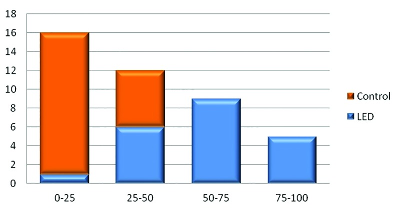
A 4-point scale evaluation by 3 observers comparing before and after pictures (baseline vs. Week 12); results statistically significant (p<0.001)
MASI. MASI score is a tool to evaluate the severity of melasma. To calculate the MASI score, the sum of the severity grade for darkness (D) and homogeneity (H) was multiplied by the numerical value of the areas (A) involved and by the percentages of the four facial areas (10–30%). Total MASI score was as follows: forehead 0.3 (D+H)A + right malar 0.3 (D+H)A + left malar 0.3 (D+H)A + chin 0.1 (D+H)A. The PBM-treated side over time went from 11.4 to 4.7 at Week 12 (p<0.001) (Figure 2).
FIGURE 2.
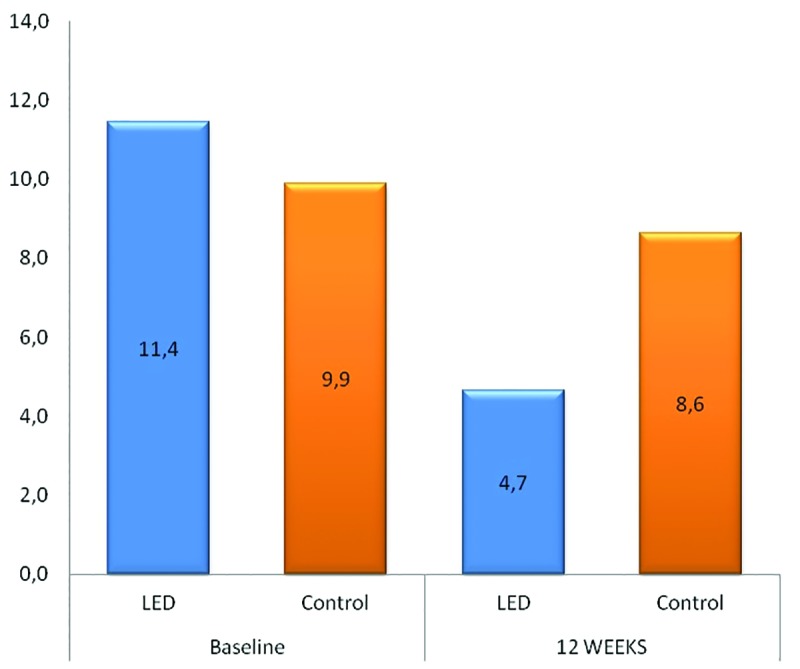
Melasma Area and Severity Index score showing a significant (p<0.001) improvement at Week 12 on the photobiomodulation-treated side
Melanin index. Although clinical improvement was clearly noticeable in the PBM-treated side, the findings of the pigment quantitative measurement (melanin index) were less impressive (Figure 3). However, the melanin index score on the PBM-treated side was significantly reduced in the majority of patients.
FIGURE 3.
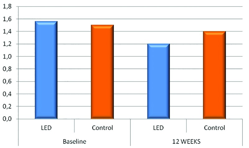
The photobiomodulation-treated side showed a 25% reduction in melanin index at Week 12 (p<0.05)
DISCUSSION
Melasma treatment remains a difficult task. Lasers in combination with topical treatments as well as chemical peels have been used in affected patients with variable results.14 A mixture of topical retinoic acid, hydroquinone, and corticosteroid applied for several weeks has been extensively used to treat melasma, showing better results than hydroquinone monotherapy.15 Unfortunately, skin irritation is very common, and long-term use might lead to exogenous ochronosis—dark blue hyperpigmentation localized where the causative agent (e.g., hydroquinone) was applied.
For deeply located dermal melasma, topical medications cannot penetrate deep enough to reach the hyperactive melanocytes. Fortunately, light-based devices such as lasers can reach the dermis. However, pigment-specific high-energy lasers might worsen melasma or induce post-inflammatory hyperpigmentation with unpredictable efficacy.16
There are many internal and external factors that determine the pigmentation dynamic process of human skin.17 Based on the premise that melasma is a dynamic acquired pigmentary disorder, as opposed to an easily treated static, benign, pigmented lesion such as lentigines, a new treatment approach is needed. In this pilot study, we selected a treatment modality (PBM) that is capable of treating dynamic processes taking place in the dermis.
Since 1968, there have been thousands of peer-reviewed articles describing PBM use.10,18 Since it promotes faster wound healing and anti-inflammatory outcomes, PBM serves as an alternative therapy through its use of specific low energy, nonthermal light parameters acting within a range of wavelengths from visible to near-infrared (NIR). PBM does not traumatize the skin, while infrared wavelengths are poorly absorbed by melanin and photons can penetrate deeply into the dermis.
Mobilization phase. Microdermabrasion. As a first step in our study, MCD was performed to open an epidermal window and mobilize a dynamic event (perivascular inflammatory infiltrate and vasodilation) prior to exposure to PBM photons (modulation phase).
Light microscopy observations on pig skin by Lee et al20 showed a reduction in the thickness and homogenization of the stratum corneum (SC) with focal compaction immediately after using a MCD device (negative pressure Al2O3 crystals) at 25cmHg (33.3KPa) for five seconds. Since we ablated the SC (corneocytes) using a stronger pressure (57Kpa=43cmHg), we probably triggered perivascular inflammatory infiltrate and vasodilation as formerly reported histopathologically following MCD.21,22 In this study, we partially removed epidermal keratinocyte layers while avoiding damage to the basal membrane layer.
Targeting signaling pathways in melanogenesis by photobiomodulation. Using cultured human melanocytes, Kim et al24 showed that NIR LED wavelengths (via PBM) effectively inhibit melanin biosynthesis and its molecular mechanisms, underlying its effects on tyrosinase, tyrosinase-related protein 1, and microphthalmia-associated transcription factor expression with an intracellular signaling pathway. These investigators showed that LED irradiation at wavelengths of 830nm, 850nm, and 940nm (96mW/cm2, 5J/cm2, 10J/cm2, and 20J/cm2; 114mW/cm2 and 1J/cm2; and 55.5mW/cm2, 5J/cm2, and 10J/cm2, respectively) effectively decreases melanin synthesis without cytotoxic effects in a normal human melanocyte monoculture and three-dimensional multiple cell type co-culture model. Based on this in-vitro study, we decided to use 940nm (to penetrate deeper in the dermis) with comparable energy parameters (fluence: 13.5J/cm2).
Another possible mechanism of action is PBM photons (at 940nm) would target p53 gene expression by downregulating hyperactive melanocytes via the p53 cell signaling pathway. The p53 cell signaling pathway plays a key role as a tumor suppressor to prevent the emergence of cancer cells. It is a transcriptional regulator and might increase deoxyribonucleic acid repair or promote apoptosis. In other words, it commands the cells to live or to die. However, it can also regulate the suntan response by p53-regulated pigmentation mechanisms, since the p53 gene is located close to the proopiomelanocortin complex.25 Since NIR PBM photons can modulate p53 gene expression,26 one can assume it could be beneficial (i.e., reduces hyperpigmentation) for an acquired pigmentary disorder like melasma.
Another p53-mediated effect, however, might have played a role in the positive response to the treatment. The effectiveness of PBM (visible and NIR) as a prophylactic measure has recently become clear.27 If nonwounded skin cells or tissues are exposed to certain wavelengths of visible and NIR light before the actual trauma, a preconditioning takes place.27 A cutaneous application of PBM, termed photoprevention, uses visible and mostly IR radiation to ready the skin for impending trauma such as UV-induced sunburn.19 Many in-vitro studies demonstrated that fibroblasts exposed to NIR light beforehand have incurred protection against future UVB damage by way of p53 cell signaling induced anti-apoptotic effects.28,29
In this study, our therapeutic goal was to use MCD and PBM to eradicate melasma. Simultaneously, we might also have preconditioned the skin with a p53 anti-apoptotic effect to better resist UV damage. In other words, we likely induced UV resistance in the skin of our subjects as we were treating them over two months. Thus, photoprevention presumably had an impact on our study, which took place in the summer months, since sun exposure is the main triggering factor of melasma.
A comparative histological study by Kang et al30 on lesional and perilesional normal skin showed the histological nature of melasma. Melanocyte numbers were comparable between lesional and perilesional skin. A total of 279 genes were found to be differentially expressed in lesional and perilesional melasma skin. They reported that vascular endothelial growth factor (VEGF) 2 was downregulated in melasma skin.
PBM has been shown to promote VEGF expression. An increase in expression was observed in the PBM-irradiated groups (904nm) in comparison within the control group in mice fibroblasts.31 Furthermore, PBM irradiation with 635nm was shown to reduce intracellular reactive oxygen species production and increase angiogenesis (enhanced VEGF expression) in human umbilical vein endothelial cells in an in-vitro oxalyl chloride-induced severe hypoxia model.32
Kang et al30 also reported that the prostaglandin metabolic process was increased in melasma. They found that prostaglandin I2 synthase and prostaglandin-endoperoxide synthase 1 were upregulated in melasma. Conversely, PBM can downregulate protein targeting to glycogen gene expression. Mizutani et al33 showed that serum prostaglandin E2 is lowered by PBM at 830nm. Additionally, Lim et al34 showed that direct PBM exposure at 635nm can inhibit the activation of pro-inflammatory mediators like prostaglandin E2 in in-vitro gingival fibroblasts. Figure 4 describes our two-step approach.
FIGURE 4.
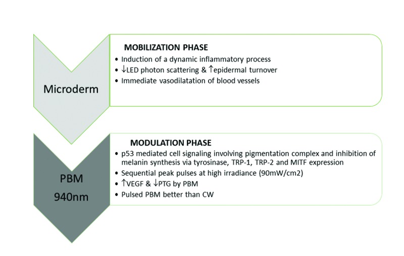
Mechanical microdermabrasion decreases photon scattering and increases epidermal turnover and blood flow (vasodilation), leading to the induction of an immediate mild inflammatory infiltrate—The mobilization phase is immediately followed by the modulation phase, since photobiomodulation works best on dynamic inflammatory processes modulating specific cell signaling pathways.
Pulsing. PBM acceptance has been slowed by the complexity of needing to choose specifics for a number of parameters, such as wavelength, dose, intensity, pulse structure, and timing of the applied light. The challenge is to gather the best combination of parameters à la carte for the condition treated. Among these, pulse structure or pulsing code seems to be an important factor to achieve optimal results.35 A review by Hashmi et al36 concluded that there is some evidence that pulsed light yields effects different from continuous wave light. In our own experience, tissue response was favorable when using sequential pulsing mode (i.e., repeated sequences of short pulse trains followed by longer intervals) rather than the continuous wave mode.37 An understanding of mitochondrial ion channels elucidates the effectiveness of the pulsing light mode as compared with the simple continuous wave in providing successful treatment. Using a transcranially positioned 810nm laser, a 10Hz pulsed mode better improved neurobehavioral recovery in patients with traumatic brain injury as compared to a continuous wave or a 100Hz pulsed mode.40 The authors of the study hypothesized that the pulse duration used in the study (100ms) is similar to a biologically occurring time period. The time period in question might be, for example, the half-life of a mitochondrial membrane ion channel or that of another membrane in the cell that responds to light.
The nitric oxide photodissociation theory might also provide a partial explanation, specifically in terms of the requirement for different pulsing patterns during LED therapy. An understanding of the light-producing mechanism of fireflies provides insight. Fireflies similarly pulse their light. A flash of light is produced when oxygen reacts with the luciferyl intermediate. The flash switches itself off though. Light dissociates nitric oxide from cytochrome c oxidase. Oxygen is then allowed to bind again. Subsequently, the mitochondria consume oxygen, enabling the luciferyl intermediate to build up until more nitric oxide arrives.41
In this study, we used a specific sequential pulsing code with a 50-percent duty cycle based on previous in-vitro studies performed in our laboratory.8,35,37 The best combination of parameters to downregulate hyperpigmentation was pulse duration (PD) set at 500µs, pulse interval (PI) at 150µs, number of pulses per pulse train (PT) set at three, and pulse train interval (PTI) at 1,550µs (Figure 5). Longer PTIs, or dark zones, might play a key role in optimizing tissue response for melasma. Some possible explanations are that several peak power pulses (at 90mw/cm2) are needed first, followed by a longer pause (PTI) not only to reduce possible tissue hyperthermia that could increase melanocytic activity but also for probable inhibition of pigment-related cell signaling pathways.
FIGURE 5.
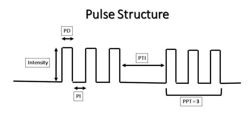
Pulsing pattern definitions—pulse duration (PD): LED on; pulse interval (PI): LED off; pulse train (PT): number of pulses/train; pulse train interval (PTI): LED off (longer interval or dark zone); in this example, PT=3
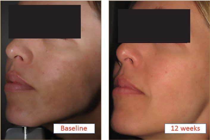
Clinical trial. This split-face pilot study with a successful response (Figure 6) to pulsed PBM brings an alternative treatment modality to melasma, which is a complex, almost untreatable, acquired pigmentary disorder. In this study, the PBM-treated side versus the control side showed statistically significant results for pigment reduction using three measurement methods (4-point scale observation by three physicians, MASI score, and melanin index score). To our knowledge, it is the first study reporting the use of PBM for melasma.
FIGURE 6.
Patient 7 at baseline and at Week 12
Limitations. There are two study limitations: the small number of patients involved and the short length of follow-up. Long-term results are key since melasma is a recurrent skin condition. For this reason, we decided to follow some patients until 12 months (n=4). For at least three of these individuals, the response was maintained (data not shown). Surprisingly, a poorly responding patient (Patient 2) during the study showed a significant comeback at Month 12, particularly on the PBM-treated side after continued (at home) weekly use of PBM on her right side. This delayed response indicates that it might take more time in some patients for improvements to show and that continued use of this treatment modality might help in building a resistance to the sun.
For practical reasons, we only performed a weekly treatment for eight weeks. However, tri-weekly treatments at home over the span of several months might be something to consider in future studies, not only to improve efficacy but also to achieve longer-term remission by treating for a longer period of time as a prophylactic modality. Even weekly treatments seemed to provide resistance to UV (photoprevention) during the course of our study, which took place in the summer.
As for wavelength, 830nm and 850nm have been shown to downregulate pigmentation equally well in vitro,23 so future studies using these wavelengths should to be considered.
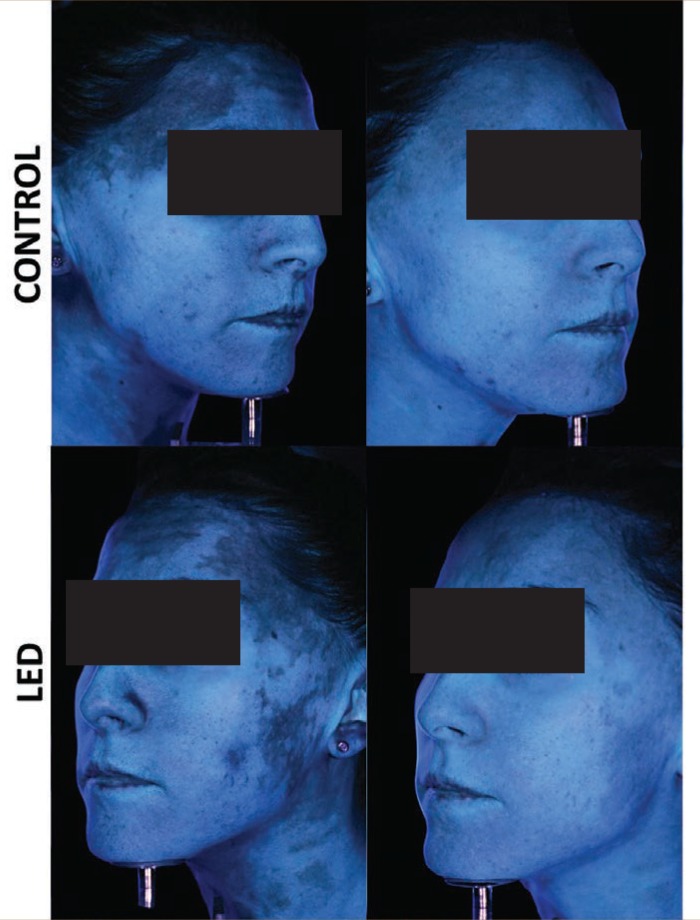
Finally, the partial improvement shown by the contralateral untreated side (Figure 7) suggests a possible response to the bilateral MCD treatment or a systemic effect of PBM. Indeed, it has been reported in the past that systemic effects of PBM might act at a site distant from the illumination site.42,43
FIGURE 7.
Patient 1 ultraviolet photography shows significant pigment reduction on the photobiomodulation-treated side (LED). This patient also obtained a partial response on the untreated control side (CONTROL), probably due to the microdermabrasion or some systemic effects of photobiomodulation.
CONCLUSION
This pilot study demonstrates that dermal melasma might be improved without tissue damage by using pulsed PBM photons. Interestingly, there might be an additional advantage to using PBM, in that it might precondition the skin, helping the patient to build a resistance to future solar UV ray exposure. This is an important finding, since the worst triggering factor in melasma is sun exposure.
In addition, the use of sequential pulsing—similar to a Morse code—seems to play a role in the response to treatment. It represents an additional way of customizing LED treatment parameters to increase tissue response and reduce dermal hyperpigmentation.
This new treatment approach is promising since, unlike topicals that are unable to reach the dermis easily, it provides a new way to reach and downregulate hyperactive dermal melanocytes without side effects. Although there are several study limitations (small sample size, n=7; no control with untreated skin; and limited long-term follow-up, n=4), the results are encouraging. Clearly, further studies are needed to validate the use of this new treatment approach and to optimize treatment parameters to ultimately better control this refractory pigmentary disorder.
ACKNOWLEDGMENTS
I am grateful to Greg Cormack for careful proofreading of this manuscript.
REFERENCES
- 1.Sofen B, Prado G, Emer J. Melasma and post inflammatory hyperpigmentation: management update and expert opinion. Skin Therapy Lett. 2016;21(1):1–7. [PubMed] [Google Scholar]
- 2.Sarkar R, Arora P, Garg VK, et al. Melasma update. Indian Dermatol Online J. 2014;5(4):426–435. doi: 10.4103/2229-5178.142484. [DOI] [PMC free article] [PubMed] [Google Scholar]
- 3.Kauvar AN. The evolution of melasma therapy: targeting melanosomes using low-fluence Q-switched neodymium-doped yttrium aluminium garnet lasers. Semin Cutan Med Surg. 2012;31(2):126–132. doi: 10.1016/j.sder.2012.02.002. [DOI] [PubMed] [Google Scholar]
- 4.Lee HM, Haw S, Kim JK, et al. Split-face study using a 1,927-nm thulium fiber fractional laser to treat photoaging and melasma in Asian skin. Dermatol Surg. 2013;39(6):879–888. doi: 10.1111/dsu.12176. [DOI] [PubMed] [Google Scholar]
- 5.Arora P, Sarkar R, Garg VK, Arya L. Lasers for treatment of melasma and post-inflammatory hyperpigmentation. J Cutan Aesthet Surg. 2012;5(2):93–103. doi: 10.4103/0974-2077.99436. [DOI] [PMC free article] [PubMed] [Google Scholar]
- 6.Letokhov VS. Laser biology and medicine. Nature. 1985;316(6026):325–330. doi: 10.1038/316325a0. [DOI] [PubMed] [Google Scholar]
- 7.Hamblin MR. Introduction to experimental and clinical studies using low-level laser (light) therapy (LLLT) Lasers Surg Med. 2010;42(6):447–449. doi: 10.1002/lsm.20959. [DOI] [PMC free article] [PubMed] [Google Scholar]
- 8.Barolet D. Light-emitting diodes (LEDs) in dermatology. Semin Cutan Med Surg. 2008;27(4):227–238. doi: 10.1016/j.sder.2008.08.003. [DOI] [PubMed] [Google Scholar]
- 9.Weiss RA, McDaniel DH, Geronemus RG, et al. Clinical experience with light-emitting diode (LED) photomodulation. Dermatol Surg. 2005;31(9 Pt 2):1199–1205. doi: 10.1111/j.1524-4725.2005.31926. [DOI] [PubMed] [Google Scholar]
- 10.Avci P, Gupta A, Sadasivam M, et al. Low-level laser (light) therapy (LLLT) in skin: stimulating, healing, restoring. Semin Cutan Med Surg. 2013;32(1):41–52. [PMC free article] [PubMed] [Google Scholar]
- 11.Weiss RA, McDaniel DH, Geronemus RG, Weiss MA. Clinical trial of a novel non-thermal LED array for reversal of photoaging: clinical, histologic, and surface profilometric results. Lasers Surg Med. 2005;36(2):85–91. doi: 10.1002/lsm.20107. [DOI] [PubMed] [Google Scholar]
- 12.Barolet D, Roberge CJ, Auger FA, et al. Regulation of skin collagen metabolism in vitro using a pulsed 660nm LED light source: clinical correlation with a single-blinded study. J Invest Dermatol. 2009;129(12):2751–2759. doi: 10.1038/jid.2009.186. [DOI] [PubMed] [Google Scholar]
- 13.aOgbechie-Godec O. A, Elbuluk N. (2017) “Melasma: an Up-to-Date Comprehensive Review.”. Dermatology and Therapy. 7(3):305–318. doi: 10.1007/s13555-017-0194-1. [DOI] [PMC free article] [PubMed] [Google Scholar]
- 14.Kang HY, Ortonne JP. What should be considered in treatment of melasma. Ann Dermatol. 2010;22(4):373–378. doi: 10.5021/ad.2010.22.4.373. [DOI] [PMC free article] [PubMed] [Google Scholar]
- 15.Kang HY, Valerio L, Bahadoran P, Ortonne JP. The role of topical retinoids in the treatment of pigmentary disorders: an evidence-based review. Am J Clin Dermatol. 2009;10(4):251–260. doi: 10.2165/00128071-200910040-00005. [DOI] [PubMed] [Google Scholar]
- 16.Ortonne JP, Passeron T. Melanin pigmentary disorders: treatment update. Dermatol Clin. 2005;23(2):209–226. doi: 10.1016/j.det.2005.01.001. [DOI] [PubMed] [Google Scholar]
- 17.Hearing VJ. The melanosome: the perfect model for cellular responses to the environment. Pigment Cell Res. 2000;13(Suppl 8):23–34. doi: 10.1034/j.1600-0749.13.s8.7.x. [DOI] [PubMed] [Google Scholar]
- 18.Mester E, Szende B, Gärtner P. The effect of laser beams on the growth of hair in mice. Radiobiol Radiother (Berl). 1968;9(5):621–626. Article in German. [PubMed] [Google Scholar]
- 19.Barolet D, Boucher A. LED photoprevention: reduced MED response following multiple LED exposures. Lasers Surg Med. 2008;40(2):106–112. doi: 10.1002/lsm.20615. [DOI] [PubMed] [Google Scholar]
- 20.Lee WR, Tsai RY, Fang CL, et al. Microdermabrasion as a novel tool to enhance drug delivery via the skin: an animal study. Dermatol Surg. 2006;32(8):1013–1022. doi: 10.1111/j.1524-4725.2006.32224.x. [DOI] [PubMed] [Google Scholar]
- 21.Freedman BM, Rueda-Pedraza E, Waddell SP. The epidermal and dermal changes associated with microdermabrasion. Dermatol Surg. 2001;27(12):1031–1033. doi: 10.1046/j.1524-4725.2001.01031.x. discussion 1033-1034. [DOI] [PubMed] [Google Scholar]
- 22.Freedman BM, Rueda-Pedraza E, Earley RV. Clinical and histologic changes determine optimal treatment regimens for microdermabrasion. J Dermatolog Treat. 2002;13(4):193–200. doi: 10.1080/09546630212345678. [DOI] [PubMed] [Google Scholar]
- 23.D’Mello SA, Finlay GJ, Baguley BC, Askarian-Amiri ME. Signaling pathways in melanogenesis. Int J Mol Sci. 2016;17(7):pii: E1144. doi: 10.3390/ijms17071144. [DOI] [PMC free article] [PubMed] [Google Scholar]
- 24.Kim JM, Kim NH, Tian YS, Lee AY. Light-emitting diodes at 830 and 850 nm inhibit melanin synthesis in vitro. Acta Derm Venereol. 2012;92(6):675–680. doi: 10.2340/00015555-1319. [DOI] [PubMed] [Google Scholar]
- 25.Oren M, Bartek J. The sunny side of p53. Cell. 2007;128(5):826–828. doi: 10.1016/j.cell.2007.02.027. [DOI] [PubMed] [Google Scholar]
- 26.Barolet D, Christiaens F, Hamblin MR. Infrared and skin: friend or foe. J Photochem Photobiol B. 2016;155:78–85. doi: 10.1016/j.jphotobiol.2015.12.014. [DOI] [PMC free article] [PubMed] [Google Scholar]
- 27.Agrawal T, Gupta GK2, Rai V, et al. Preconditioning with low-level laser (light) therapy: light before the storm. Dose Response. 2014;12(4):619–649. doi: 10.2203/dose-response.14-032.Agrawal. [DOI] [PMC free article] [PubMed] [Google Scholar]
- 28.Menezes S, Coulomb B, Lebreton C, Dubertret L. Non-coherent near infrared radiation protects normal human dermal fibroblasts from solar ultraviolet toxicity. J Invest Dermatol. 1998;111(4):629–633. doi: 10.1046/j.1523-1747.1998.00338.x. [DOI] [PubMed] [Google Scholar]
- 29.Frank S, Menezes S, Lebreton-De Coster C, et al. Infrared radiation induces the p53 signaling pathway: role in infrared prevention of ultraviolet B toxicity. Exp Dermatol. 2006;15(2):130–137. doi: 10.1111/j.1600-0625.2005.00397.x. [DOI] [PubMed] [Google Scholar]
- 30.Kang HY, Suzuki I, Lee DJ, et al. Transcriptional profiling shows altered expression of wnt pathway- and lipid metabolism-related genes as well as melanogenesis-related genes in melasma. J Invest Dermatol. 2011;131(8):1692–1700. doi: 10.1038/jid.2011.109. [DOI] [PubMed] [Google Scholar]
- 31.Martignago CC, Oliveira RF, Pires-Oliveira DA, et al. Effect of low-level laser therapy on the gene expression of collagen and vascular endothelial growth factor in a culture of fibroblast cells in mice. Lasers Med Sci. 2015;30(1):203–208. doi: 10.1007/s10103-014-1644-y. [DOI] [PubMed] [Google Scholar]
- 32.Lim WB, Kim JS, Ko YJ, et al. Effects of 635nm light-emitting diode irradiation on angiogenesis in CoCl(2)-exposed HUVECs. Lasers Surg Med. 2011;43(4):344–352. doi: 10.1002/lsm.21038. [DOI] [PubMed] [Google Scholar]
- 33.Mizutani K, Musya Y, Wakae K, et al. A clinical study on serum prostaglandin E2 with low-level laser therapy. Photomed Laser Surg. 2004;22(6):537–539. doi: 10.1089/pho.2004.22.537. [DOI] [PubMed] [Google Scholar]
- 34.Lim W, Choi H, Kim J, et al. Anti-inflammatory effect of 635 nm irradiations on in vitro direct/indirect irradiation model. J Oral Pathol Med. 2015;44(2):94–102. doi: 10.1111/jop.12204. [DOI] [PubMed] [Google Scholar]
- 35.Barolet D, Duplay P, Jacomy H, Auclair M. Importance of pulsing illumination parameters in low-level-light therapy. J Biomed Opt. 2010;15(4):048005. doi: 10.1117/1.3477186. [DOI] [PubMed] [Google Scholar]
- 36.Hashmi JT, Huang YY, Sharma SK, et al. Effect of pulsing in low-level light therapy. Lasers Surg Med. 2010;42(6):450–466. doi: 10.1002/lsm.20950. [DOI] [PMC free article] [PubMed] [Google Scholar]
- 37.Barolet D. Pulsed versus continuous wave low-level light therapy on osteoarticular signs and symptoms in limited scleroderma (CREST syndrome): a case report. J Biomed Opt. 2014;19(11):118001. doi: 10.1117/1.JBO.19.11.118001. [DOI] [PubMed] [Google Scholar]
- 38.Pogue BW, Lilge L, Patterson MS, et al. Absorbed photodynamic dose from pulsed versus continuous wave light examined with tissue-simulating dosimeters. Appl Opt. 1997;36(28):7257–7269. doi: 10.1364/ao.36.007257. [DOI] [PubMed] [Google Scholar]
- 39.Sterenborg HJ, van Gemert MJ. Photodynamic therapy with pulsed light sources: a theoretical analysis. Phys Med Biol. 1996;41(5):835–849. doi: 10.1088/0031-9155/41/5/002. [DOI] [PubMed] [Google Scholar]
- 40.Ando T, Xuan W, Xu T, et al. Comparison of therapeutic effects between pulsed and continuous wave 810-nm wavelength laser irradiation for traumatic brain injury in mice. PLoS One. 2011;6(10):e26212. doi: 10.1371/journal.pone.0026212. [DOI] [PMC free article] [PubMed] [Google Scholar]
- 41.Trimmer BA, Aprille JR, Dudzinski DM, et al. Nitric oxide and the control of firefly flashing. Science. 2001;292(5526):2486–2488. doi: 10.1126/science.1059833. [DOI] [PubMed] [Google Scholar]
- 42.Lima AA, Spínola LG, Baccan G, et al. Evaluation of corticosterone and IL-1beta, IL-6, IL-10 and TNF-alpha expression after 670-nm laser photobiomodulation in rats. Lasers Med Sci. 2014;29(2):709–715. doi: 10.1007/s10103-013-1356-8. [DOI] [PubMed] [Google Scholar]
- 43.Michel F, Barolet D. Presented at: SPIE BiOS. San Francisco: CA; Feb, 2016. A new visual analog scale to measure distinctive well-being effects of LED photobiomodulation; pp. 13–18. [Google Scholar]


