Clinical Image
Recent reports described that low-signal intensity (LSI) rims along the cerebral cortex and U-fibers adjacent to the white matter lesions of progressive multifocal leukoencephalopathy (PML) on susceptibility-weighted imaging (SWI) might be a potential new characteristic finding.1 We herein present a case of PML in which the SWI finding of LSI rims provides a clue to the early diagnosis.
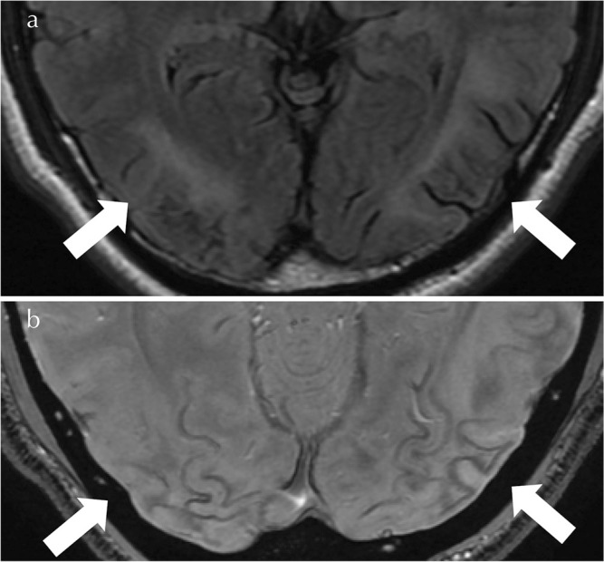
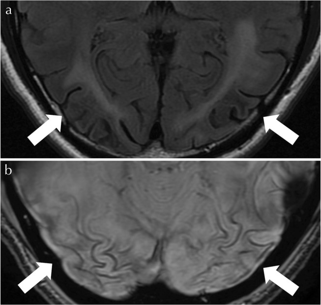
A previously healthy male in his 40s visited a nearby hospital because of acute visual impairment. Initial brain MRI revealed high signal intensities in the subcortical white matter of the bilateral occipital lobes on T2-weighted imaging (T2WI), fluid-attenuated inversion recovery (FLAIR) imaging and diffusion-weighted imaging (DWI). He was referred to a neurologist in our hospital due to his exacerbation of visual impairment. MRI at 3T (Signa Pioneer, GE Healthcare Japan, Hino, Japan) showed relatively symmetrical hyperintense lesions on T2WI and FLAIR imaging in the subcortical white matter of the bilateral occipital lobes (Fig. 1a). The lesions showed LSIs on T1-weighted imaging (T1WI) and peripheral hyperintensities on DWI. SWI revealed LSI rims along the cerebral cortex and U-fibers adjacent to the lesions (Fig. 1b), which suggested PML. No mass effect was detected in these lesions. Serological blood tests revealed human immunodeficiency virus (HIV) infection. Definitive diagnosis of PML was made by the detection of John Cunningham virus DNA in the cerebrospinal fluid. Although highly active anti-retroviral therapy was performed, visual impairment progressed from 20/500 vision to sensus luminis. MRI at 3T 2 months after the second MRI showed expanded hyperintense lesions on T2WI and FLAIR imaging (Fig. 2a), and expanded LSI rims on SWI, which showed lower signal intensities than the previous MRI. These findings were associated with exacerbation of visual impairment. Also, the sulci of the brain around the lesions expanded with time, which reflected mild progression of the lesions’ atrophy (Fig. 2b).
Fig. 1.
(a) A fluid-attenuated inversion recover (FLAIR) image (TR/TE/TI, 9600/90/2531 ms; slice thickness, 5 mm) shows relatively symmetrical hyperintense lesions in the subcortical white matter of the bilateral occipital lobes. (b) A susceptibility-weighted imaging (SWI) susceptibility-weighted angiography (SWAN) image (TR/TE, 55/22 ms; FA, 10; slice thickness, 1.4 mm) clearly reveals low-signal intensity (LSI) rims along the cerebral cortex and U-fibers adjacent to the lesions.
Fig. 2.
(a) A fluid-attenuated inversion recover (FLAIR) image shows expanded hyperintense lesions in the subcortical white matter of the bilateral occipital lobes. (b) A susceptibility-weighted imaging (SWI) image apparently reveals expanded low-signal intensity (LSI) rims along the cerebral cortex and U-fibers adjacent to the lesions. These LSI rims became lower signal intensities than the previous MRI. The sulci of brain around the lesions expanded over time, which reflected slight development of the lesions’ atrophy.
Hodel et al. reported that these susceptibility changes were observed in all patients at the chronic stage, and in relatively high frequency (75%) even at the pre-symptomatic stage.2 Umino et al. reported that LSI rims could be observed infrequently in infarct and encephalitis.3 Sensitivity, specificity and underlying cause of LSI rims on SWI remain unclear. Further investigation must be done to reveal these important issues.
In conclusion, characteristic LSI rims on SWI was identified in our case of PML. These findings apparently progressed with disease progression. Although further investigation with a larger-scale study is needed, these new findings may suggest an early diagnosis of PML and can be a helpful clue in follow-up and appropriate clinical practice.
Footnotes
Conflicts of Interest
The authors declare that they have no conflicts of interest.
References
- 1.Miyagawa M, Maeda M, Umino M, Kagawa K, Nakamichi K, Sakuma H, Tomimoto H. Low signal intensity in U-fiber identified by susceptibility-weighted imaging in two cases of progressive multifocal leukoencephalopathy. J Neurol Sci 2014; 344:198–202. [DOI] [PubMed] [Google Scholar]
- 2.Hodel J, Outteryck O, Verclytte S, et al. Brain magnetic susceptibility changes in patients with natalizumab-associated progressive multifocal leukoencephalopathy. AJNR Am J Neuroradiol 2015; 36:2296–2302. [DOI] [PMC free article] [PubMed] [Google Scholar]
- 3.Umino M, Maeda M, Ii Y, Tomimoto H, Sakuma H. Low-signal-intensity rim on susceptibility-weighted imaging is not a specific finding to progressive multifocal leukoencephalopathy. J Neurol Sci 2016; 362:155–159. [DOI] [PubMed] [Google Scholar]


