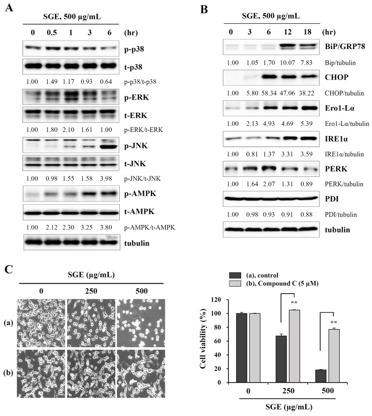
Figure 2. SGE increases the phosphorylation of MAPK and AMPK and induces ER stress.
(A) CT-26 cells were treated with 500 μg/mL SGE for 0.5, 1, 3, and 6 h, and the levels of total and phosphorylated p38, ERK, JNK, and AMPK were examined by Western blotting. (B) The levels of ER stress-related proteins were measured by Western blotting in CT-26 cells after treating with 500 μg/mL SGE for 3, 6, 12, and 18 h. The data are representative of three independent experiments, and the relative band intensities were calculated using ImageJ software after normalizing to tubulin expression. (C) Cells pretreated with or without compound C (5 μM) for 1 h were treated with 250 and 500 μg/mL SGE. After incubation for 24 h, cell viability was assessed by the CCK assay, and cell morphology was observed under an inverted microscope. **p < 0.01 vs. untreated control.

