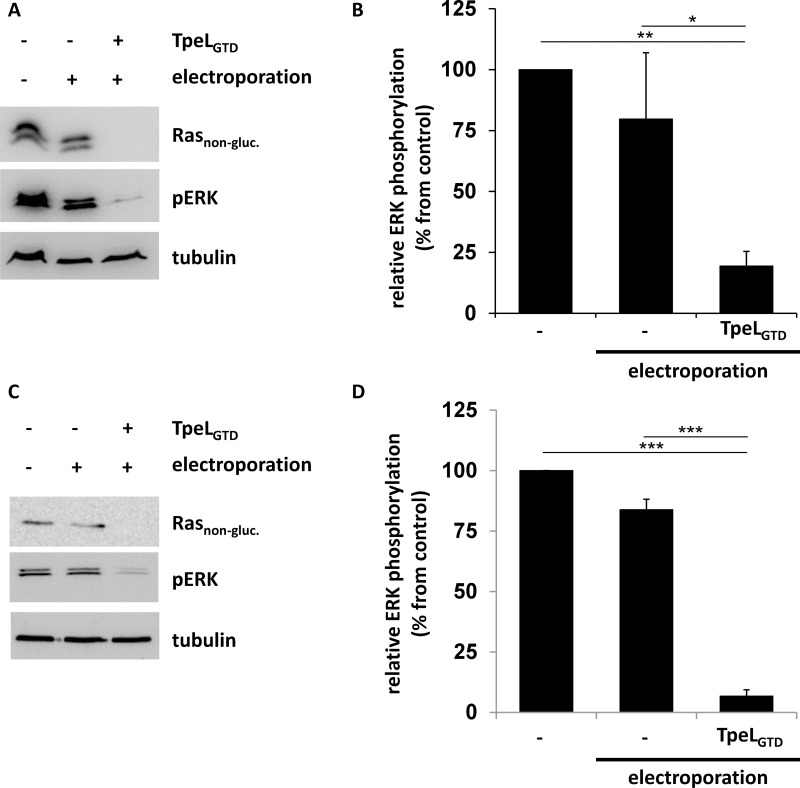
Figure 4. TpeLGTD translocated into the cytosol of host cells by electroporation is sufficient to inactivate Ras.
(A) HCT-116 cells were electroporated in the presence of TpeLGTD and plated for 2 h before lysis. Rasnon-gluc., pERK, and tubulin were probed and a representative blot of three independent experiments is shown. (B) Statistical analysis of phosphorylated ERK following electroporation treatment as presented in A. (C) Capan-2 cells were electroporated in the presence of TpeLGTD and plated for 2 h before lysis. Rasnon-gluc., pERK, and tubulin were probed and a representative blot of three independent experiments is shown. (D) Statistical analysis of phosphorylated ERK following electroporation treatment as presented in C.

