Abstract
In general, acute lower limb ischaemia is caused by embolic, thrombotic or traumatic phenomena. Here, we describe the case of a 67-year-old woman in an emergency room setting who was initially assessed for paralysis and numbness of her lower left limb. On physical examination, the abdomen was distended and non-compressible. An abdominal AngioScan showed complete occlusion of the left iliac artery by extrinsic compression of the dilated small intestine. After a review of the literature, no case was found describing a lower limb ischaemia by extrinsic vascular compression secondary to a compartment syndrome caused by small bowel obstruction. The treatment of this case required surgical decompression of the abdomen which led to an instantaneous reperfusion of the left leg. Unfortunately, the patient deceased a few hours after the surgery due to haemodynamic deterioration.
Keywords: general surgery, vascular surgery
Background
Considered as a medical emergency, acute ischaemia of the lower limb is defined as: ‘a sudden loss of lower limb perfusion’.1 A patient’s limb should be reperfused as quickly as possible to avoid amputation. Patients usually show signs of the notorious ‘five Ps’: pain, pallor, paraesthesia, paralysis and pulselessness. According to some sources, a sixth ‘P’, poikilothermia, can be added to the list. Pain is predominantly the symptom which prompts patients to seek emergency care. The most common causes of this medical emergency are embolism, thrombosis, trauma and complications related to peripheral aneurysms. Despite its rareness, extrinsic compression of the iliac artery is also a cause of acute lower limb ischaemia. The following text describes an acute case of ischaemia of the lower limb secondary to extrinsic compression of the iliac artery by a distended intestine in a patient with small bowel obstruction.
Case presentation
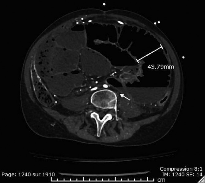
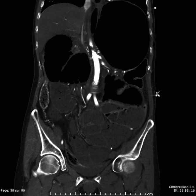
A 67-year-old woman consulted the emergency department for numbness in her left leg that had begun late in the afternoon. Her medical history includes a subarachnoid cyst along with tuberous sclerosis which had required the implantation of a ventricular shunt that was repositioned several times. She had also undergone a laparoscopic cholecystectomy. The patient had come to the emergency room suffering from paralysis, decreased sensation and paraesthesia of the lower left limb. She had waited several hours before seeking emergency care. Initial vital signs were stable. The left leg was cold and pale, and no Doppler signal was present at the dorsalis pedis, posterior tibial and femoral popliteal arteries. The abdomen was considerably distended and non-compressible and showed numerous scars, but remained insensitive on palpation nevertheless. Laboratory findings revealed increased levels of creatinine (132 µmol/L) and lactate (16.3 mmol/L). An abdominal AngioScan performed thereafter revealed the sudden stop of the opacification of the left iliac artery (figure 1) due to extrinsic compression by the dilated small intestine (figures 1 and 2). The celiac trunk was not opacified, but the formation of small collaterals nearby suggested that this could be a chronic phenomenon. The right renal artery was filiform. There was also a significant intestinal and gastric distension without a clearly identified transition point. No evidence of intestinal suffering was identified. The general surgery team was urgently contacted. Given the gravity of the findings on the AngioScan and haemodynamic deterioration of the patient, an intensive fluid resuscitation protocol and a heparin infusion were immediately started while the operating room was being prepared for immediate surgical intervention.
Figure 1.

Non-opacified left iliac artery and small bowel distention.
Figure 2.

Small bowel distention.
Investigations
See the case presentation section.
Treatment
The patient was rushed to the operating room, where the vascular surgeon joined alongside the rest of the team. Acute ischaemia of the lower left limb secondary to compression of the left iliac artery due to abdominal compartment syndrome was the diagnosis. A midline laparotomy was performed revealing an impressive distension of the bowel loops and stomach despite the insertion of a nasogastric tube. The opening of the abdominal wall was insufficient to decompress the abdominal compartment that was full of adhesions. The patient continued to deteriorate haemodynamically which prompted a decompressive gastrotomy, thereby restoring the haemodynamic stability of the patient and the perfusion within the left leg about 6 hours from the onset of symptoms. A lysis of adhesions had been undertaken to relieve the obstruction. Secondary to distension, the intestinal wall was very fragile. The surgery was complicated by an accidental enterotomy which allowed the digestive contents to spill into the abdominal cavity. The leakage was quickly controlled. The enterotomy was then used to clear the bowel of its contents, thus helping to reduce the intestinal distension. The patient also had a second episode of haemodynamic deterioration due to a secondary malignant cardiac arrhythmia. Cardioversion was performed, thus restabilising the patient. Given the precarious clinical condition of the patient, both the enterotomy and gastrotomy were primarily closed and the abdominal cavity was then irrigated thoroughly. The distal end of the ventriculoperitoneal shunt catheter was externalised at the subcutaneous passage at the upper thorax by the neurosurgical team. A temporary abdominal closure dressing was placed. Our plan was to return to the operating room for a secondary exploration, following the stabilisation of the patient. No vascular procedure or left leg fasciotomy was possible to perform due to the contamination of the surgical field by the faecal spill.
Outcome and follow-up
The patient was transferred to the intensive care unit. She subsequently developed a severe multiorgan failure showing clear signs of hyperkalemia, metabolic acidosis, hypothermia, acute renal failure, acute respiratory failure, liver failure and disseminated intravascular coagulation. After discussing with the patient’s family, treatment was discontinued given the poor prognosis and the serious consequences that might have been ensued if the patient had continued through this catastrophic episode. Unfortunately, the patient deceased a few hours after the surgery.
Discussion
An extensive review of the literature revealed very few case reports describing patients presenting acute ischaemia of the lower extremity secondary to an extrinsic compression of the iliac artery or the aorta. One particular report, however, describes a young patient who had suffered an acute aortic occlusion secondary to the extrinsic compression of a dilated intestinal loop (closed loop) contained in peritoneal encapsulation.2 This isolated situation is, in many aspects, similar to that of the 67-year-old woman. The perfusion of his lower limbs was restored after resection of the closed loop. Other rare cases of extrinsic vascular compression by external bodies include lymphangioma,3 neurofibroma,4 faecal impaction5 and leiomyoma.6
Interestingly, one other case of lower limb ischaemia secondary to an abdominal compartment syndrome has already been described. However, in this instance, significant ascites secondary to pancreatic cancer was responsible for such outcome and was quickly treated with ascites drainage.7
Nevertheless, the pathogenesis and clinical presentation of the described situation here are very unique. Abdominal compartment syndrome secondary to intestinal obstruction from abdominal adhesions, leading to the compression of the iliac artery causing acute ischaemia of the lower limb, has never been described. This case is also unique from a clinical perspective, where it is unusual for a patient to consult so late in the course of an occlusive phenomenon. Pain and paresis of the left leg were the predominant symptoms prompting the patient to seek emergency care. Therefore, we ask ourselves, why was not the abdominal issue more prominent in this patient? Could there be a link to her neurological history that would have reduced her pain threshold?
Although it was a difficult diagnosis, the intervention was quickly directed towards resolving the main cause: the abdominal compartment syndrome. The decompression of the abdomen allowed the quick restoration of vascular permeability. However, this caused a significant systemic release of inflammatory mediators and products related to ischaemia. This inevitable phenomenon was the most likely cause of haemodynamic deterioration and multiorgan failure which was subsequently the cause of death of this patient.
In conclusion, rare extrinsic compression of the iliac artery should be part of the differential diagnosis for clinicians’ assessment of patients with acute lower limb ischaemia. Regardless of the cause, abdominal compartment syndrome requires immediate emergency surgical intervention. It is important to recognise this condition as quickly as possible while not delaying the decompression of the abdomen, as this is likely the only method to restore perfusion of the ischaemic limb.
Learning points.
Acute lower limb ischaemia is generally caused by embolic, thrombotic or traumatic phenomena, but extrinsic compression of the blood vessels supplying the leg may occur.
Intestinal occlusion can cause abdominal compartment syndrome leading to the occlusion of the common iliac artery.
Acute lower limb ischaemia secondary to abdominal compartment syndrome is a medical emergency requiring surgery.
Footnotes
Contributors: All authors contributed to the conception and design of the case, the acquisition of data as well as analysis and interpretation of the data available. F-CM and PA drafted the report and F-CM, VB and VL revised the article critically for important intellectual content. All authors gave final approval of the version published.
Funding: This research received no specific grant from any funding agency in the public, commercial or not-for-profit sectors.
Competing interests: None declared.
Patient consent: Parental/guardian consent obtained.
Provenance and peer review: Not commissioned; externally peer reviewed.
References
- 1.Brunicardi F, Andersen D, Billiar T, et al. Schwartz’s principles of surgery. New York: McGraw-Hill Education/Medical, 2015. [Google Scholar]
- 2.Silva MB, Connolly MM, Burford-Foggs A, et al. Acute aortic occlusion as a result of extrinsic compression from peritoneal encapsulation. J Vasc Surg 1992;16:286–9. 10.1016/0741-5214(92)90120-W [DOI] [PubMed] [Google Scholar]
- 3.Mora R, Pozo C, Barria C, et al. Compression of the common femoral artery by a lymphangioma causing intermittent claudication. Ann Vasc Surg 2009;23:412.e11–4. 10.1016/j.avsg.2008.08.005 [DOI] [PubMed] [Google Scholar]
- 4.Pulathan Z, Imamoglu M, Cay A, et al. Intermittent claudication due to right common femoral artery compression by a solitary neurofibroma. Eur J Pediatr 2005;164:463–5. 10.1007/s00431-005-1688-x [DOI] [PubMed] [Google Scholar]
- 5.Hoballah JJ, Chalmers RT, Sharp WJ, et al. Fecal impaction as a cause of acute lower limb ischemia. Am J Gastroenterol 1995;90:2055–7. [PubMed] [Google Scholar]
- 6.Zinn HL, Abulafia O, Sherer DM, et al. Lower extremity claudication resulting from uterine leiomyoma-associated common iliac artery compression. Obstet Gynecol 2010;115:468–70. 10.1097/AOG.0b013e3181cb8f6c [DOI] [PubMed] [Google Scholar]
- 7.Maeda A, Wakabayashi K, Suzuki H. Acute limb ischemia due to abdominal compartment syndrome. Catheter Cardiovasc Interv 2014;83:141–3. 10.1002/ccd.25083 [DOI] [PubMed] [Google Scholar]


