Summary
Background
Snapping scapula syndrome, also known as scapulothoracic crepitus or bursitis, is a manifestation of a mechanical abnormality of the scapulothoracic joint. In addition to characteristic findings on physical examination, magnetic resonance imaging (MRI) exquisitely reveals soft tissue changes such as muscle edema and scapulothoracic bursitis.
Case Report
We present a case of a 10-year-old boy who had snapping scapula syndrome of the right scapula that was associated with edema of the serratus anterior muscle at the scapulothoracic interface and with scapulothoracic, specifically supraserratus, bursitis on MRI.
Conclusions
MRI in snapping scapula syndrome, which is a clinical diagnosis, exquisitely reveals soft tissue changes such as muscle edema and scapulothoracic bursitis. Such soft tissue findings of snapping scapula syndrome need to be kept in mind while evaluating routine shoulder and/or scapular region MRI, especially in the absence of relevant clinical information at the time of the imaging study.
Keywords: Bursitis, Child, Scapula
Background
Snapping scapula syndrome, a possibly underrecognized entity [1, 2], is a manifestation of abnormal biomechanics of the scapulothoracic joint. In the disease, soft tissues repetitively impinge between the scapula and the ribs, and the disorder has been predominantly reported in adults [1–10]. However, snapping scapula can also be seen in pediatric patients [2, 3, 10]. Among the underlying causes of snapping scapula syndrome are frequent trauma in adults and overuse injury in pediatric patients [3, 4]. Plain radiography [5], fluoroscopy [5, 6], computed tomography (CT) [5, 7], ultrasonography (US) [5, 9], and magnetic resonance imaging (MRI) [5] have been used in the assessment of snapping scapula. As regards pediatric patients, the imaging techniques used to evaluate snapping scapula syndrome are plain radiography and CT [3]. We describe a child with snapping scapula syndrome and abnormalities of the scapulothoracic joint and soft tissue changes on MRI. To our knowledge, MRI features of snapping scapula, not related to osteochondroma, have not been reported in pediatric patients before [10].
Case Report
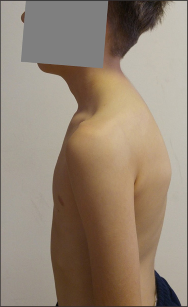
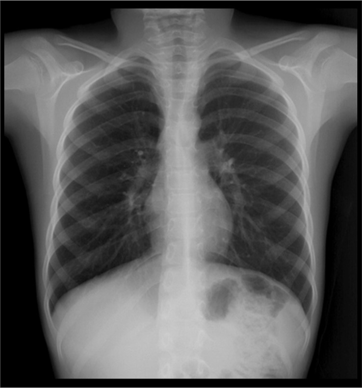
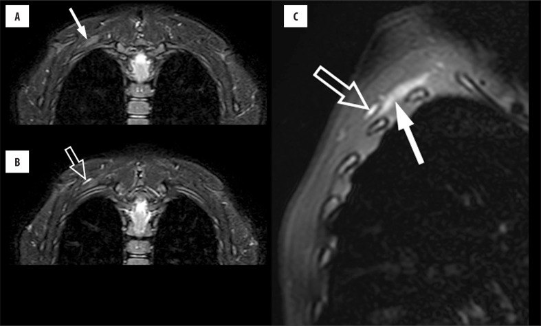
A 10-year-old boy presented with symptoms of snapping scapula syndrome of the right scapula for the last 3–4 months. Before the occurrence of snapping, the boy had reportedly started to tilt his head sideways occasionally for a couple of months to relieve a “feeling of uneasiness” at the base of the right neck–back junction. Once he started to snap his right scapula by rotating his right shoulder while his right arm remained adducted, he stopped the habit of tilting his head which was initially considered by his parents to be a tic. The boy reported that he did the snapping “from time to time” to ease the “fullness” at the base of the right neck– back junction. His medical history was otherwise unremarkable. He was right-handed and doing well in school. He played soccer almost daily. Physical examination revealed an audible and palpable crepitus during snapping, which the boy himself readily elicited (see the movie presented as online supplementary material). He had a full range of shoulder motion bilaterally. His posture was moderately disturbed, with the head tilted forwards (kyphotic) (Figure 1). Frontal chest radiograph was unremarkable and showed normally positioned scapulae (Figure 2). MRI revealed mild edema at the superior portion of the right serratus anterior muscle, supraserratus bursitis (Figure 3), and mildly increased thoracic kyphosis. His scapulae and ribs were bilaterally normal, and his cervicothoracic spinal column was otherwise unremarkable on MRI, which also showed symmetrical cervical and thoracic paraspinal muscles, with normal signal intensity, and symmetrical and otherwise normal thoracic wall musculature. In the absence of local pain and motion restriction of the right upper extremity, the patient was started on a physical therapy regimen consisting of postural correction to be followed by periscapular strengthening if necessary. The postural correction program involved manual stretching of the scalene, sternocleidomastoid, pectoral, and upper trapezius muscles, manual thoracic mobility techniques, and diaphragmatic muscle training, which was performed twice a week. Moreover, self-stretching of the same muscles and training of the diaphragm were taught to the parents as a home exercise program.
Figure 1.
The patient’s posture whilst standing is characterized by head leaning forwards (kyphotic).
Figure 2.
Frontal chest radiograph shows symmetrical and normally positioned scapulae.
Figure 3.
Coronal STIR (A, B) and oblique sagittal fat-saturated T2-weighted (C) MR images show edema-like signal (solid arrows, A and C) within the right serratus anterior muscle between the scapula and ribs, and supraserratus bursitis (open arrows, B and C) between the serratus anterior and subscapularis muscles.
Discussion
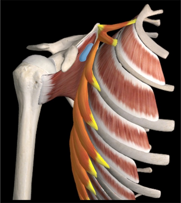
Snapping scapula is the result of bony or soft tissue abnormalities at the scapulothoracic interface (Figure 4) that hinder normal gliding of the scapula along the chest wall. In the absence of an osteochondroma as the causative factor, radiographs and CT are usually normal [5], although three- [2] or four-dimensional CT [7] may show the snapping site. US may not only show scapulothoracic bursitis [8], which may be associated with snapping scapula syndrome, but may also provide guidance for steroid/anesthetic injections of the involved bursa [8]. MRI is the imaging modality of choice to show soft tissue changes such as muscle edema and/or scapulothoracic bursitis [5], as in our case, and to depict potential osteochondromas. Scapulothoracic bursitis in snapping scapula syndrome may occur as an overuse injury from repetitive friction along the thoracic wall muscles and the subscapularis muscle that is located just anterior to the scapular blade [1, 5, 8]. Muscle edema on MRI, which was also present in our case, is likely caused by the same mechanism. Scapulothoracic bursitis is characterized by fluid distention of one or more of the several bursae (e.g., supraserratus, infraserratus bursae) that normally facilitate gliding of the scapula along the scapulothoracic joint.
Figure 4.
Illustration depicts parts of the scapulothoracic joint relevant to the case reported herein. The external intercostal muscles are between the ribs. The muscle group highlighted in orange-yellow is the serratus anterior. Superiorly, between the serratus anterior and the subscapularis, which is anteriorly adjacent to the scapula, is the supraserratus bursa (blue). (Adapted from Essential Anatomy 5 App with permission of 3D4Medical.com).
Moderate to severe forward head position, suggesting increased kyphosis, is associated with scapulothoracic bursitis and snapping scapula [9]. Our patient also had a moderately kyphotic posture, and correcting the postural abnormality was planned as the first-line physical therapy regimen. Treatment of snapping scapula syndrome in children and adolescents also involves scapular stabilization and periscapular strengthening with physical therapy and home exercises for a minimum of three months [3]. Most patients respond well to non-operative management, and surgical treatment is reserved for patients with persistent pain and snapping scapula after a trial of physical therapy [3]. Surgical approach entails arthroscopic lysis of adhesions or mini-open lysis of adhesions with scapular superomedial corner excision and bursectomy [3].
Conclusions
MRI in snapping scapula syndrome, which is a clinical diagnosis, exquisitely reveals soft tissue changes such as muscle edema and scapulothoracic bursitis. Such soft tissue findings associated with snapping scapula syndrome need to be kept in mind while evaluating routine shoulder and/or scapular region MRI, especially in the absence of relevant clinical information at the time of imaging.
Conflict of interest
The authors declare that they have no conflict of interest.
References
- 1.Warth RJ, Spiegl UJ, Millett PJ. Scapulothoracic bursitis and snapping scapula syndrome: A critical review of current evidence. Am J Sports. 2015;43:236–45. doi: 10.1177/0363546514526373. [DOI] [PubMed] [Google Scholar]
- 2.Kuhne M, Boniquit N, Ghodadra N, et al. The snapping scapula: Diagnosis and treatment. Arthroscopy. 2009;25:1298–311. doi: 10.1016/j.arthro.2008.12.022. [DOI] [PubMed] [Google Scholar]
- 3.Haus B, Nasreddine AY, Suppan C, Kocher MS. Treatment of snapping scapula syndrome in children and adolescents. J Pediatr Orthop. 2016;36:541–47. doi: 10.1097/BPO.0000000000000486. [DOI] [PubMed] [Google Scholar]
- 4.Pearse EO, Bruguera J, Massoud SN, et al. Arthroscopic management of the painful snapping scapula. Arthroscopy. 2006;22:755–61. doi: 10.1016/j.arthro.2006.04.079. [DOI] [PubMed] [Google Scholar]
- 5.Lazar MA, Kwon YW, Rokito AS. Snapping scapula syndrome. J Bone Joint Surg Am. 2009;91:2251–62. doi: 10.2106/JBJS.H.01347. [DOI] [PubMed] [Google Scholar]
- 6.Oizumi N, Suenaga N, Minami A. Snapping scapula caused by abnormal angulation of the superior angle of the scapula. J Shoulder Elbow Surg. 2004;13:115–18. doi: 10.1016/s1058-2746(03)00178-2. [DOI] [PubMed] [Google Scholar]
- 7.Bell SN, Troupis JM, Miller D, et al. Four-dimensional computed tomography scans facilitate preoperative planning in snapping scapula syndrome. J Shoulder Elbow Surg. 2015;24:83–90. doi: 10.1016/j.jse.2014.09.020. [DOI] [PubMed] [Google Scholar]
- 8.Huang CC, Ko SF, Ng SH, et al. Scapulothoracic bursitis of the chest wall: sonographic features with pathologic correlation. J Ultrasound Med. 2005;24:1437–40. doi: 10.7863/jum.2005.24.10.1437. [DOI] [PubMed] [Google Scholar]
- 9.Conduah AH, Baker CL, 3rd, Baker CL., Jr Clinical management of scapulothoracic bursitis and the snapping scapula. Sports Health. 2010;2:147–55. doi: 10.1177/1941738109338359. [DOI] [PMC free article] [PubMed] [Google Scholar]
- 10.Frost NL, Parada SA, Manoso MW, et al. Scapular osteochondromas treated with surgical excision. Orthopedics. 2010;33:804. doi: 10.3928/01477447-20100924-09. [DOI] [PubMed] [Google Scholar]


