Abstract
Aims:
Both fluoroscopic water-soluble contrast swallow (FWSCS) and CT water-soluble contrast swallow (CTWSCS) are widely performed as a routine in the post-esophagectomy patient to assess for anastomotic leak. Several prospective studies have compared FWSCS and CTWSCS; however, no synthesis of the data exists.
Materials and Methods:
Systematic review and meta-analysis of diagnostic test accuracy studies comparing FWSCS and CTWSCS in the adult patient following esophagectomy for malignancy was performed in accordance with PRISMA guidelines.
Results:
Three diagnostic test accuracy studies met the inclusion criteria, directly comparing FWSCS and CTWSCS in 185 patients. FWSCS demonstrated high specificity (98%), but low sensitivity (64%). CTWSCS can be categorized as normal, mediastinal gas without contrast leak, or leakage of oral contrast. Visible leakage of oral contrast demonstrated high specificity (98%) but low sensitivity (56%). The presence of mediastinal gas increased sensitivity (84%), but reduced specificity (85%). The higher sensitivity of CTWSCS over FWSCS failed to reach significance (P = 0.125).
Conclusion:
CTWSCS shares the high specificity of FWSCS. Its higher sensitivity increases its utility as a rule-out test in the postoperative period. Additional factors that may influence decision-making are described.
Keywords: Anastomotic leakage, computed tomography, esophagectomy, fluoroscopy
Introduction
Esophageal cancer affects over 450,000 people worldwide each year, ranking eighth among cancer incidence. The incidence of primary esophageal cancer continues to increase.[1] The role and choice of imaging in the postoperative period remains unclear. The primary imaging modalities for assessment of anastomotic integrity are fluoroscopic water-soluble contrast swallow (FWSCS), and more recently computed tomography water-soluble contrast swallow (CTWSCS). It remains uncertain whether FWSCS or CTWSCS represents a superior test. Both tests have been well described separately, with similar sensitivities and specificities and without clear superiority of either modality. Several studies have directly compared diagnostic test accuracy, however, to date no synthesis of available randomized controlled trials is available to guide decision-making. The aim of this study was a meta-analysis of available comparative diagnostic test accuracy studies, comparing CT and fluoroscopic swallow in the assessment of post-esophagectomy anastomotic leak.
Materials and Methods
A systematic review was performed to investigate the relative diagnostic test accuracies of both commonly performed post-esophagectomy imaging modalities. Preferred reporting items for systematic reviews and meta-analyses (PRISMA) was selected as a structured approach to the systematic meta-analysis of randomized trials.[2] The population was adult patients following esophagectomy for primary esophageal malignancy. The intervention was CTWSCS compared with conventional FWSCS. The outcomes of interest were sensitivity and specificity for esophageal anastomotic leak. Inclusion criteria were prospective diagnostic test accuracy studies comparing both FWSCS with CTWSCS. Studies where the two examinations were performed in each patient were included. Studies where only one examination was performed were excluded. Studies of Centre for Evidence Based Medicine (CEBM) evidence level 4 or lower (such as case reports) were excluded.[3] Studies where a published full text English-language translation was not available were excluded. Nonpublished studies were excluded, and authors were not contacted for nonpublished information. Data from all studies were extracted from each publication, and pooled for analysis, which was performed using Prism 7 (version 7.0; GraphPad Software, La Jolla, CA, USA).
Results
Search results
MEDLINE PubMed search was performed using the MESH terms esophagectomy, fluoroscopy CT, and anastomotic leak at study initiation in May 2015, and repeated prior to analysis in 2016. Of 10,961 publications addressing esophagectomy, two studies met all search terms and adhered to the inclusion and exclusion criteria.[4,5] Reverse citation tracking and alternate engines using identical search terms (Google Scholar, TRIP, Cochrane Database, and ClinicalTrilas.gov) revealed one further study adhering to the inclusion and exclusion criteria [Figure 1].[6]
Figure 1.
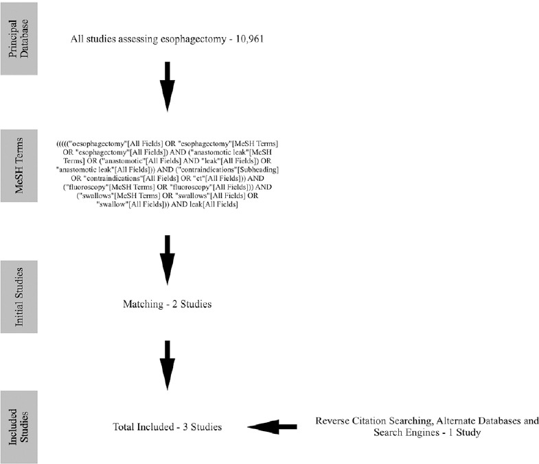
Search Strategy
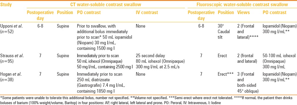
Study technique
In all studies, both tests were performed on each patient at day 7, with scan technique as described [Table 1]. All studies reported binary results for the presence of leakage on FWSCS. With regard to CTWSCS, one study did not permit identification of cases where mediastinal gas alone was identified, and while included in the overall analysis, was excluded from certain subgroup analyses.[4] The remaining studies allowed for further categorization of CTWSCS as normal, mediastinal gas without contrast leakage, or frank contrast leakage. All readers were blinded. Each study was analyzed separately, and pooled data were analyzed. Pooled data assessed the presence or absence of leak on FWSCS. Pooled data for CT assessed the presence or absence of leak. Furthermore, the presence and absence of mediastinal gas on CT was also assessed. This permitted analysis of three CT subgroups describing the commonly encountered scenarios: (1) Neither mediastinal air or gas was seen [normal study], (2) Mediastinal gas without leakage of oral contrast [abnormal study], and (3) Anastomotic leakage of oral contrast.
Table 1.
Study technique
Study results
The eligible studies enrolled 187 patients. 74.3% were males (n = 139) and 25.7% were females (n = 48). Of these 187 patients, two patients dropped out (gender not specified), with 185 patients undergoing both FWSCS and CTWSCS following esophagectomy for malignancy. Mean patient age was 64.5 years (range 28–85). 15.5% (n = 20) of esophagectomies were cervical anastomoses and 84.5% (n = 129) were mediastinal anastomoses. The level of anastomoses was not specified in the remaining 36 patients. Anastomotic leakage rate was 13.5% (25 of 185). Individual study data and forest plots are described for all studies and pooled data [Figure 2]. Summary statistics were calculated [Table 2].
Figure 2.
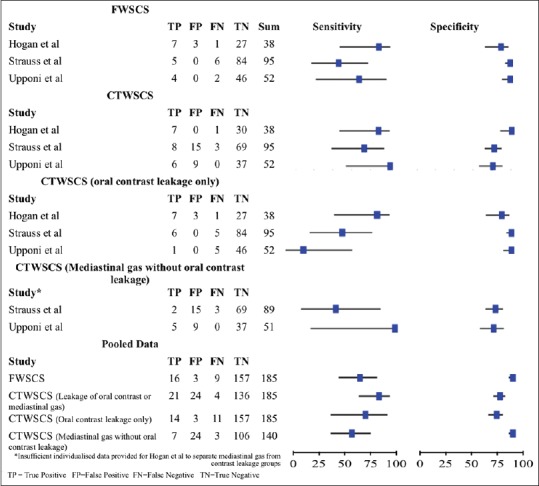
Plot of individual study results. *Insufficient individualised data provided for Hogan et al to separate mediastinal gas from contrast leakage groups. TP: True Positive, FP: False Positive, FN: False Negative, TN: True Negative, FWSCS: Fluoroscopic Water Soluble Contrast Swallow, CTWSCS: CT Water Soluble Contrast Swallow
Table 2.
Comparative results of diagnostic test accuracy

When the presence of either mediastinal air or contrast leakage on CTWSCS was considered positive for leakage, the sensitivity of CTWSCS (84%) was 20% higher than FWSCS (64%); however, this failed to reach significance (one-tailed McNemar test, P = 0.125). The specificity of FWSCS (98%) was 13% higher than that of CTWSCS (85%), which was significant (P = 0.000025).
Leakage of oral contrast on CTWSCS, irrespective of mediastinal gas, demonstrated 98% specificity for anastomotic leakage, not significantly different from that of FWSCS at 98% (P = 0.125). Using only the leakage of oral contrast for diagnosis reduced the sensitivity of CTWSCS from 84% to 56%; this was also not significantly different from that of FWSCS (P = 0.250).
When mediastinal gas was seen on CTWSCS without leakage of oral contrast, sensitivity and specificity for the detection of anastomotic leak were 70 and 82%, respectively. Among the 31 patients with mediastinal gas but no leakage of oral contrast on CTWSCS, there were 7 anastomotic leaks, of which 4 were evident on FWSCS. Thus among patients with mediastinal gas but no contrast leak, the incidence of anastomotic leak was 22.6%, greater than the overall incidence of 13.5%. Where gas is seen on CTWSCS, additional performance of a FWSCS revealed 57.1% of anastomotic leaks.
Risk of bias
A structured assessment of bias was performed in accordance with the QUADAS2 (Quality Assessment of Diagnostic Accuracy Studies) guidelines.[7] All studies stated consecutive prospective enrolment, and diagnostic tests were performed within one day of each other, at 7 days from surgery. Each patient underwent both tests, thus acting as their own control. Inclusion and exclusion criteria were homogenous. In each case, the diagnostic imaging was interpreted by a blinded radiologist, without knowledge of clinical status or of biochemical, endoscopic or alternative imaging results. Regardless of radiologist blinding to patients status, several secondary signs of clinical status cannot be blinded: patient's posture, the presence of additional hardware (such as central lines, tubes, and drainage catheters), atelectasis, or adjacent lung findings. These are all hallmarks of the unwell patient seen on both FWSCS and CTWSCS. As a result, the authors attribute a low-to-medium risk of bias in this regard. While further bias on behalf of the surgeon may have been present, it was considered unlikely to significantly influence the results of this diagnostic test accuracy study.
The reference standard in all studies was clinical exclusion of leak following resumption of oral feeding and successful patient discharge. In certain cases endoscopy and thoracotomy was used. This introduces a risk of bias as and potentially present subtle anastomotic leaks could have been missed on imaging. These would be, by definition, not clinically significant however. Several patients in all studies were initially enrolled, but due to clinical deterioration some required accelerated imaging or thoracotomy (3.6%, n = 7), hence these were excluded. As the studies were undertaken to compare the accuracy of routine post-esophagectomy imaging in ruling out leakage, these patients are not relevant to the clinical question. Given the small numbers, and doubtful relationship to the study question, the risk of bias is considered low.
Patient cohort, underlying pathology, and operative outcomes may vary between institutions. All studies were single-center large tertiary referral high-volume centers in Western European or American patients, and true leakage rates were narrowly distributed. While such variations may introduce heterogeneity, the risk of bias was felt to be low.
Technical parameters for diagnostic examinations varied; the oral contrast regimes, the use of intravenous contrast at CT, and the number of projections viewed at fluoroscopy varied [Table 1]. Only one study described CT acquisition kV and mA.[5] Despite this, overall techniques remain broadly aligned between studies, with a resulting low risk of bias.
Discussion
Following esophagectomy for malignancy, the presence of contrast leakage on routine (day 7) postoperative FWSCS and CTWSCS is 98% specific for anastomotic leakage in both tests. Contrast leakage lacks sensitivity however, 64% on FWSCS and 56% on CTWSCS. CT allows for the assessment of mediastinal gas, which is not typically appreciable on fluoroscopy. The presence of mediastinal gas alone on CTWSCS, even in the absence of leakage of contrast, is 70% sensitive and 82% specific. Combining assessment for contrast leakage or mediastinal gas allows for a sensitivity of 84% and a specificity of 85%.
Among the 31 patients with mediastinal gas but no leakage of contrast on CTWSCS, there were 7 anastomotic leaks, of which 4 were evident on FWSCS. Thus among patients with mediastinal gas, the incidence of anastomotic leak was 22.6%, greater than the baseline incidence of 13.5%. If gas is seen on CTWSCS, subsequent performance of a FWSCS reveals 57.1% (4/7) of these occult leaks.
The role of routine imaging in the postoperative period is as a rule-out test, to exclude an anastomotic leak where absent prior to commencement of feeding, and to facilitate early intervention when present. Where CTWSCS was normal, without either mediastinal gas or leakage, only 2.7% (3/109) of patients demonstrated subsequent anastomotic leak. FWSCS was also negative in these three cases. The routine performance of FWSCS in addition to negative CTWSCS does not appear to add value.
Encompassing the above results, an algorithm for the radiological screening of anastomotic leakage is proposed [Figure 3]. Following this algorithm permitted confident diagnosis in 79% of cases where CTWSCS showed either a clear contrast leak, or no mediastinal air, with only three false positives and no false negatives. In the remaining 21% of patients, representing those with mediastinal gas but no leakage of contrast on either CTWSCS or FWSCS, there is an 11.1% leak rate. In this group, the possibility of anastomotic leakage must remain a consideration, requiring a persisting high degree of clinical suspicion, and expectant management with biochemical, endoscopic or clinical correlation may be an appropriate strategy.
Figure 3.
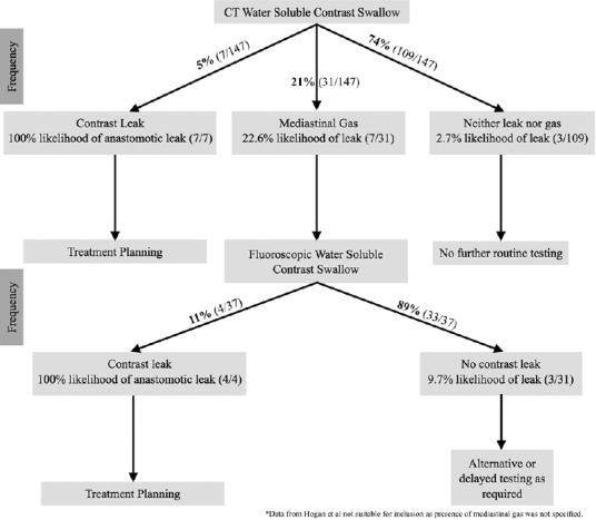
Suggested algorithm for the exclusion of anastomotic leakage. [Patients from Strauss et al (n=95) and Upponi et al (n=52) included, totalling 147 patients. Reported data from Hogan et al (n=38) did not permit individual case assessment]
Post-esophagectomy imaging is primarily performed to identify anastomotic leaks, in order to identify those patients who may require more intensive management or additional intervention. As such, the higher sensitivity of CTWSCS to leakage and its resulting higher negative predictive value suggest it is a more useful test in permitting the radiologist to exclude an anastomotic leak. A caveat remains that even with the more sensitive CTWSCS, 17.6% of anastomotic leaks were radiologically occult, demonstrating neither contrast leakage nor mediastinal gas.
False positive detection of contrast leakage was uncommon, reflecting the relatively specific appearance of iodinated contrast leakage into the mediastinum. Upponi et al. reported one case of hyperdense perianastamotic hematoma mimicking iodinated contrast, however, this was subsequently recognized and correctly diagnosed. Mediastinal gas alone lacks specificity, while perianastamotic gas in the late preoperative period is suggestive of leakage, the rate of gas resorption from surgery varies and even at one postoperative week, a cohort of patients with an intact anastomosis will still have mediastinal air, which is therefore presumed operative. Clearly the presence of an associated leakage of contrast strongly favors a true positive result. False negative results occurred in patients with no radiological leakage, but a leak became clinically apparent or was identified on alternate modalities, including endoscopy and thoracotomy. While the exact cause for missed leakage is uncertain, potential causes include diagnostic error, a small leak where the volume of oral contrast may be undetectable on imaging, a transient leak which may remain closed for a small bolus of fluid but open for a larger/solid bolus, or delayed anastomotic breakdown occurring after imaging in the postoperative course.
Prior studies have described a greater cervical leakage rate in cervical anastomoses as compared with mediastinal (intrathoracic) anastomoses (12.3 versus 9.3%).[8] While all three analyzed studies reported the type of anastomosis, individual results by anastomosis type were not presented, preventing subgroup analysis.
Recent separate studies by Cools-Lartigue et al.[9] and Tirnaksiz et al.[10] refuted the role for post-esophagectomy fluoroscopic imaging; however, this conclusion is based on poorer sensitivities then demonstrated in the included studies (40.4 and 45.5%, respectively). Given the higher pooled sensitivity described above with the use of CTWSCS (84%), the associated higher negative predictive value may justify the use of this screening test, given the reasonably common occurrence of post-esophagectomy anastomotic leakage (n = 25, 16%).
The use of CTWSCS is associated with a higher rate of false positives (15%), predominantly gas-related. It is important that the reporting radiologist is aware of this high rate of false positives and communicates this along with any abnormal report. In contemporary practice, these findings are often followed with endoscopy or delayed repeat imaging as dictated by the condition of the patient, particularly in the absence of clinical and biochemical signs of thoracomediastinal sepsis. This lower specificity of CT is likely due to a combination of factors. Certain patients will demonstrate residual air at 7 days without an associated true leakage, which may be misinterpreted as a possible leak. Additionally, one case of hyperdense extramural hematoma was initially interpreted as possible contrast leakage on CTWSCS, although it was recognized at the time.[5] This is rarely a diagnostic dilemma, due to typically differing densities of hematoma and contrast. In cases of doubt, where there is concern of a small leakage of contrast mixing in a perianastamotic fluid collection, or where contrast is too dilute approaching hematoma density, a noncontrast phase of CT or FWSCS can be considered for confirmation.
Further factors may influence choice of imaging modality [Table 3]. Many institutions will routinely perform CT scans of the thorax and upper abdomen in patients where a leak has been demonstrated or is strongly suspected, as this provides additional information on the presence of drainable collections, or fistulation into the adjacent organs. With the increasing use of CT-guided drainage, covered stent insertion and advanced endoscopic therapy, there is a greater scope for conservative management, and this additional CT-provided information is valuable in informing any such decisions. Whilst this is not reflected in the presented diagnostic test accuracy data, it is an advantage of CT.
Table 3.
Nondiagnostic differences between tests
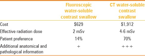
Upponi et al.[5] demonstrated a patient preference for CTWSCS in 70% of patients, with only 14% of patients preferring FWSCS, and 16% expressing no preference. Similarly, Strauss et al.[6] reported greater patient tolerability and acceptability in the CTWSCS. The reason for this preference was not further investigated, possible causes may include the ability of CT to be performed supine, the smaller volumes of contrast required, and the shorter duration of a CT scan. The radiation dose is higher with CTWSCS (4–6 mSv) than with FWSCS (2 mSv), which is a further consideration particularly in younger patients, although the total doses are within the same order of magnitude.[11,12] Furthermore, CT of the thorax and upper abdomen costs an average of $1,912 in the US, while upper abdominal contrast study cost is $629.[13] Clearly, this is subject to wide variation across sites, however, this increased cost of CT is a further consideration for healthcare providers and patients.
The primary limitation of this study is the number of enrolled patients across all studies; 185 patients underwent both tests contemporaneously with a reference standard. Analysis of the combined results reached significance in several measures (including specificity); importantly, however, trends in sensitivity failed to reach significance. Separating a lack of study-power from and a lack of significance is important, and the study authors note that current meta-analysis falls short of statistical significance in several key areas.
Among post-esophagectomy patients, the presence of contrast leakage on routine FWSCS and CTWSS is highly specific for leakage (both 98%). The sensitivity of FWSCS (64%) renders it suboptimal as a screening test. The sensitivity of CTWSCS (84%), while higher than FWSCS, failed to reach significance (P = 0.125), which may reflect low patient numbers. The high negative predictive value of CTWSCS (97%) suggests that where a test is required post-esophagectomy to exclude anastomotic leak, the trend toward greater sensitivity favors this is test.
Financial support and sponsorship
Nil.
Conflicts of interest
There are no conflicts of interest.
References
- 1.Zhang Y. Epidemiology of esophageal cancer. World J Gastroenterol. 2013;19:5598. doi: 10.3748/wjg.v19.i34.5598. [DOI] [PMC free article] [PubMed] [Google Scholar]
- 2.Moher D, Liberati A, Tetzlaff J, Altman DG. Preferred reporting items for systematic reviews and meta-analyses: The PRISMA statement. BMJ. 2009:339. [PMC free article] [PubMed] [Google Scholar]
- 3.Howick J CI, Glasziou P, Greenhalgh T, Heneghan C, Liberati A, Moschetti I, OCEBM Table of Evidence Working Group et al. The 2011 Oxford CEBM Levels of Evi-dence (Introductory Document, Background Document, Table) Oxford Centre for Evidence-Based Medicine. 2011 [Google Scholar]
- 4.Hogan BA, Winter DC, Broe D, Broe P, Lee MJ. Prospective trial comparing contrast swallow, computed tomography and endoscopy to identify anastomotic leak following oe-sophagogastric surgery. Surg Endosc. 2008;22:767–771. doi: 10.1007/s00464-007-9629-6. [DOI] [PubMed] [Google Scholar]
- 5.Upponi S, Ganeshan A, D'Costa H, Betts M, Maynard N, Bungay H, et al. Radiologi-cal detection of post-oesophagectomy anastomotic leak—a comparison between multidetec-tor CT and fluoroscopy. Br J Radiol. 2008;81:545–8. doi: 10.1259/bjr/30515892. [DOI] [PubMed] [Google Scholar]
- 6.Strauss C, Mal F, Perniceni T, Bouzar N, Lenoir S, Gayet B, et al. Computed tomog-raphy versus water-soluble contrast swallow in the detection of intrathoracic anastomotic leak complicating esophagogastrectomy (Ivor Lewis): A prospective study in 97 patients. Ann Surg. 2010;251:647–51. doi: 10.1097/SLA.0b013e3181c1aeb8. [DOI] [PubMed] [Google Scholar]
- 7.Whiting PF, Rutjes AW, Westwood ME, Mallett S, Deeks JJ, Reitsma JB, et al. QUADAS-2: A revised tool for the quality assessment of diagnostic accuracy studies. Ann Internal Med. 2011;155:529–36. doi: 10.7326/0003-4819-155-8-201110180-00009. [DOI] [PubMed] [Google Scholar]
- 8.Kassis ES, Kosinski AS, Ross P, Jr, Koppes KE, Donahue JM, Daniel VC. Predictors of anastomotic leak after esophagectomy: An analysis of the society of thoracic surgeons general thoracic database. Ann Thorac Surg. 96:1919–26. doi: 10.1016/j.athoracsur.2013.07.119. [DOI] [PubMed] [Google Scholar]
- 9.Tirnaksiz MB, Deschamps C, Allen MS, Johnson DC, Pairolero PC. Effectiveness of screening aqueous contrast swallow in detecting clinically significant anastomotic leaks after esophagectomy. Eur Surg Res. 2005;37:123–8. doi: 10.1159/000084544. [DOI] [PubMed] [Google Scholar]
- 10.Cools-Lartigue J, Andalib A, Abo-Alsaud A, Gowing S, Nguyen M, Mulder D, et al. Routine contrast esophagram has minimal impact on the postoperative management of pa-tients undergoing esophagectomy for esophageal cancer. Ann Surg Oncol. 2014;21:2573–9. doi: 10.1245/s10434-014-3654-1. [DOI] [PubMed] [Google Scholar]
- 11.Lungren MP, Yoshizumi TT, Brady SM, Toncheva G, Anderson-Evans C, Lowry C, et al. Radiation dose estimations to the thorax using organ-based dose modulation. AJR Am J Roentgenol. 2012;199:W65–73. doi: 10.2214/AJR.11.7798. [DOI] [PubMed] [Google Scholar]
- 12.Wambani JS, Korir GK, Tries MA, Korir IK, Sakwa JM. Patient radiation exposure during general fluoroscopy examinations. Med Imaging. 2014;15:4555. doi: 10.1120/jacmp.v15i2.4555. [DOI] [PMC free article] [PubMed] [Google Scholar]
- 13.MDSave.com. [website] MDSave Incorporated. Brentwood, Tennessee: [Last accessed on 2016 Nov 30]. Updated 30/11/16. [Google Scholar]


